The most common is diagnostic testing outside of pregnancy, which can be carried out using several methods:
- Transvaginal. Examination of the uterus and other organs using this method allows you to diagnose the female genital area as accurately as possible. It is carried out by introducing a special sensor into the vagina.
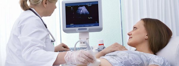
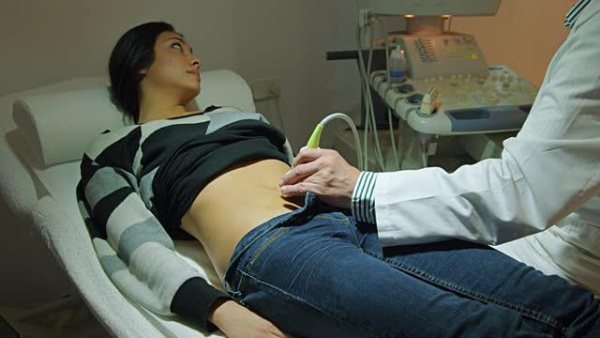
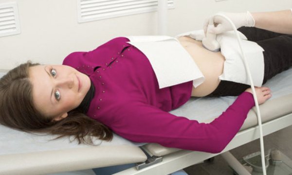
- Transabdominal. The doctor prescribes this test for a woman of any age with an intact hymen. With its help, serious pathology of the pelvic organs is detected. It is carried out through the abdominal wall.
- Transrectal. Diagnosis of women is carried out through the rectum. Allows you to obtain no less comprehensive information than with the transvaginal method. It is carried out mainly in virgins.
There is another type of diagnosis - folliculometry.
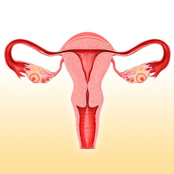
In this case, only the ovaries are examined for the maturation of follicles in them. Folliculometry in almost all cases is performed transvaginally. An ultrasound of the uterus is also performed during pregnancy, during which both the fetus itself and the condition of the uterus, cervix, fallopian tubes and appendages are examined. In the 1st trimester of pregnancy it is performed transvaginally, and in the remaining trimesters – transabdominally. The price of each method depends on its complexity.
On what day of the cycle is an ultrasound of the uterus and appendages performed?
Typically, an ultrasound of the female reproductive system is performed in the absence of menstruation. First of all, because it will be much more difficult for a diagnostician to examine the uterine cavity and assess the condition of the endometrium due to bloody clots. Various neoplasms are difficult to see during menstruation. Accordingly, the diagnosis may be incorrect.
An echography of the uterus and its adnexal structures is prescribed, as a rule, on days 5–10 of the menstrual cycle, when the vast majority of women have already had their periods. A new mucous membrane is formed in the uterus. At the time of the examination, it has not yet hardened, and therefore it can be examined qualitatively, pathological formations can be excluded or confirmed.
Another reason why it is advisable to postpone a planned examination of the organs of the reproductive system until the end of menstruation is psychological discomfort - it is unpleasant for a woman to see a doctor on “special” days.
Not least important is the issue of hygiene. In addition, during a transvaginal examination, the insertion of a special sensor may cause some pain, while on other days the manipulation does not provoke pain.
Ultrasound of the pelvic organs. When should a woman perform it and how to prepare?
Ultrasound of the pelvic organs is a mandatory, annual and safe procedure for women, which allows you to assess the condition of the organs quickly and accurately!
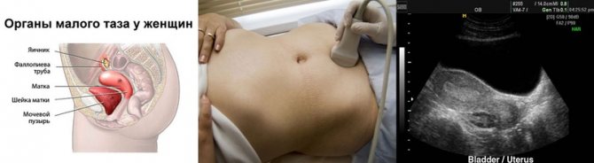
Diseases of the pelvic organs can already manifest themselves in adolescence, so if you have complaints, you should immediately sign up for an ultrasound examination of the pelvis. An ultrasound diagnostic doctor evaluates and describes the condition of the uterus, cervix, ovaries, fallopian tubes, the presence and determination of the gestational age.
What complaints should be addressed to an ultrasound diagnostic doctor:
1. Menstrual irregularities
2. Painful menstruation, intermenstrual spotting, that is, in the middle of the cycle, or spotting before and after menstruation
3. If pregnancy is suspected, that is, absence of menstruation at the usual rate
4. Pain in the lower abdomen
5. Uterine bleeding
6. Suspicion of formations in the ovaries (cysts), fallopian tubes, uterus (fibroids, adenomas, etc.), cervix (cysts)
7. After surgical interventions, after childbirth, after abortions.
For examination of the pelvic organs, ultrasound diagnostics is the most informative, fast and relatively inexpensive service. But for a gynecologist, this is simply an irreplaceable procedure for clarifying the diagnosis and prescribing treatment!
What is the best way to prepare for research?
There are several ways to examine the pelvic organs using ultrasound:
• The first and most common is transvaginal (through the vagina).
It allows for the best ultrasound. Before it, you need to empty your bladder and the recommended day of the menstrual cycle is from 3 to 7 days of m.c.
• The second method is transabdominal - through the anterior abdominal wall.
This method is usually used for young girls who are not yet sexually active. Here, on the contrary, the study must be performed with a full bladder, that is, before the study it is better to drink 1 liter of water
• The third method is often found with obstetric ultrasound - screening ultrasound during pregnancy, here the indications are a certain stage of pregnancy (in the first, second and third trimester) in specified weeks. You can also drink some water.
How is the research going?
Before any ultrasound, it is better to empty the intestines and try not to eat gas-forming foods, so that the doctor can see better.
Ultrasounds are not performed during menstruation. The results of the study depend largely on the day of the menstrual cycle.
Before the study, it is necessary to pre-register by phone. 8-952-277-71-74, since not all medical doctors do ultrasound of the pelvic organs.
You should take your passport to the examination to complete medical documentation.
During the examination, you lie on the couch on your back (we only use disposable diapers), so you do not need to bring a diaper with you. With transabdominal, the woman frees her lower abdomen, and with transvaginal, she undresses below the waist.
The doctor lubricates the sensor with gel (during transvaginal ultrasound, a disposable condom is always used) and begins the ultrasound, which lasts from 5 to 20 minutes, on average 15 minutes. The procedure is absolutely painless, with the exception of acute inflammatory diseases in the ovaries and fallopian tubes. The patient then wipes off the gel and awaits his conclusion. The doctor prints out a report on the computer and gives recommendations on whether or not to see a doctor. Since the protocol is just a conclusion, and the final diagnosis is made by a gynecologist.
What can we see with ultrasound diagnostics?
- the size of the organs that are located in the pelvic cavity (uterus, ovaries, fallopian tubes, cervix). The size of the uterus depends on the number of pregnancies and births a woman has.
- location of these organs (correct and not, for example, the bend of the uterus)
— developmental anomalies (for example, bicornuate uterus, etc.)
- thickness of the endometrium and its structure
- the number of dominant follicles, their size - important when planning pregnancy (sometimes the doctor prescribes folliculometry - this is a description of the follicles in different phases of the menstrual cycle)
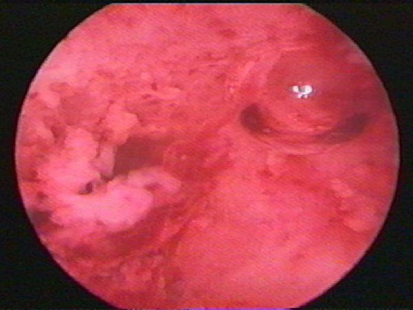
- benign or malignant formations (cysts, polyps, fibroids)
-Features of fetal development during pregnancy, gestational age
- presence of intrauterine contraceptives
Ultrasound of the pelvic organs is a very important diagnostic procedure! Therefore, dear women, if the doctor recommends it, do not refuse and follow the doctor’s instructions!
Where can I get an ultrasound of the pelvic organs?

In the medical office at the address: Ulyanovka village, st. Pobeda, house 39, Leningrad region, Tosnensky district.
Discounts are provided to pensioners, disabled people, mothers of many children in the amount of 3%. There is also a discount system for accumulating discounts from 3% to 7%.
This study is performed by a gynecologist, ultrasound diagnostics doctor Elena Vladimirovna Gubarkova,
Sycheva Maria Sergeevna
Exceptions to the general canons
They do not always wait until days 5–10 of the cycle to conduct an ultrasound examination. Moreover, when treating a number of gynecological ailments, ultrasound data obtained on other days is necessary. The examination cannot be delayed either in extreme situations when a woman’s life is in danger.
Indications for ultrasound during bleeding:
- uterine hemorrhage or heavy discharge during menstruation - may indicate dangerous pathologies of the endometrium;
- risk of miscarriage;
- acute inflammatory processes of the reproductive organs;
- suspected tubal pregnancy;
- complication after abortion;
- suspicion of uterine fibroids or hyperplasia, polyps, cysts (up to 10 mm in size) - ultrasound is indicated on days 1 - 3 of the cycle;
- injuries, burns of the pelvic organs;
- intense pain of unknown origin.
If a woman is carrying a child, the appearance of bloody discharge may indicate a threat of miscarriage, and therefore an ultrasound in this situation is performed urgently. Echography provides a complete assessment of the condition of the fertilized egg and the reliability of its implantation into the mucous membrane of the inner wall of the uterus.
An ultrasound is performed during menstruation if folliculogenesis needs to be examined. The procedure helps to record and evaluate all stages of follicle maturation and its migration from the ovary. The study is prescribed on days 1–4 of the cycle and continued until the 15th day. This data is necessary for the diagnosis and treatment of female infertility and cycle disorders.
How to prepare for an ultrasound examination of the ovaries?
The main thing that needs to be done is to properly prepare for an ultrasound examination of the ovaries. You need to follow a diet and fill your bladder 2 hours before the examination. More detailed recommendations below.
| 3 days before the procedure | The evening before the event | Day | |
| From your daily diet you need to exclude foods that can contribute to increased gas formation: fatty fish and meat, carbonated drinks, brown bread, sweet products, dairy products, juices, legumes, vegetables and fruits. | Light dinner, last meal no later than 20:00. You should not eat meat and fish products, even if they are dietary. | In cases where the examination is scheduled for the morning, breakfast on the day of admission is excluded | |
| Meals should be fractional, in small volumes, 4-5 times a day | If there is a stable tendency to constipation, then it is imperative to take a laxative orally no later than 16:00. | The examination is after 15:00, then a light breakfast is possible, but no later than 11:00 | |
| Meals should be fractional, in small volumes, 4-5 times a day | If the patient’s body does not tolerate laxatives well, then you can use a Besacodyl suppository (constipation suppositories) | 1-2 hours before the procedure, fill your bladder by drinking 1-2 liters of water | |
| On the recommendation of a doctor, various types of adsorbents can be prescribed if the patient experiences flatulence | 2 days before the test, you need to do an enema to cleanse the intestines. | IMPORTANT: Before the procedure itself, you should not smoke, take antispasmodics, chew gum, suck lollipops or other sweets. | |
| Permitted for use: durum grain porridge, lean poultry, lean fish, cheese, tea, coffee. | |||
Sign up for an ovarian ultrasound
Make an appointment
Ultrasound of other organs during menstruation
If there is a need to undergo a diagnostic examination of non-genital organs, the day of the menstrual cycle is not taken into account. You can undergo an examination and get a reliable result at any time, including during monthly bleeding, which will in no way interfere with the diagnosis or affect the result of the study.
Most often, using an ultrasound machine, the following structures of the body are viewed:
- heart, blood vessels;
- The lymph nodes;
- liver, gall bladder;
- kidneys;
- joints;
- thyroid gland.
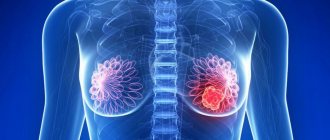
For a routine examination of the mammary glands, which are considered part of the reproductive system, the doctor will suggest that the woman appear on days 8–12 of the cycle, when the breasts are not swollen and its lobules and ducts can be qualitatively examined. If an urgent reason arises, the date may also be adjusted.
Preparation for the procedure (ultrasound of the genital organs)
Ultrasound examination of the female reproductive apparatus is carried out in two main ways - transabdominal (through the abdominal wall) and transvaginally (through the vaginal wall). They are often combined.
If the doctor examines the reproductive organs through the abdominal wall, the woman will be asked to lie on her back and bend her legs at the knee joints. The lower abdomen is bare. The procedure requires preliminary preparation: the woman must appear in the ultrasound room with a full bladder (to be agreed with the doctor). A conductive gel-like agent is applied to the device’s sensor, the tip is moved over the abdomen and the necessary organs are examined.
The transvaginal method involves inserting a sensor into the vagina and examining the uterus and appendages. There is no need to drink water before the procedure, you cannot douche - hygienic washing is enough. The woman must have a bowel movement. A regular condom is put on the sensor, the gel is applied, and inserted into the vagina. The echo image is displayed on the device monitor.
So, to the very frequently asked question “Is it possible to do an ultrasound during menstruation?”, the answer can be given in the affirmative, if we are talking about the study of non-genital organs, as well as when studying folliculogenesis and in all the emergency situations listed above.
It is advisable to plan a preventive examination of the reproductive system on days 5–10 of the cycle, when the uterus is sufficiently cleared of blood clots and remnants of exfoliated mucosa, and there will be no interference with a quality examination.
You should not interpret the result yourself, or self-medicate if you do not have a medical education. With the received conclusion of the ultrasound examination, you need to contact a specialized specialist - he will give a competent assessment of the data and prescribe therapy if necessary.
Pros and cons of transvaginal ultrasound
Doctors in Moscow recommend undergoing a transvaginal examination of the pelvis, both independently and in conjunction with a conventional ultrasound through the abdominal wall for a comprehensive study of the desired area.
If we compare these types of studies, we get the following table:
| characteristic | Abdominal ultrasound (through the abdominal wall) | Transvaginal ultrasound (with a vaginal sensor) |
| safety | 100% | 100% |
| painlessness | 100% | sometimes minor discomfort |
| complications | No | No |
| restrictions on holding | No | allergy to latex |
| visualization | may be reduced for various reasons (for example, obesity) | increased due to tighter contact of organs with the sensor |
| review | general | detailed |
| early diagnosis | the image is not informative enough | the image is informative |
| during pregnancy | before the baby is born | up to 12 weeks |
| virginity | it is possible to carry out | impossible to carry out |
| inflammatory diseases | it is possible to carry out | may be difficult to carry out |
As you can see, a gynecological ultrasound examination undoubtedly gives a clearer picture of the condition of the organs, but has some contraindications. In any case, the method is determined by the doctor at a preliminary consultation.