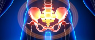
Computed tomography of the pelvic organs is a study that is used to identify urological, gynecological and oncological diseases in this part of the human body.
This method is characterized by high accuracy, efficiency, financial accessibility and the ability to identify a wide range of pathologies of various origins.
A CT scan of the pelvic organs is not accompanied by painful or uncomfortable sensations, except that during the procedure the patient hears noise from the operating tomograph and before the procedure he will need special preparation.
You can get a CT scan of the pelvic organs in Moscow in one of the most modern medical centers - the Yusupov Hospital. Performing a CT scan of the pelvic organs, a CT scan of the pelvic bones on the latest generation tomograph, which the clinic is equipped with, allows us to build a three-dimensional model of the structure under study. Before undergoing the examination, it is necessary to strictly study the indications and contraindications for its use.
CT scan of the pelvic organs: the essence of the procedure
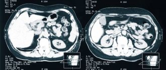
CT scanning of the abdominal cavity and small pelvis is based on the different speeds of transmission of X-rays through the tissues of internal structures, which is recorded by the sensor.
To process the data and form a visual image, the radiologist uses a special program. The radiologist’s conclusion helps the specialized specialist (oncologist, urologist, gynecologist, proctologist, etc.) establish the correct diagnosis and plan an effective treatment regimen. A CT scan of the pelvis allows you to identify abnormalities in the pelvic organs, as well as the vessels and lymph nodes surrounding them.
In some cases, a CT scan of the pelvic organs with contrast is prescribed. How accurate the examination results will be depends to a large extent on the patients themselves, in particular, on their compliance with the doctor’s recommendations for preparing for the procedure.
The results of a computed tomography scan are usually ready in one and a half to two hours. The patient receives not only printed images and a report, but also images in digital format, which can be useful for making another copy or sending it to the doctor by e-mail.
How is the procedure done?
The examination procedure using a tomograph takes place in a special room where the device is located. Before sitting on the couch, the patient must remove all metal objects. During a pelvic CT scan, the patient should lie still while the machine takes pictures. The specialist will inform the patient that they should hold their breath and not move for approximately 10-15 seconds while the organs are being scanned. The entire procedure lasts 20 minutes, with the patient lying on a special table that slides into the ring of the device. Throughout the entire time, the laboratory assistant will be in the next room, but you can communicate with him via speakerphone.
CT scan of the pelvic organs with contrast
CT scan of the pelvic organs with contrast is prescribed for the diagnosis of cancer. Typically, the contrast agent is administered intravenously or orally. The drug is safe and is eliminated from the body within one and a half days. If a CT scan of the abdominal cavity and pelvis is performed with the introduction of contrast, it is possible to view the arteries and veins that lead to the organs. In addition, the use of contrast will allow specialists to detect inflammatory processes and other pathological changes. It is very important before conducting diagnostics with the introduction of a contrast agent to consult a doctor and determine possible individual intolerance to the drug.
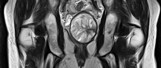
What does a pelvic CT scan show in women and men?
Pelvic CT is an effective diagnostic method that is prescribed for both men and women. It ensures timely detection of circulatory disorders, inflammatory processes and infections, pathogenic neoplasms of various etiologies, degenerative changes in soft tissues and bones, as well as congenital anomalies of the pelvic organs.
During the procedure, the condition of the following organs is assessed:
- ureter;
- Bladder;
- rectum;
- vas deferens, testicles, prostate gland (men);
- uterus, fallopian tubes, vagina, appendages (women).
In some cases, patients require an enhanced procedure (using a contrast agent), which provides a detailed study of the blood supply and those areas that are not sufficiently visualized using conventional computed tomography.
Alternative methods for diagnosing the pelvic organs
Computed tomography, as mentioned above, is usually prescribed to clarify the results of other research methods.
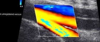
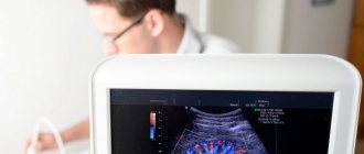
Ultrasound does not provide a complete picture of the disease, and the image on the ultrasound specialist’s computer monitor is only in one plane. With CT, a whole series of layer-by-layer images is obtained in different projections;
MRI in some cases provides more information than CT because CT and MRI images are from different angles. Doctors prefer to combine these diagnostic methods, which complement each other perfectly.
Indications for CT scan of the pelvis
CT scan of the pelvic organs is prescribed for patients with the following diseases:
- congenital anomalies of the structure of the pelvic organs (if they are incorrectly located, underdeveloped, etc.);
- oncological diseases of the lower gastrointestinal tract and reproductive system;
- recurrent cancer;
- acute and chronic inflammatory processes;
- problems with conceiving a child (infertility);
- urolithiasis;
- filling internal cavities with blood;
- internal ulcers (abscesses).
Contraindications for pelvic CT scanning
CT scanning of the pelvic organs is contraindicated for certain categories of patients:
- in case of serious renal dysfunction, hormonal imbalances (if a CT scan with a contrast agent is necessary);
- if pregnancy is suspected or present;
- during breastfeeding;
- in childhood (computed tomography of the pelvic organs is not performed on children under 14 years of age);
- if the patient has mental illness and epilepsy;
- patients suffering from hyperkinesis;
- with a body weight of more than 150-160 kg (due to the limited size of the tomograph).
In what cases is CT of the pelvic vessels prescribed?
Computed tomographic angiography of the pelvic vessels is indicated for suspected development of vascular diseases and to determine the etiology of dysfunction of internal organs. The examination helps in the differential diagnosis of the following pathological conditions:
- Leriche syndrome;
- thrombosis, thromboembolism of the iliac vessels;
- atherosclerosis of the pelvic arteries;
- neoplasms developing in nearby organs and tissues and compressing the vessel;
- phlebitis, vasculitis;
- vascular tumors;
- abdominal injuries;
- aneurysm;
- disturbances in the functioning of the pelvic organs of unknown etiology;
- congenital vascular anomalies;
- impotence in men;
- infertility.
CTA is prescribed in case of low effectiveness of other examination methods, if necessary, to clarify the diagnosis and select effective therapy.
In the process of preparing for surgical treatment, computed tomography is recommended to determine the location and extent of the upcoming intervention. After surgery, a study is prescribed to monitor the restoration of circulatory system function and tissue regeneration.
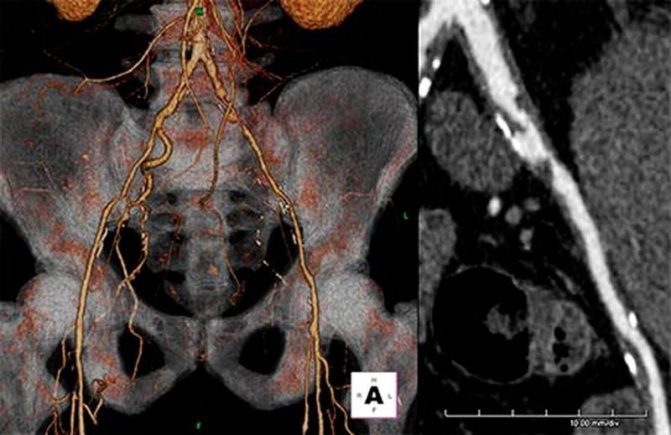
Peripheral arteries of the pelvis, three-dimensional image (A)
If a tumor is suspected in the pelvic area, vascular CT helps determine the direction of further treatment, predict the possible consequences of surgical intervention, and adjust the scope of radiation and chemotherapy.
Preparing for a pelvic CT scan
Preparation for a CT scan of the pelvic organs depends on whether the injection of a contrast agent is planned during the study. If contrast is not required, the patient does not need any special measures, except for sedative medications if he is very nervous before the procedure.
Immediately before the CT scan, the patient should drink about 500 ml of purified water and refrain from urinating.
Performing an amplification procedure requires special preparation, the rules of which will be explained in advance by the attending physician or radiologist.
Preparing men for a pelvic CT scan
To obtain the clearest images, the patient needs to avoid eating foods and drinking drinks for a couple of days, which can lead to bloating. If necessary, they are recommended to take special medications that reduce gas formation.
Immediately before screening (6-8 hours before the test), you need to completely stop eating.
The bladder should be full at the start of the study.
Preparing women for a pelvic CT scan
Preparing women for a pelvic CT scan is no different from preparing men. If there are problems with defecation, the attending physician will prescribe the use of laxatives or a cleansing enema before the study.
Indications and contraindications for CT of the pelvic vessels
MSCT is performed using ionizing radiation, so scanning is done only if indicated. CT angiography is recommended for patients if they have the following symptoms:
- pain in the legs of unknown etiology, numbness, swelling, cramps in the area of large muscles;
- signs of pelvic organ dysfunction;
- abdominal pain in the absence of objective reasons;
- signs of internal bleeding in the pelvic area;
- violation of reproductive function against the background of changes in the nature of the blood supply to the organs of the genitourinary system;
- post-traumatic conditions when injuries are localized in the pelvic area.
A referral for tomographic angiography of the pelvic vessels is given by the attending physician: surgeon, phlebologist, gynecologist, urologist and other specialized specialists.
The use of modern equipment when performing MSCT angiography allows us to reduce the radiation dose to the patient. But the combination of side effects of X-ray scanning and the administration of a contrast solution determines a number of contraindications to this examination method:
- pregnancy in the first trimester;
- conditions and diseases that exclude the possibility of scanning organs and systems using ionizing radiation;
- early childhood (due to difficulties with catheter installation and the need to remain still during the study);
- allergy to iodine preparations;
- severe renal failure;
- diabetes mellitus during therapy with Metformin and analogues;
- hyperfunction of the thyroid gland.
A relative contraindication for CT angiography of the pelvic vessels is a patient weight of more than 150 kg or a chest/abdominal circumference of more than 150 cm, which is associated with the size of the annular part of the tomograph and the load capacity of the scanner table. If there are one or more restrictions, the doctor will select another method of hardware examination of the bloodstream.
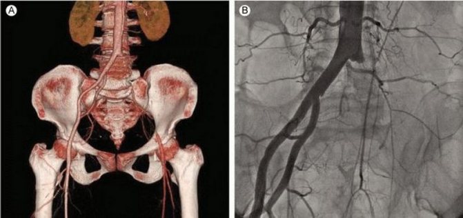
MSCT angiography of the pelvic vessels: acute arterial thrombosis with 3D reconstruction (A) and on frontal images (B)
How is a pelvic CT scan performed?
CT scan results may vary depending on the class and power of the scanner. The powerful tomograph that the Yusupov Hospital is equipped with allows you to obtain better and more accurate images with a large number of sections for maximum detail.
A CT scan of the pelvis at the Yusupov Hospital is performed by a highly professional radiologist. Before the procedure, the patient must lie on the mobile table of the tomograph, which is moved into the ring of the device using a control panel.
The scanning is accompanied by noise and knocking, which disappear at the end of the procedure when the mobile table returns to its original position.

CT scan of the pelvis with contrast: price, goals of the procedure
Computed tomography of the pelvic organs using a contrast agent is prescribed in order to obtain the most detailed information about the disease, assess its extent, the site of development and the condition of nearby blood vessels.
As a rule, an enhanced CT scan of the pelvis is recommended for people suffering from cancer and patients before undergoing surgical treatment.
Patients are preliminarily prescribed allergy tests to exclude the possibility of anaphylactic shock caused by the administration of a contrast agent.
Pelvic CT or MRI: which study is better?
Often, patients make comparisons between CT and magnetic resonance imaging, trying to figure out which is better. Each of these methods has its own advantages and disadvantages, which are determined by different ways of influencing the human body - X-rays or electromagnetic fields.
CT of the pelvic organs is used to most accurately determine oncological and other pathological processes in bone structures and hollow organs (if a contrast agent is used).
The duration of the study ranges from two to three to twenty minutes, thanks to which doctors have the opportunity to quickly establish the correct diagnosis and begin therapy. However, the scope of application of pelvic CT is somewhat narrowed due to contraindications for pregnant women, children and people with excessive body weight.
Magnetic resonance imaging requires more time. This study is contraindicated for patients who have built-in metal or electromagnetic devices in their bodies. However, MRI allows you to diagnose any pathological processes in soft tissues and blood vessels. The number of MRI procedures is unlimited. The disadvantages of this diagnostic method include its rather high cost - the price of magnetic resonance imaging is, on average, 10% higher than the price of computed tomography.