When is the best time to undergo a gynecological ultrasound?
Gynecological ultrasound is best performed 3-5 days after the end of menstruation, but no later than 7-10 days of the cycle, if you count from the beginning of menstruation from the first day. Patients ask why gynecological ultrasound is best done in the first phase of the menstrual cycle. It's simple: during this period, the endometrium, which is the mucous membrane lining the inside of the uterus, is quite thin. If there are any pathologies of the uterine cavity or endometrium, for example, fibroids, hyperplasia, polyps, it is easier to examine them on ultrasound with a thin endometrium than with a thick one. The endometrium thickens in the second phase of the cycle and small polyps can hide in its thickness, remaining unnoticed.
From the second half of the cycle, a follicle begins to mature in one of the ovaries, which explains the appearance of cysts with a diameter of 2-3 cm. These cysts are normal physiological structures and are either a corpus luteum cyst or a follicle that should soon ovulate. In the first 3-5 days from the beginning of menstruation and at the very end of the menstrual cycle, small cysts with a diameter of up to 7-12 mm are distinguished in the ovaries. This is considered the norm. However, it is almost impossible to distinguish them by their external structure from pathological cysts that need to be removed.
Indications for a gynecological ultrasound in the middle or second half of the menstrual cycle are observations of follicle maturation in order to confirm the fact of ovulation. This study is indicated for women seeing a specialist for infertility.
Ultrasound of the pelvic organs: what you need to know?
The ultrasound procedure is performed by an ultrasound diagnostician with special training.
Ideally, the conclusion is issued not by a general sonologist, but by a gynecologist or obstetrician-gynecologist with additional specialization in ultrasound diagnostics.
Methods
Transabdominal: the pelvic organs are examined with a probe through the anterior abdominal wall. Before the diagnostic procedure, it is recommended to drink a large amount of fluid and fill the bladder to the point of “I can’t stand it anymore.”
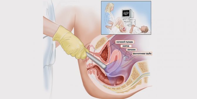
Transvaginal: a special sensor with a condom pre-attached to it is inserted into the vagina. Before the examination, the bladder, on the contrary, is emptied.
For ultrasound during pregnancy , there are no strict recommendations for preparation, unless the doctor gives any special instructions.
General preparation rules
In several days:
* if you are prone to the formation of gases or constipation, it is recommended to take medications such as Mezim Forte, Espumisan or activated carbon;
* do not consume foods that increase gas formation: legumes, vegetables containing coarse fiber, fermented milk.
Stages of gynecological ultrasound
Gynecological ultrasound can be performed either as an independent examination or as part of an examination by a gynecologist. Very often, it is the gynecologist who performs the ultrasound examination. In this case, the ultrasound machine will be located in the gynecological examination room.
- Before the test, your doctor will ask you to empty your bladder if you are not a virgin. Before starting the procedure, you need to remove some of your clothes and lie down on the couch.
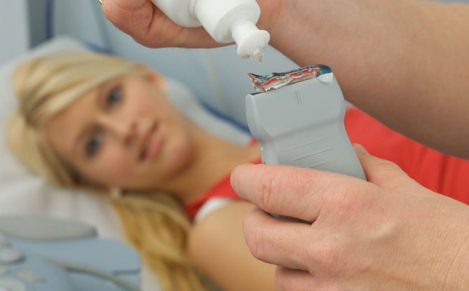
- A gynecological ultrasound is performed using a vaginal probe called an abdominal probe. This sensor is an elongated cylinder with a diameter of 2-2.5 cm. Before inserting the sensor into the vagina, the doctor will put a special nozzle or condom on it and apply a special gel that facilitates the passage of ultrasonic waves.
- After this, the sensor will be inserted into the vagina. This procedure is completely painless and should not frighten the patient. A gynecological ultrasound lasts only 10-20 minutes.
- At the end of the study, the doctor gives the patient a conclusion. Thanks to modern equipment, it is possible to shoot short films and take photographs, recording them on digital media. These images and films can be subsequently transferred to another doctor to review the patient’s medical history and obtain another independent opinion for a more complete picture of the condition of the female reproductive system.
At the Norma medical center, every woman will be able to undergo a gynecological ultrasound and learn everything about the state of her health. The clinic employs experienced specialists using modern equipment that allows for a complete diagnosis of diseases of the reproductive organs, based on its results, making an objective diagnosis and, if necessary, prescribing effective treatment.
Ask your question on the forum
Ultrasound: what is the essence of the method?
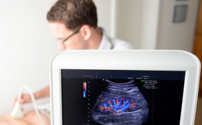
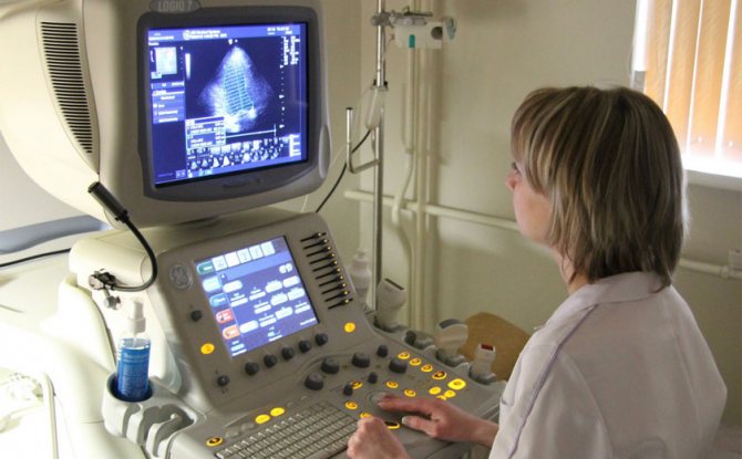
The ultrasound machine sensor (the doctor moves it over the area under study) sends a sound wave that reaches organs or tissues of different densities.
Ultrasound is reflected from the achieved target in different ways, and the receiving sensor records the changes, translating them into an image. The resulting “picture” is redirected to the monitor screen and gives an idea of what is happening inside.
Ultrasound is a method with “broad powers”
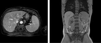
Using ultrasound, the kidneys, pancreas, uterus, ovaries, heart, thyroid gland and many other organs are examined.
The examination helps to identify most diseases in the early stages - when the patient is not worried about anything and the changes are minor. With a good and competent approach to conducting research, the reliability of the data approaches almost 100%.
The information content of the method is expanded when using modern ultrasound systems (3D and 4D ultrasound). The qualifications, specialization and experience of the doctor performing the diagnostic procedure are of great importance.
Is ultrasound dangerous?
No. Ultrasound does not expose the patient to radiation and is considered harmless. If necessary, the study is performed an unlimited number of times (sometimes even several times within one day).
Is it possible for everyone or not?
Ultrasound is a simple, accessible and has no contraindications method.
Temporary and conditional restrictions:
* Ultrasound of the pelvic organs during menstruation, but in urgent cases the examination is carried out;
* the study is difficult to perform or is not very informative in the presence of postoperative scars, bandages, obesity, or severe bloating.
Ultrasound during pregnancy
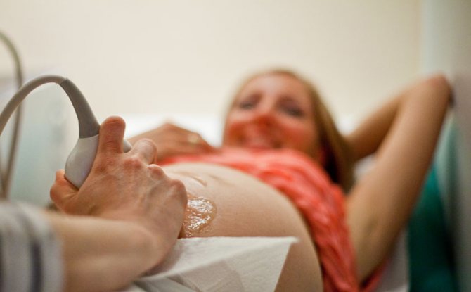
Each expectant mother undergoes several ultrasound examinations at different stages to monitor the course of pregnancy and the intrauterine development of the baby.
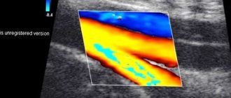
Doppler ultrasound is also performed - measuring the speed of blood flow in the vessels of the placenta and uterus (starting from 32 weeks).
Both ultrasound and Dopplerometry can be prescribed quite often (the studies are harmless) if there are indications (outside of screening). For example, on the part of the baby - developmental delay or insufficient oxygen supply (fetal hypoxia), the expectant mother - gestosis of pregnant women or premature placental abruption.
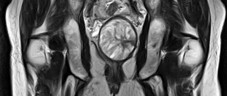
Ultrasound of the symphysis pubis
During pregnancy, under the influence of certain factors and the hormone relaxin, the ligaments of the pubic symphysis (the pubic area) relax - the pelvic bones prepare for childbirth.
In this case, sometimes inflammation, swelling and softening occur locally, the joint becomes excessively “loose” and the bones move apart—symphysitis develops.
You should be wary of:
* the appearance of shooting and pulling pain in the pubic area, pain during sexual intercourse;
* increased pain during movement, especially when abducting the hip to the side;
* reduction or disappearance of pain at rest;
* change in gait - “duck” or “waddle”.
An ultrasound of the symphysis pubis is performed both during pregnancy and after childbirth.
Indications for ultrasound
The purpose and functions of ultrasound of the pelvic organs in women are wide, ranging from standard pregnancy monitoring to the diagnosis of acute conditions of the patient.
A gynecologist may order a test for:
- Confirmation of inflammatory diseases;
- Clarification of diagnoses characterized by the inflammatory process of the urinary system;
- Assessment of the condition of the reproductive organs before IVF;
- Confirmation and assessment of tumor processes (cancer, fibroids);
- Diagnosis and assessment of the severity of emergency conditions (rupture of the fallopian tube, tubal abortion, accumulation of purulent exudate);
- Checking the patency of the fallopian tubes;
- Identifying the causes of pain in the lower abdomen, cycle disorders, abnormal discharge;
- Clarification of the causes of reproductive disorders, inability to conceive and bear a child.
Ultrasound in gynecology
It is performed using both transvaginal and transabdominal methods - the doctor makes the decision individually depending on the situation.
Allows you to evaluate the structure, position and size of the pelvic organs:
* ovaries with follicles;
* uterus (endometrium - inner layer, myometrium - muscle layer) and cervix;
* bladder, parts of the ureters, rectum, retrouterine space;
* fallopian/fallopian tubes with changes (normally not visible during the study).
To whom and when is the study indicated?
The examination must be ordered by a doctor. However, no matter how experienced the doctor is, he will not be able to help you if you do not tell about yourself and what worries you. Be careful and listen to yourself. Even a small “trifle” can serve as a signal of a rather serious illness.
Alarming symptoms:
* complaints of pain in the lower abdomen (acute/aching) associated or unrelated to the menstrual cycle;
* painful sexual intercourse;
* menstrual cycle disorders: delay, shortening, intermenstrual bleeding (even minor ones such as “spotting”);
* menstruation lasts less than three and more than seven days;
* uterine bleeding, heavy menstruation;
* frequent and/or painful urination;
* urinary incontinence or loss of urine during exertion (sneezing, coughing);
* the appearance of any bloody vaginal discharge (from spotting to bleeding) in postmenopausal women.
Ultrasound of the pelvic organs is also prescribed:
* to control the position of the intrauterine device;
* when planning pregnancy or infertility (including folliculometry - assessment of ovarian function and endometrial condition);
* for diagnosing early pregnancy, including ectopic;
* for control after surgical interventions.
What helps to identify?
Many ailments and conditions: ovarian cysts, inflammatory processes, endometriosis (proliferation of the uterine mucosa in other areas), tumors of the uterus and ovaries, endometrial polyps, fibroids, fluid in the retrouterine space, congenital developmental anomalies and others.
When is it held?
For menstruating women, the day of the ultrasound is determined by the doctor, depending on the expected disorders and diagnosis. The timing may vary for each individual woman, depending on the length of the cycle.
Usually, ultrasound is recommended to be performed in the first phase of the cycle - 5-7 days from the start of the last menstruation. After receiving the results, the doctor decides on the need and timing of the following studies. Sometimes the phase of the cycle is not taken into account, and the diagnostic procedure is performed when the patient consults a doctor - for example, in case of acute and severe pain or uterine bleeding.
In postmenopausal women , the day of the study does not matter.
How to prepare for an ultrasound examination
Preparation depends on how the ultrasound is performed - transvaginally (performed through the vagina), transabdominally (through the abdominal wall) or transrectally (through the rectum). The uzologist must tell you in advance how the procedure will be carried out, because only the vaginal method does not require preparation.
Preparation for transabdominal ultrasound:
- You should not eat gas-forming foods. It is better to eat cereals, steamed vegetables, lean fish and meat;
- If the diet does not bring results, then to get rid of gases, you can take activated charcoal a couple of days before the ultrasound;
- In the morning, on the day of the examination, breakfast is prohibited. You are allowed to have dinner the night before and also do an enema;
- an hour before the ultrasound, the patient drinks 1.5-2 liters of water so that the bladder is full during the examination.
Preparation for transrectal examination
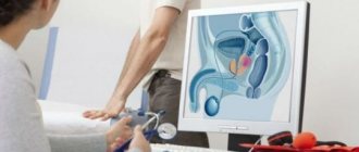
Before a rectal examination (about 2-3 hours in advance), you need to do an enema. If a study of the functioning of the prostate is carried out, the search for the causes of erectile dysfunction and infertility in the bladder should be complete. To do this, you should drink 800-1000 ml of water an hour before the ultrasound.
Transvaginal research method
With the vaginal method, the bladder does not need to be filled. The study can be performed on any day of the cycle (except for the days of menstruation), but it will be more effective immediately after the end of the discharge. To assess the correct functioning of the ovaries, as well as to ensure that the follicles are maturing, an ultrasound scan can be prescribed on different days of the cycle.