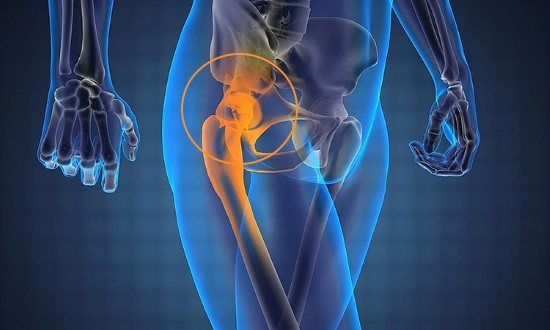
Indications for radiography of the femur
A hip x-ray may be prescribed in the following cases:
- Injury. In this case, there may be aching or acute pain in the thigh, visually noticeable deformation, swelling and redness, limited ability to move the leg or complete immobility.
- The need to monitor surgical treatment. After fractures, it is important to evaluate how correctly the bone fragments are juxtaposed and how quickly they heal.
- If you suspect a tumor of the femur and soft tissues, as well as metastases.
- If you suspect the presence of purulent, inflammatory diseases of bone tissue: osteomyelitis, periostitis.
- If degenerative diseases of the femoral bone are suspected (aseptic necrosis of the femoral head).
What does a hip x-ray reveal?
X-ray allows you to reliably identify the following pathological situations:
- fractures, cracks of the femur;
- the presence of bone fragments and their displacement;
- tumors in the femur and surrounding soft tissues;
- osteomyelitis;
- periostitis;
- deformation of the hip bone due to illness or injury.
Types of X-ray examinations of bones and joints
Depending on the indications and symptoms, the following types of X-ray examinations are performed:
- X-ray of the elbow, wrist, knee and hip joints;
- X-ray of tubular bones
- X-ray of the sternum, collarbone;
- X-ray of the hand and foot;
- X-ray of the pelvis;
- X-ray of the sacroiliac joints.
1 X-ray of the hand in MedicCity using the ITALRAY CLINODIGIT COMPACT device in MedicCity
2 X-ray of the hand in MedicCity using the ITALRAY CLINODIGIT COMPACT device in MedicCity
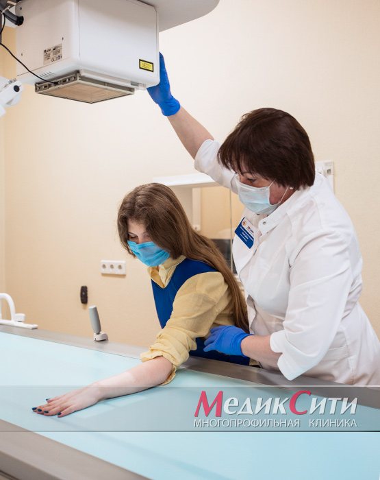
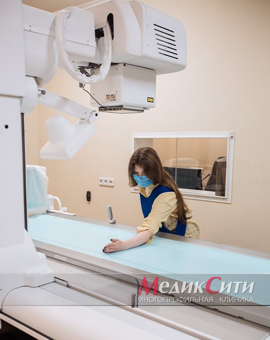
How is the procedure done?
The patient lies down on the work table of the X-ray machine, the markings of which indicate the area of exposure to ionizing rays. Parts of the body that are not subject to examination (especially the thyroid gland and genitals) are protected with lead aprons.
To perform direct projection shooting, the subject lies on his back with his legs extended forward. For the lateral projection, he must assume a position lying on his side with his body tilted back and the other leg. You must maintain this position until the end of the study. The entire diagnostic procedure lasts no more than 5-7 minutes.
Decoding the results
When interpreting the image, the radiologist determines the presence of markers of a particular pathology:
- a fracture is indicated by bone fragments, sometimes displaced;
- with dislocation, displacement of the joints is observed;
- when bone density decreases, osteoporosis can be suspected;
- dysplasia of the femoral head is indicated by its pathological development, incorrect position in the hip joint;
- neoplasms that are visualized on X-ray images of the femur as dark lesions indicate tumor diseases (including enostoma).
A description of the images is given by a radiologist
. It does not establish a specific diagnosis, but only describes the features of the anatomical structure of the bone and its relationship with other adjacent bone elements. The radiologist also indicates the presence of pathological neoplasms, if the patient has them. With a ready-made research protocol, the patient is sent to the attending physician, who, based on the provided description and the results of other diagnostic procedures, makes a diagnosis and prescribes treatment.
Normal indicators and detectable pathologies
- Normally, the obtained X-ray images of the femur should not show any pathological shadows. If a person is healthy, the outlines of his bones are clear and their structure is uniform.
- The images are used to evaluate the location of the femur bone, as well as its size. Thickening or, conversely, a decrease in bone size indicates a developing pathology.
- With cancer in the hip area, areas of destruction of the upper layer of bone tissue may be observed.
- If the images show shadowed areas in the white areas of the bones, osteoporosis may be diagnosed.
Osteoma
Treatment of osteoma in Rostov-on-Don
Often the word “neoplasm” in a diagnosis sounds like a death sentence for many. However, let's figure out what you really should be wary of and how it is advisable to act if you suspect an osteogenic tumor
.
Osteogenic tumors can be benign - these are osteoma and osteoid-osteoma; malignant - osteosarcoma, and occupying an intermediate position - osteoblastoma.
is a type of benign osteogenic neoplasm built from compact bone tissue. Osteoma that occurs on the surface of the bone is known as exostosis, and if it develops in the bone marrow cavity, it is known as enostosis. Synonyms: osteoma - “ivory exostosis”; enostosis - bone island.
In most cases, osteomas occur in bones formed as a result of intramembranous ossification - the roof and bones of the facial skull, rarely outside the skull. Enostoses usually occur in the epi- and metaphyses of long bones, pelvic bones and vertebral bodies. Histologically, compact and spongy osteomas are distinguished. They are built from lamellar bone tissue, and in spongy bone tissue, with increased restructuring activity, areas resembling osteoblastoma can be found. Consists of highly differentiated bone tissue. It has a favorable course, without aggressive growth. The tumor does not become malignant, does not affect neighboring tissues (not counting the possibility of mechanical compression during tumor growth) and does not metastasize. It usually appears between the ages of 5-20 years.
The doctors at our center have many years of experience in treating diseases of the musculoskeletal system, including those of a tumor nature.
| Where to begin? It is necessary to contact an orthopedic traumatologist or oncologist for a correct diagnosis. | What examinations are needed? X-rays, MRI, clinical tests and other examinations - as indicated |
Osteoma and osteoid osteoma are usually diagnosed using X-rays, MRI, CT, and radioisotope scanning. To exclude malignancy of the tumor, the doctor performs a biopsy of a bone tissue sample with histological examination.
Differential diagnosis is carried out with osteosarcoma, Koenig's disease, osteoperiostitis, sclerosing osteomyelitis, chronic Brody's abscess.
| X-ray of the femur with osteoma | CT scan of the frontal bone with osteoma |
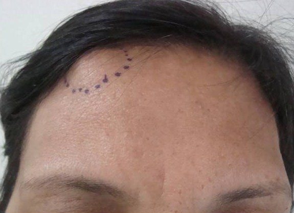
Photo
frontal bone osteomas
Symptoms
. Osteoma grows very slowly, causes virtually no concern and is discovered by chance, especially in the early stages of development.
- found on flat, tubular bones, vertebrae, in the walls of the paranasal sinuses, on the surface of the skull;
- upon palpation - does not hurt when pressed
- motionless, dense;
- has a smooth surface; with clear boundaries;
You should consult a doctor and undergo an examination if the patient experiences similar symptoms
:
- pain;
- if the tumor is in the nasal paranasal sinuses, drooping of the eyelid (ptosis), blurred vision, etc.
- memory problems, epilepsy (if located on the inner surface of the skull);
- lameness (if localized on the bones of the legs);
- nosebleeds, difficulty breathing (if the tumor is located in the maxillary sinus area).
Symptoms of osteoma vary depending on the location:
Posterior wall of the frontal sinus - persistent headaches, increased intracranial pressure
The lower wall of the frontal sinus is a protrusion of the eyeball visible to the naked eye.
Nasal cavity - difficulty breathing through the nose, lack of smell, double vision, drooping eyelid, protrusion of the eyeball, blurred vision
· Paranasal sinuses - blurred vision, pain.
· Frontal bone - headaches, memory impairment, increased intracranial pressure, convulsions.
· Occipital bone - frequent headaches, epileptic seizures.
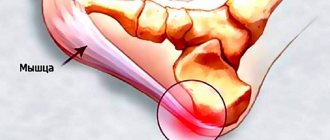
· Femur, talus, tibia - unusual gait, swelling of the legs, muscle pain while walking.
· Knee joint – changes in gait, dysfunction.
· Rib - pain behind the sternum.
· Vertebrae – curvature of the spine, scoliosis.
· “Turkish sella” – hormonal disorders.
Causes of the disease
1. Genetic factor (probability of occurrence in children – up to 50%)
2. The presence of a trigger factor – injury, especially repeated
3. Some inflammatory or infectious diseases (rheumatism, gout, syphilis);
4. Hypothermia
5. Disorders of calcium metabolism
Classification
benign osteogenic tumors
1. Heteroplastic - formed from connective tissue. They are found not only on bones, but in soft tissues and organs - in the diaphragm, tendon attachment points, membranes of the heart, etc. These include osteophytes.
2. Hyperplastic – formed from bone tissue. This group includes osteoid osteomas and osteomas.
Internal and external osteophytes
.
· Internal ones are called “enostoses”, they grow into the bone marrow canal. Usually solitary, except for osteopoikilosis. They do not produce symptoms and are usually discovered incidentally when a person has an x-ray.
· External ones are called “exostoses”. They grow on the surface of the bones and can appear for no reason or due to certain pathological processes. Causeless exostoses usually appear on the bones of the skull, facial and pelvic bones. They may not cause symptoms and may only be an aesthetic defect or put pressure on neighboring organs. In some cases, a fracture of the exostosis leg occurs and bone deformation occurs.
Osteoma
– does not differ in structure from bone tissue, usually found on the facial and cranial bones, including the paranasal sinuses. Bone osteoma is diagnosed twice as often in men, in the area of the facial bones – three times more often. These are almost always single tumors, but with Gardner's disease multiple tumors can grow on long bones. They are painless and do not cause symptoms, but when adjacent structures are compressed, various symptoms appear. Separately, there are multiple congenital osteomas of the cranial bones, which are combined with other developmental defects.
- Osteoid osteoma
is a highly differentiated bone tumor. However, it has a different structure - it consists of vascular-rich elements of osteogenic tissue, zones of bone tissue destruction, and bone beams. Usually there is no pain 1 cm in diameter. It is a common disease, accounting for approximately 12% of all benign bone tumors.
For us, each patient is an individual who has certain external and internal influences on the body, so treatment is selected specifically for each individual.
Treatment
Osteoma can be symptomatic and surgical.
Symptomatic treatment is aimed at reducing pain and discomfort and includes analgesics and topical NSAIDs.
Surgery is performed according to indications:
appearance of pain
· significant discomfort
· compression of adjacent organs and tissues
organ dysfunction
· cosmetic defect causes significant psychological discomfort
In the last decade, minimally invasive methods for removing the focus of benign osteogenic tumors have become increasingly widespread: reaming the focus under CT control, laser photocoagulation, cryotherapy and CT-guided percutaneous radiofrequency ablation.
Our clinic specializes in problems related to diseases of the musculoskeletal system. The doctors working in our center have many years of experience in treating diseases and injuries of the musculoskeletal system, both surgically and mainly non-surgically. We will restore health to your joints.