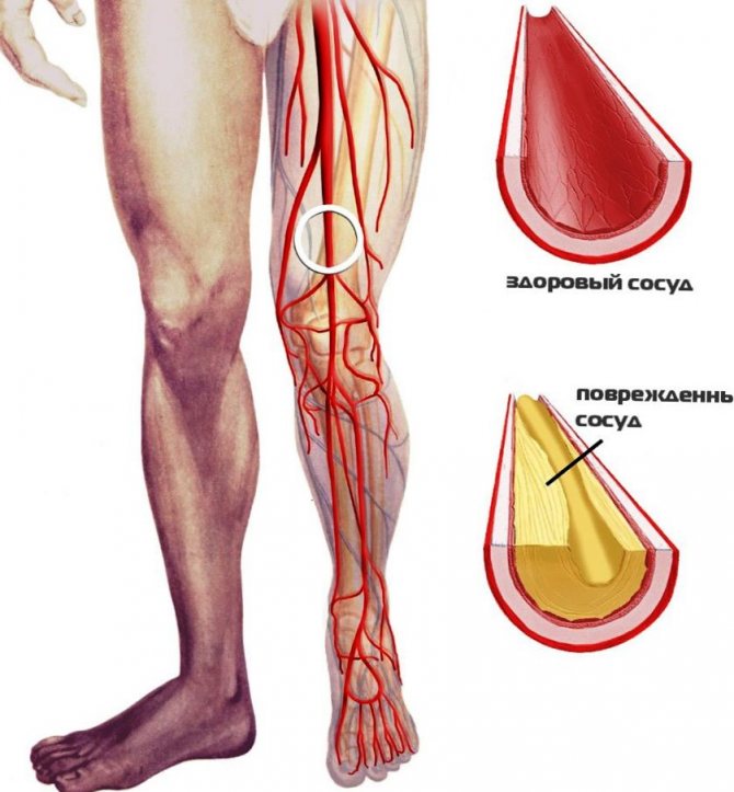
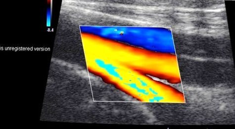
Dopplerography (USDG) of the vessels of the lower extremities makes it possible to study the extent of damage to the great vessels of the legs. The technique helps determine the condition of the vascular walls, the artery and the speed of blood flow. It is based on the Doppler effect, modified and supplemented in accordance with the requirements for modern diagnostics. The device detects and then registers the difference between the ultrasonic pulse transmitted to the organ tissue and the echo signal reflected from the red blood cells.
In what cases is the examination carried out?
What distinguishes this study from conventional ultrasound is the ability to study moving objects, and not just the structure of organs. The device perfectly helps to understand how actively blood moves through the vessels. The diagnostic procedure is carried out if there are signs of circulatory disorders in the vessels of the legs.
These include the following signs
constant swelling of the legs, bruises on the skin that occur with minor injuries, numbness, weakness, cramps in the lower extremities, pain that intensifies when walking, cold temperatures, poor skin, signs of venous insufficiency (cyanotic skin, spider veins, dilatation, deformation of the saphenous veins) trophic ulcers.
This diagnostic procedure is prescribed when signs of chronic and acute vascular insufficiency appear in the lower extremities.
Why is ultrasound of the iliac vessels performed?
The abdominal aorta at the very bottom is divided into two branches - the iliac veins, which pass in the pelvis to the lower extremities, where they are divided into small arteries up to the feet. The iliac veins are responsible for blood circulation in the genitals and legs.
Ultrasound helps identify any abnormalities in blood flow by reflecting ultrasound from the area being examined and recording the resulting signals on a monitor. With this type of diagnosis, you can scan the internal activity of the veins and detect circulatory problems caused by compression, stenosis or blockage of the arteries. The doctor can accurately determine the size, patency of the vessel and the exact location of the sections of the vein where blood flow is obstructed. If blood clots are present, their size and density can be determined and the effect of therapy on them can be analyzed.
Indications and contraindications for Dopplerography of vessels and veins of the lower extremities
Indications for the study are the suspicion of the attending physician that the patient has insufficiency of venous or arterial circulation in the lower extremities, which has arisen acutely or chronically in the patient. It is necessary to find the cause of these conditions when making a diagnosis.
Ultrasound scanning of blood vessels in the legs is prescribed to confirm the following pathologies:
pathological vascular spasm of various origins; thrombosis; obliterating narrowing of the arteries; atherosclerosis; varicose veins of the legs; arterial aneurysms; thrombophlebitis; pathological anastomosis between veins and arteries.
Vascular diagnostics are prescribed to monitor the effectiveness of treatment for diseases of the lower extremities. Doppler ultrasound is easily tolerated and is a painless and safe type of examination. Therefore, there are practically no contraindications. It is not recommended if you are allergic to the gel, or if the integrity of the skin in the area where the manipulation is performed is compromised.
Indications for ultrasound examination of the veins of the lower extremities
An appointment for ultrasound examination of the veins of the lower extremities is given by a phlebologist after examining the patient and collecting an anamnesis.
Primary ultrasound examination of the veins of the lower extremities is prescribed when the patient complains of pain in the legs, swelling, the appearance of spider veins and skin thickening over the superficial veins. Sometimes the reason for the examination may be seizures. Changes in skin color on the legs, protruding tubercles and vein nodules, fatigue, numbness, heaviness in the legs at the end of the day, non-healing wounds on the legs - all these symptoms are the cause and an ultrasound scan should be performed.
Repeated ultrasound examination of the veins of the lower extremities is prescribed after therapeutic treatment or surgery in order to track the positive or negative dynamics of the treatment.
Ultrasound scanning of the veins of the lower extremities is recommended for people who are at risk: cooks, waiters, hairdressers, transport cashiers, salespeople and other specialists who spend most of their working time on their feet. The standing position is the most unfavorable, as it increases the force of gravity, which prevents the blood from rising to the heart.
Also, ultrasound examination of the veins of the lower extremities is recommended for pregnant women with suspected varicose veins.
For people at risk, ultrasound examination of the veins of the lower extremities is the price of their future health.
Preparation for ultrasound examination of veins and arteries
No special preparation is required for this study. However, before performing the procedures, it is recommended to remove hair from the skin of the lower extremities. The patient is prohibited from smoking, drinking strong coffee, tea, or energy drinks immediately before the examination.
Algorithm for performing ultrasound of the lower extremities
Before the procedure, the patient is asked to remove clothing from the lower extremities. According to the decision of the functional diagnostics doctor, the patient is examined lying on the couch or standing.
Diagnosis is carried out as follows
the patient is placed on his stomach on the surface of the skin, the gel for research is evenly applied, then the sensor moves gradually, following the course of the great vessels, then the patient is placed on his back, the gel is applied, the study of arteries and veins is continued during a dynamic study, compression tests are performed, the gel is washed off at the end of the procedure.
After completing the session, the doctor describes and analyzes the data obtained from studying the vessels.
Preparing for an ultrasound
Preparation for ultrasound diagnostics is required for a certain type of research.
Abdominal ultrasound
- Carried out on an empty stomach
- Do not drink liquid 2 hours before the ultrasound
- Do not eat food for 6-8 hours
- The dinner on the eve of the study does not contain gas-forming foods: bread, legumes, vegetables, fruits, milk
Ultrasound of the bladder
- Performed with a full bladder
- 1.5 hours before the ultrasound you need to drink 4-6 glasses of still water
Ultrasound of the mammary glands
- It is carried out from 5 to 14 days of the menstrual cycle
- If you have any complaints - any day!
Ultrasound of the pelvic organs
- External sensor. It is carried out with a full bladder, 1 liter of water is drunk in 1.5 hours
- Internal (vaginal) sensor. It is necessary to empty your bladder before the procedure.
Diagnostic results
In pathologies, the technique demonstrates a narrowing of the vascular lumen. It is due to the following problems:
the presence of atherosclerotic plaques, assessment of the speed of blood movement, study of blood filling of vessels, narrowing of the vascular lumen due to thickening of the walls, presence, localization of wall thrombi, assessment of the functional ability of venous valves, assessment of the phasing of blood flow during exhalation, inhalation, pathological tortuosity, vasospasms, aneurysms, various damage to the arterial wall.
The technique is completely reliable, harmless and painless. This allows you to repeat the procedure as necessary.
USDG
Ultrasound Dopplerography (Doppler ultrasound) of the veins and arteries of the lower extremities is a vascular research technology that gives quick and accurate results. Detects problems with the vascular system without x-rays or injections, so it has no contraindications.
Doppler ultrasound is based on the Doppler effect and the repulsion of ultrasound waves from blood cells in motion. The test is used to measure the volume of blood flowing through arteries and veins, assessing the speed of blood movement, and helps detect blood flow abnormalities in arteries and blood vessels.
Unlike ultrasound machines, which produce waves at equal intervals of time and receive the signal back, Doppler produces waves at one speed and receives them at another.
The difference between the fluctuations helps to analyze the movement of blood through the vessels, including its speed.
Doppler sonography is a safe and painless procedure that does not require complex preparation. The effectiveness of the study depends on the professionalism of the doctor and the quality of the device, therefore, ultrasound examination of the lower extremities should be performed in certified medical centers. When you contact EVERMEDIC, you can be sure that the test results will be accurate.
Sign up for ultrasound examination in Moscow
The procedure usually lasts 20–60 minutes and no preparation is required. During the test, the doctor may ask the patient to lie down, sit down, or stand up. Before the examination begins, a non-allergenic gel is applied to the corresponding area of the skin.
Advantages of the method:
- high degree of information content;
- the ability to detect pathological changes at an early stage;
- painlessness;
- safety;
- no contraindications;
- ease of implementation;
- affordable price.
The RGNCC uses modern diagnostic equipment to perform the study, which allows you to clearly visualize tissues and vessels. If violations are detected, patients can consult a specialist and undergo treatment.
The center is located in Moscow at the address: 1st Leonova Street, building 16. To clarify prices for services and make an appointment for examination and treatment, call the numbers listed on the website or use the online form.
Indications for ultrasound examination
Examination of the vessels of the head and neck may be required if there are symptoms indicating vascular pathologies. This:
- cognitive impairment;
- sleep disorders;
- headaches, migraine;
- dizziness, fainting;
- episodic speech disorders;
- ringing, noise in the ears.
The study also helps to assess the impact of diseases of the musculoskeletal system and nervous system on the circulatory system, so the procedure can be prescribed in the presence of osteochondrosis, stroke, or traumatic brain injury.
Indications for ultrasound scanning of extremity vessels:
- local decrease or increase in temperature;
- numbness;
- muscle pain;
- change in skin tone;
- swelling;
- visually noticeable changes in veins;
- spider veins;
- different strength and frequency of pulsation of the veins on the left and right sides of the body.
As a preventive measure, regular ultrasound scanning is recommended for patients over 40 years of age to monitor age-related changes. The study is often included in the preoperative preparation plan.
Advantages of vascular ultrasound (arteries and veins) at the Rassvet clinic
The Rassvet clinic performs a full range of ultrasound examinations, including vascular ultrasound (arteries and veins).
The diagnostic department of the clinic is equipped with high-resolution ultrasound machines manufactured by GE (USA) and Esaot (Italy), which allow one to obtain an accurate picture of changes in blood flow speed and timely detect blockage of blood vessels and the formation of blood clots.
Ultrasound diagnostic doctors at Rassvet are certified specialists, have extensive practical experience in Doppler ultrasound, and provide a quick and high-quality assessment of the study result.
When is it appointed?
Ultrasound and Dopplerography of the genitourinary system is performed for both women and men. For non-pregnant women, the indications for the procedure are:
- menstrual irregularities;
- pain in the lower abdomen;
- inability to get pregnant within a year;
- repeated miscarriages;
- detection during examination and routine ultrasound of a neoplasm in the uterus or bladder, changes in the thickness of the walls of these organs;
- urinary disorders.
Pregnant women undergo duplex scanning:
- on routine ultrasound (such a study is recommended at 20-25 weeks and mandatory at 30-34 weeks);
- when the fetus lags behind in development or gains weight very quickly;
- in case of Rh conflict;
- in case of multiple pregnancy;
- if the fetus is in abnormal position on the eve of birth.
Ultrasound ultrasound during pregnancy is required in the presence of diseases that contribute to the development of pathology in the veins and arteries:
- diabetes mellitus;
- hypertension;
- renal failure;
- liver diseases.
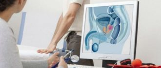
For men, the study is prescribed for:
- infertility;
- erectile dysfunction;
- changes in the testicles (reduction, enlargement, appearance of compactions in them);
- prostate diseases.