Often, an external examination is not enough to identify diseases of the knee joint. In this case, it is necessary to study its internal state. These may be diagnostic methods such as MRI, radiography or ultrasound. Most often, preference is given to the latter, because it combines high efficiency, safety and convenience.
How to prepare for an ultrasound of the knee joint?
No special preparation is required for an ultrasound examination of the knee joint. It is only recommended to refrain from intra-articular injections three to four days before the examination.
The patient lies with his back on the couch. After some time, the doctor asks him to bend or straighten his knees, then roll over onto his stomach. For comparison, an ultrasound scan of both the diseased and healthy joint is performed. In general, the procedure takes no more than 20 minutes.
The method of performing an ultrasound of the knee joint consists of sequentially studying it in different projections.
- Front projection. Allows you to study the quadriceps femoris muscle, patella, superior inversion of the knee joint, patellar ligament, patellar bursa. Examination is performed in a supine position with knees straight.
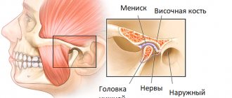
- Rear projection. Needed to visualize the posterior horn of the meniscus, head of the gastrocnemius muscles, tendon, and cruciate ligament. To do this, the patient should lie on his stomach with his knees bent. The doctor uses a sensor at this moment to examine the popliteal fossa.
- Medial projection. Necessary for studying the menisci and ligaments inside. From this angle, the doctor can assess the level of joint fluid, the condition of the bones and hyaline cartilage. In this case, the patient lies on his back with his knees straight, and the doctor examines them from the inside.
- Lateral projection. It makes it possible to examine the external meniscus, lateral ligament, tendons, and lateral compartment. It is also performed in a supine position, on your back. In this case, the knee should be bent at an angle of about 45 degrees. The surface of the joint is examined from the side in the longitudinal direction.
Ultrasound of the soft tissues of the knee joint
In parallel with the cartilage, an ultrasound of the soft tissues of the knee joint is also performed. Unlike X-rays, ultrasound is very good at visualizing the structure of soft tissues and its changes.
Ultrasound of the knee joint
Ultrasound of the knee joint is an informative method for studying the musculoskeletal system. It is completely safe for the patient, does not cause pain or discomfort, and at the same time reflects in detail the condition of the different structures of the knee.
Description of the knee joint
Ultrasound of the knee joint
The body pays close attention to the health of the knee joint. The knee is a joint with a complex structure that combines the patella, femur, and tibia. It is quite large and often suffers from injuries, inflammatory and degenerative diseases.
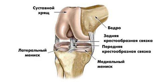
The ligaments responsible for the integrity and stability of the joint are the anterior and posterior cruciate, medial and lateral, patellar and transverse ligaments. They are located in the knee cavity and prevent the leg from moving excessively forward or backward. The kneecap, or patella, is connected to the thigh muscles by tendons.
The articular surfaces of the bones are covered with hyaline cartilage; the menisci, crescent-shaped cartilages located between the tibia and femur, are also built from similar tissue. The knee joint has several synovial bursae (bursae) filled with fluid.
Why is ultrasound prescribed to examine knees?
An ultrasound diagnostic method, or ultrasound of the knee joint, is a popular type of examination that involves the use of a special ultrasound machine.
Ultrasound becomes the first method of examining a patient with complaints of pain and other unpleasant symptoms in the knee joint. It is considered more popular than CT, MRI, and radiography.
The reason for the demand for knee ultrasound is its high efficiency and ease of implementation. Ultrasound has no obvious contraindications and meets the requirements for diagnosing diseases of the musculoskeletal system.
A person receives results almost immediately after execution; their decoding is minimal in time.
Ultrasound can be performed even on pregnant or breastfeeding women - the procedure will not harm the baby. Even children with an injury or other problem can be examined without consequences or crying, because it is carried out quickly and easily. Considering the low price compared to MRI and CT, ultrasound is used everywhere, helping to examine soft tissues, cartilage in detail and identify pathologies at any stage of their development.
What knee diseases can be detected using knee ultrasound?
Who should do this research and why?
The knee joint undergoes serious loads every day when walking and other movements, while it has to constantly withstand body weight, which can be significant.
This area of the musculoskeletal system is subject to severe wear and tear. Pathological changes in some patients begin in young and middle age.
Early detection of problems is possible using ultrasound, although this type of examination also reflects advanced stages of degenerative processes.
They carry out examinations both routinely and for any indication. Routine (preventive) examinations are needed for athletes and people who experience heavy loads on their knees.
Diagnostic sessions for athletes help prevent joint pathologies in the future. The examination is also carried out before competitions, after heavy loads, or if an injury is suspected. It is shown even in childhood when playing professional sports.
The main purposes of knee ultrasound are:
- diagnostic measures;
- determination of professional suitability;
- preoperative preparation;
- identifying the consequences of injuries;
- assessment of ongoing therapy;
- postoperative control.
Most often, an ultrasound scan is prescribed to patients who have suffered domestic or sports injuries to the lower extremity, therefore this technique is widely used in emergency rooms and during the initial appointment in traumatology departments.
The study will reveal:
- bruise - damage to soft tissue;
- hemarthrosis – bleeding into the joint;
- complete or partial damage to ligaments;
- tear, rupture of tendons;
- meniscus injuries;
- fracture of the patella, tibia condyles;
- displaced fractures;
- combined injuries;
- foreign body in the joint.
Ultrasound of the knee joint is recommended if symptoms regularly appear:
- palpable lumps and tumors;
- swelling in the morning or evening;
- stiffness and decreased mobility of the knee;
- local redness;
- hyperemia and hyperthermia.
When interpreting an ultrasound of the knee joint, you can find the cause of pain, deformation of bones and articular surfaces. Most often, such signs become part of the symptom complex of bursitis, synovitis, arthritis, arthrosis, and tendonitis.
The technique is indicated during operations, punctures, and arthroscopy. If Doppler ultrasound is additionally used, it is possible to simultaneously study the function of blood vessels, identify problems with blood flow, defects in the walls of veins and arteries, and a tendency to develop blood clots in the knee area.
If a patient develops joint pathology, the following data are revealed:
- the presence of free fluid in the intra-articular cavity, an increased volume of fluid in the bags (bursae), including those mixed with blood and pus;
- the presence of a foreign body (usually due to trauma), bone fragments;
- changes in the length, width, thickness, volume of various structures - joint space, cartilage, fat accumulations, folds, connective tissue elements;
- violation of the integrity of the ligaments - partial rupture of individual fibers or even complete rupture of the ligament;
- the presence of neoplasms - cysts, bone growths, tumors.
As a rule, all pathologies can be combined into the following groups:
- Injuries of the tendon-ligamentous apparatus (this is a rupture of the patellar ligament, and an injury to the tendon of the quadriceps femoris muscle or the lateral collateral ligament, etc.). Injuries of this kind occur if the joint was suddenly loaded due to improper sudden movement. There may even be a complete rupture with the formation of hematomas. As a rule, this is clearly manifested on an ultrasound of the knee in the form of a violation of tissue integrity, the formation of hypoechoic zones and other signs of serious injury. If the tendon ruptures only partially, its contours are clearly visible on ultrasound, then the site of damage will be designated as a hypoechoic area. So, if, for example, the patella is broken, then hypoechoic areas are clearly visible inside the bone fragments.
- Damage to the meniscus, as well as postoperative condition. A meniscal injury may not always be detected. As a rule, to understand whether it is damaged or not, they look at its outline. When its lines are distorted, hypogenic stripes form in parts of the organ, the leg itself swells, displacing the ligaments to the side, and the articular cavity accumulates fluid, it’s time to change the meniscus.
- Degenerative tissue change. Knee joints, like other groups of cells, can be subject to deformation. This can be easily determined. So, if a patient has arthrosis deformans, in the ultrasound image of the knee joint the hyaline cartilage looks very damaged, and the width of the joint space itself decreases.
- Visualization of cysts in the form of cavities with a certain fluid.
- Dysplastic process.
- Tendinitis, the peculiarity of which is a decrease in the echo density of the tendons to the maximum.
- Inflammation of the synovial membrane and increase in its size.
Ultrasound of the knee joint is indicated for suspected other joint problems:
- various injuries;
- meniscopathies;
- hemorrhages;
- inflammatory pathologies;
- dysplasia;
- neoplasms.
Using ultrasound, specialists can make a prognostic calculation: for example, based on the height of cartilage and the volume of intra-articular fluid, it is possible to determine the presence of initial signs of degeneration and recommend preventive measures. The procedure is indispensable for damage to the knee area; it will help differentiate sprained ligaments from their rupture, bone fractures from injuries to the menisci and kneecaps.
The study will allow us to draw a conclusion about the benignity or malignancy of the tumor process and identify a Baker's cyst, which is characteristic of sports injuries. It can be done as often as required. Repeat sessions are usually needed to evaluate the results of the treatment.
Ultrasound of the knee joint is also prescribed for:
- frequent fractures, dislocations in this area;
- in children with congenital dysplasia, structural anomalies;
- increased body weight, severe obesity;
- rupture of Baker's cyst.
Direct indications for ultrasound of the knee joint:
- Suspicion of defects or structural anomalies in adults or children, suspicion of birth injury.
- Chronic or acute pain (including if the knee hurts only on palpation, but in a calm state does not bother the patient).
- Unsteadiness of gait, development of flat feet for no apparent reason, feeling of instability in the joint.
- Shortening or lengthening of one limb (in this case, it is recommended to simultaneously do an MRI of the pelvic area).
- Destabilization of the knee joint (its movement out of the groove, visible to the naked eye) during physical activity.
- The patient’s ability to independently move (displace) the joint (this is abnormal).
- Development of tumor neoplasms in the joint area of any consistency (hard, soft, containing liquid).
- The feeling of heat in the knee is both subjective (felt only by the patient) and objective (felt by the doctor during palpation).
Diagnosis of the knee joint using ultrasound is indicated for different groups of patients of all ages and genders. Most often, the technique is prescribed to people in the older age group and athletes.
Preparation and technique for ultrasound examination of the knee joint
No preparation is required from patients, with the exception of those receiving intra-articular injections. The last injection should be given no later than 5 days before the examination of the knee joint. Otherwise, visualization may deteriorate. You should definitely take with you the results of previous procedures and a doctor’s prescription (referral).
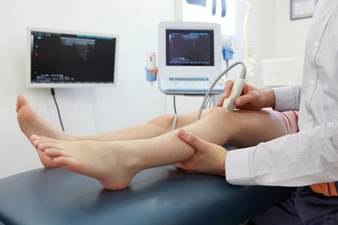
Ultrasound is most often performed in the supine position. The person is asked to lie down on a couch, and the doctor applies a special conductive gel to the skin, after which he moves the sensor over the desired anatomical area. To study the condition of the knee in more detail, the patient is asked to change the position of the body and legs several times during one study.
Four approaches are usually practiced, which help to study the structure of the knee joint in different ways:
- Anterior access. Provides information about the thigh muscles, patella with ligaments, patellar bursae, and fatty tissue. At this time, the patient lies on his back with his leg straightened.
- Rear access. Allows you to visualize the menisci, nerves and vessels of the popliteal zone, tendons, calf and calf muscles, and the cruciate ligament. For the examination you need to lie on your stomach.
- Medial access. Helps identify all problems of the joint capsule, collateral ligaments, inner part of the meniscus, articular surfaces of bones, cartilage and synovial fluid. From a lying position on your back, you need to extend your leg straight.
- Lateral access. Detects pathologies of the fascia lata, large tendons, external meniscus, external collateral ligament, and joint capsule. For the procedure, the leg should be bent at the knee 30-40 degrees.
If there are unpleasant symptoms in one limb, the doctor in any case examines the condition of both knee joints. The information is displayed on the screen, after which the data is entered into a protocol, printed and handed to the subject. The duration of an ultrasound of the knee joint in a medical setting usually does not exceed 15-20 minutes.
How is an ultrasound of the knee joint performed?
As already mentioned, there is no need to prepare for the procedure. It is carried out quickly, the doctor records the results along the way and reports them to the patient. Then the doctor fills out a protocol (conclusion) of the ultrasound of the knee joint, with a detailed description of what he saw. And then the radiologist determines further treatment. But that's it in a nutshell. Let's consider the procedure itself in more detail.
- First, the patient is placed on his back and his limbs are straightened. This is necessary to take a picture of the anterior and lateral parts of the joint. If you need to examine the meniscus, the legs are bent. Then they move on to the back of the knee - for this the patient is asked to lie on his stomach. To assess what the degenerative changes are and how they compare with the norm, pictures of the healthy leg are taken at the same time.
- To obtain complete information from an ultrasound examination of the knee joint, filming is done in many projections (described in more detail below). Thus, the patella, its bursa and the presence of effusion in it are examined in the anterior projection. And just below are the contours of the tibia. If you rotate the transducer to a transverse view, you can examine the contours of the femur, suprapatellar bursa, hyaline cartilage, etc.
- If the joint cavity is examined, there should be no effusion inside it, and the capsules themselves should not be changed. Hyaline cartilage should be echo-homogeneous, with clear and even contours. In this case, the synovial membranes are not visible, the patellas are located strictly in the center, without displacement, and the menisci remain intact, with the correct structure.
Advantages and disadvantages of knee ultrasound
In addition to ultrasound, there are other ways to examine joints. Ultrasound is leading in popularity due to a number of advantages. The strengths of this study are:
- no need for preparation;
- complete absence of pain, discomfort, skin irritation during the procedure;
- hypoallergenicity of the gel used;
- safety of the method for children, pregnant and lactating women, as well as people with severe chronic diseases;
- absence of invasive manipulations, radiation exposure, speed and ease;
- no contraindications for implementation;
- high speed of obtaining results;
- sufficient information content for most joint diseases;
- the ability to detect pathologies at the earliest stages;
- low cost;
- availability of the service in most clinics, hospitals, private clinics;
- detection of problems in all joint structures, nerves and blood vessels.
The disadvantage of the ultrasound method is that it does not reflect the condition of the bones in too much detail, so in some cases it is necessary to carry out further examination in the form of radiography or CT.
You can always make an ultrasound appointment with an ultrasound doctor in Sochi at the medical ]VegaMed[/anchor], ultrasound prices start from 400 rubles, you can visit an ultrasound doctor on the same day as the clinic has free rooms for receiving patients.
You can make an appointment by calling +7 (862) 295-00-95 We also invite you to follow us on Instagram You can use WhatsAPP Make an appointment without a phone call to the medical center “Click here to sign up”
Interpretation of ultrasound of the knee joint
The patient receives the examination protocol in his hands after the procedure. The ultrasound protocol of the knee joint is deciphered by the ultrasound specialist or the attending physician who referred for the examination. The absence of pathologies is evidenced by smooth, clear contours, the absence of neoplasms, and a homogeneous structure.
What pathologies can be seen on an ultrasound of the knee joint?
We can talk about any pathology of the knee joint if there are some deviations from the norm in the ultrasound protocol (the presence of fluid, the presence of foreign bodies, neoplasms, changes in the integrity, structure and size of the ligamentous apparatus, hyaline cartilage, ligaments and torsions). A sign of arthritis, synovitis or bursitis is fluid in the joint cavity. The presence of foreign bodies may indicate an intra-articular fracture. You can also distinguish a cyst or tumor in the images.
What does an ultrasound of the knee joint show?
Carrying out this instrumental study makes it possible to diagnose various dysfunctions of the joint.
- Joint injury. Changes can be of any type. For example, in the case of damaged ligaments, sonography can often detect an area of significant hardening or hemorrhage.
These changes are reflected in the structure of the ligament, which is especially noticeable during a paired examination of the joints. Thus, a minor injury can provoke an exacerbation of rheumatoid arthritis, which until now, for a long time, has not bothered the patient.
- Rheumatic disease. Often, if rheumatism is suspected, ultrasound is the only correct indicative examination.
This disease is always characterized by the presence of inflammatory processes, the extent of which can be determined by the amount of accumulated fluid. If there is a lot of fluid in the knee joints, this indicates extensive inflammation, which is clearly visible on ultrasound.
- Various inflammations in soft tissues. Some inflammatory processes occur in ligaments or muscles. There are a number of diseases for which ultrasound examination is mandatory. This includes synovitis or arthritis of the joints. In addition, ultrasound is used to diagnose bursitis, muscle tendonitis, Baker's cyst and other diseases.
- Various pathologies potentially dangerous to joints. The development of flat feet is the most common of them. Today, with a noticeable decrease in the age of the disease, almost every third person is susceptible to this disease. It is dangerous to ignore this disease, since as a result of deformation of the arch of the foot, a disease such as osteoarthritis can develop over time, which can lead to disability.
It is thanks to the timely detection of inflammation of the knee joints by ultrasound that subsequent complications can be avoided.
Alternative methods for examining the knee joint
In addition to ultrasound of the knee joint, there are other equally effective research methods: radiography and magnetic resonance imaging.
Radiography
If you have pain in the knee joint, limited mobility, swelling or changes in skin color, your doctor may order an x-ray
. This can be either a regular X-ray or arthroscopy - an X-ray with contrast. The photographs can very well determine the pathology of bones and joints (cracks, changes in the width between bones and joints, changes in their shape, etc.). The disadvantage of this study is the inability to visualize the soft tissues of the joints. Also, due to harmful radiation, the diagnosis cannot be repeated after a short period of time in order to track the dynamics of treatment.
MRI
Thanks to MRI research
, many diseases can be detected at a very early stage. The procedure makes it possible to see the slightest changes in joints, bones and adjacent soft tissues. In terms of information content, MRI is the most effective examination method. However, it has a number of contraindications. If the patient has a built-in pacemaker or insulin pump, metal pins and fixed prostheses, then this study will be contraindicated. In addition, compared to ultrasound diagnostics, it is also expensive.
Indications and contraindications for ultrasound of the knee joint
Indications for ultrasound of the knee joint:
- impaired motor activity, lameness;
- pain in the joint that is constant;
- swelling of the tissue around the joint, swelling and redness, local increase in temperature;
- knee injury or bruise;
- crunching in the knee, which is accompanied by pain.
The doctor prescribes a study if there is a suspicion of arthritis, arthrosis, bursitis, synovitis, ligament rupture or damage to the integrity of cartilage and bones. Ultrasound is also performed during treatment to assess its effectiveness.
The ultrasound examination method is considered safe for the patient, since no side effects from the procedure have been identified, so diagnostics can be performed as often as necessary. The examination is prescribed for patients in serious condition, pregnant women and children from the 1st day of life.
Relative contraindications include conditions in which the skin in the affected area is damaged. First you need to treat the rash or wound, and then go for examination.
What does an ultrasound of the knee joint reveal?
Ultrasound of the knee joints is used to detect the following pathologies:
- ✔ Osteoarthrosis.
- ✔ Knee arthrosis.
- ✔ Arthritis, synovitis, tendinitis.
- ✔ Disease associated with the meniscus.
- ✔ Ligament damage.
- ✔ Open and closed fracture.
- ✔ Congenital disorders of the joint.
- ✔ Various neoplasms.
Diagnosis of the knee joint makes it possible to determine specific treatment methods, understand the need for intra-articular injections and therapeutic or diagnostic arthroscopic puncture.
Ultrasound of the knee joint for a child at SM-Clinic
Ultrasounds at SM-Clinic are performed by doctors of the highest, first and second qualification categories. The clinical experience of our specialists exceeds 16 years. Our doctors not only conduct a high-quality examination, but also treat every little patient with care, find a common language with everyone and make the procedure as comfortable as possible for the little ones.
Our medical centers in St. Petersburg are equipped with advanced equipment made in Italy. High-precision technology allows you to obtain reliable information, identify pathologies in the early stages and begin treatment in a timely manner.
To find out the price for an ultrasound of the knee joints for a child or to make an appointment for a consultation, please contact us by phone or through the electronic feedback form. Our clinic’s website also contains a price list with the current cost of basic diagnostic and treatment services.
How is an ultrasound of the knee performed?
Ultrasound of the knee joint does not require special preparation.
The patient exposes the knee area and lies down on the couch. The doctor applies a water-based gel to the skin to improve the conductivity of ultrasound and applies an ultrasound probe. The device sends ultrasonic waves that penetrate into tissues and are repelled from them at different frequencies. The sensor records the result and transmits it to the computer, as a result the doctor sees on the monitor a visual image of the knee joint, blood vessels, nerves, ligaments and tendons.
The doctor takes measurements and evaluates the contours, density and integrity of the tissues. The doctor records the data obtained in a research protocol, which is given to the patient immediately after the ultrasound.
During the diagnosis, the doctor will ask you to change the position of the body, bend or straighten the leg at the knee joint in order to study the articulation in different planes. There are 4 projections of the knee joint:
- Anterior access. When viewed from the front, the patient is positioned supine with the leg straight. The doctor evaluates the condition of the patellar ligaments, popliteal and patellar bursa, and quadriceps muscle.
- Medial access. When viewed from the side, the patient lies on his back with his legs straightened. It is possible to examine the joint capsule from the inside, lateral ligaments, menisci, and estimate the amount of joint fluid.
- Lateral access. The patient lies with the knee bent, the transducer is moved longitudinally from the side to evaluate the collateral ligament, fascia lata, and tendons. You can also see the menisci from above and the joint capsule from the side.
- Rear access. The patient lies on his stomach with his legs straight. The sensor is placed under the knee to examine the vessels and nerves, the heads of the calf muscles, the cruciate ligament, tendons and menisci at the back.
After the ultrasound is completed, the patient wipes the remaining gel from the skin and gets dressed. The whole procedure takes 10–15 minutes.