Otolaryngologist for adults and children
Manevich
Igor Semenovich
Experience 26 years
Otorhinolaryngologist of the highest category, member of the European Rhinologic Society
Make an appointment
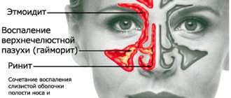
The bones of the human skull have cavities called sinuses or paranasal sinuses. Sinusitis is the general name for inflammation of these sinuses. The disease has several varieties. One of the most severe forms of sinusitis is frontal sinusitis. This is an inflammation of the mucous membrane of the frontal sinus. It is sometimes called frontal sinusitis.
Sinus diseases are very common. They account for about 24-32% of all cases when a patient is sent to a hospital for treatment due to pathologies of the ENT organs. About 14% of the adult population suffers from various forms of sinusitis, of which 3-5% are due to frontal sinusitis. It is more common between the ages of 16 and 35, with approximately 60% of the total number of patients being men.
Frontal sinusitis is a dangerous disease, so it should not be treated like a common cold. The disease can lead to serious complications, including:
- meningitis;
- sepsis;
- osteomyelitis (purulent melting of the frontal bone);
- orbital abscess with transition to the orbit;
- periostitis (inflammation of the tissues surrounding the bone).
The development of phlegmon is dangerous due to complete loss of vision and even death. This occurs if an infection has entered the blood and caused it to become infected. Also, with frontal sinuses, inflammation can spread to other sinuses, causing sinusitis, ethmoiditis or labyrinthitis.
Symptoms and signs of frontal sinusitis
Depending on which frontal sinus is inflamed, frontal sinusitis can be unilateral or bilateral. More often it is the left- or right-sided form, when the left or right sinus is affected. In this case, pain and swelling appear only on one side of the face. For unilateral frontal sinusitis there are other characteristic signs:
- body temperature 37.3-39°C;
- mucopurulent and mucous discharge from the nose;
- deterioration of nasal breathing and sense of smell;
- moderate headache in the superciliary area;
- a feeling of pain and discomfort in the inner corner of the eye.
With bilateral frontitis, the patient feels much worse than with unilateral frontitis. He is deprived of free breathing from both nostrils at once. Because of this, pain is more pronounced, as well as a feeling of fullness in the forehead. Discharge also occurs from both nostrils.
In mild forms of frontal sinusitis, predominantly local symptoms appear. In the case of a moderate course, signs of intoxication appear: the temperature rises, weakness and malaise appear, loss of appetite and worsening sleep are observed. With severe frontal sinusitis, more severe symptoms occur:
- severe, sometimes unbearable pain in the forehead;
- reactive swelling of the eyelids;
- swelling and redness of the skin in the projection area of the frontal sinus;
- puffiness of the face;
- photophobia;
- lacrimation.
Painful sensations
Pain in the forehead increases when tilting or moving the eyeballs. It can radiate to the temporoparietal region. If the patient sleeps on his back, then in the morning he experiences sharp exacerbations of pain. This is due to the accumulation of inflammatory masses in the frontal sinuses.
Discharge
Initially, the patient experiences a thin, mucous discharge. They are transparent, but over time they become viscous and acquire a yellow tint. The development of a purulent process is indicated by green discharge with an unpleasant odor. With unilateral frontal sinusitis, they come out of only one nostril, and with bilateral sinusitis, they come out of both nostrils at once.
Acute frontal sinusitis
Acute frontal sinusitis is characterized by a sharp onset and persistence of symptoms for up to 12 weeks. If the patient receives high-quality treatment, then complete recovery without residual signs is possible. The main symptoms of acute frontal sinusitis include:
- pressing or aching pain in the forehead (intensified by tapping);
- discharge of purulent sputum from the nose in the morning;
- sharp pain in the inner corner of the eye when pressing;
- feeling of fullness in the area between the eyebrows;
- elevated temperature.
Chronic frontal sinusitis
The transition of frontal sinusitis to a chronic form is associated with the lack of treatment or incorrectly selected therapy. A chronic process is said to occur when a person does not recover for more than 12 weeks. Symptoms characteristic of acute frontal sinusitis become less pronounced in the chronic form. The patient is concerned about the following manifestations:
- malaise that prevents you from doing your usual things;
- sometimes the temperature rises to 37°C;
- nasal congestion in the morning;
- persistent cough that is not helped by antitussive medications;
- swelling of the walls of the eye sockets in the morning;
- deterioration of sense of smell;
- feeling of heaviness behind the eyes.
Breathing problems in chronic frontal sinusitis are associated with the growth of polyps in the nasal cavity. Discharge in chronic frontal sinusitis may be absent. The first sign of an exacerbation of the disease is a feeling of “heat” or “flush” in the forehead. Only then does pain appear in this area. It intensifies with bending over and physical activity, and is sometimes accompanied by pulsation.
Catarrhal
The term “catarrhal” means “superficial.” This type of frontitis is accompanied by pain in the area of the brow ridges, which often torments the patient at night and 1 hour after waking up. It radiates to one or both eyes and jaw. Around lunchtime the pain goes away.
The catarrhal form is considered the first stage of the inflammatory process in the frontal sinuses. In addition to pain, the disease causes the following symptoms:
- temperature increase;
- photophobia;
- sleep problems;
- increased tearfulness;
- decreased performance and fatigue;
- swelling of the mucous membrane of the sinuses themselves, which leads to nasal congestion.
Purulent
One of the most dangerous forms of the disease is acute purulent frontal sinusitis. It can quickly lead to inflammation of the periosteum, cellulitis and osteomyelitis. This is due to the fact that the source of inflammation is located near vital organs. The purulent-necrotic process can spread to brain tissue and craniofacial nerves, which is dangerous for the development of sepsis, meningitis, and sometimes even death. Purulent frontal sinusitis can be recognized by the following symptoms:
- temperature 39-40°C;
- purulent nasal discharge;
- throbbing pain in the forehead, which intensifies when tilting and turning the head;
- cough at night and in the morning;
- Strong headache;
- photophobia;
- severe nasal congestion;
- tearfulness;
- tension and swelling in the bridge of the nose.
Do you have symptoms of frontal sinusitis?
Only a doctor can accurately diagnose the disease. Don't delay your consultation - call
Symptoms of chronic sinusitis
· Persistent runny nose that does not respond to treatment with conventional methods.
· Frequent headaches, mainly localized in the frontal and temporal regions.
· Decreased sense of smell.
· Morning swelling of the eyelids, brow ridges.
· Cough that cannot be treated with antitussive drugs for a long time.
· Fatigue, lethargy, decreased performance.
Only a specialist can make a diagnosis after examination and a series of studies.
Causes of frontal sinusitis
Frontal sinusitis rarely develops as an independent pathology. In most cases, it develops against the background of other diseases, most often of the ENT organs. This group of causes of frontal sinusitis includes the following pathologies:
- tonsillitis;
- sinusitis;
- pharyngitis;
- adenoids;
- chronic, allergic or vasomotor rhinitis;
- tubootitis.
With such diseases, there is a potential source of infection in the body, which can spread to the frontal sinuses. The causes of frontal sinusitis also include immunodeficiency states due to AIDS, malignant tumors, and if the patient receives radiation therapy.
Inflammation of the frontal sinuses is caused by various traumatic injuries or operations in the skull area, if they lead to deformation or blockage of the excretory duct or narrowing of the frontal sinus. Traumatic causes also include:
- deviated nasal septum;
- deformation of the ethmoid labyrinth and middle turbinate;
- other anomalies of the structure of the nose;
- fractures or displacement of the nasal septum.
Even improper nose blowing, when a person blows both nostrils at the same time, can act as a trigger for the onset of inflammation. Depending on the cause, frontal sinusitis is divided into several types:
- allergic;
- bacterial (the disease is caused by staphylococci, hemophilus influenzae, streptococci and other bacteria);
- viral (inflammation of the frontal sinuses occurs against the background of acute respiratory viral infections, influenza, measles, rubella, damage by adenoviruses, etc.);
- fungal;
- mixed.
In children, frontal sinusitis may occur no earlier than 5 years of age. Until this age, the child’s frontal sinuses are not yet fully developed. They cannot become inflamed, and therefore frontal sinusitis does not develop. In childhood, the disease most often occurs due to an allergic runny nose or foreign bodies entering the nasal cavity. Also, frontal sinusitis in children can be caused by the same factors as in adults.
Reasons for the development of inflammation
· Frontal sinusitis can be a complication of ARVI, rhinitis, and influenza. If these diseases are insufficiently or incorrectly treated, pathogenic flora can enter the frontal sinuses.
· Mechanical obstructions in the nasal cavity. For example, polyps, cysts, adenoids, even small ones, can impede the communication of the sinuses with the nasal passages, disrupting the outflow of contents and predisposing infectious agents to the existence and reproduction there.
· Injuries to the nose and skull bones. Post-traumatic changes in the nasal septum, frontal bone, and turbinates lead to disruption of the aeration of the sinus, the outflow of mucus from it, and the maintenance of a constant inflammatory process in it.
· Congenital anatomical features of the structure of the facial skeleton. Congenital deviated septum, tight or tortuous nasal passages, too narrow frontonasal canals disrupt nasal breathing and contribute to the easy development of infection in the sinus.
· Chronic diseases of the upper respiratory tract, such as pharyngitis, tonsillitis, laryngitis. The constant presence of pathogenic flora in the respiratory tract is dangerous due to its spread to surrounding structures, especially to the sinuses.
Sinus inflammation can be acute or chronic. Each of these forms of the disease has a clinical picture.
Diagnosis of frontal sinusitis
To make a diagnosis, the patient is subjected to a comprehensive examination. At the first stage, the doctor carries out:
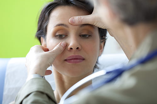
- physical examination. The doctor notes the characteristic symptoms of frontal sinusitis: swelling, pain and redness in the bridge of the nose. The specialist applies pressure to this area to see if the pain gets worse;
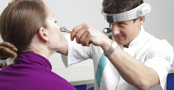
- examination of the nasal cavity. The test is called rhinoscopy. It is necessary to detect purulent exudate in the nose, which drains from the front of the nasal passage. The doctor also notes that there is redness and swelling of the mucous membrane.
If necessary, the nasal cavity can be examined using an endoscope. This is a special device with a flexible tube, which is inserted into the nose to examine the mucous membrane. Additionally, frontal sinusitis diagnostics includes:
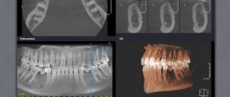
- X-ray of the paranasal sinuses. This diagnostic method is considered the leading one in detecting frontal sinusitis. To get the most complete picture of the condition of the paranasal sinuses, doctors take x-rays in 3 projections;
- computed tomography of the paranasal sinuses. If the X-ray does not produce results, then doctors do a tomogram. On it you can clearly see all the changes in the bone structures and the entrance channel of the sinuses;
- bacteriological research. It is necessary to detect the causative agent of the disease. Before bacterial culture of the patient, the doctor prescribes broad-spectrum antibiotics to the patient, and after receiving the test results, he adjusts the therapy by prescribing a medicine against a specific pathogen. For research, samples of sinus tissue obtained by puncture or nasal discharge are used.
In acute frontal sinusitis, making a diagnosis is not difficult. The difficulty may lie only in detecting the pathogen. That is why specialists study the patient’s nasal discharge.
How are x-rays taken of the paranasal sinuses?
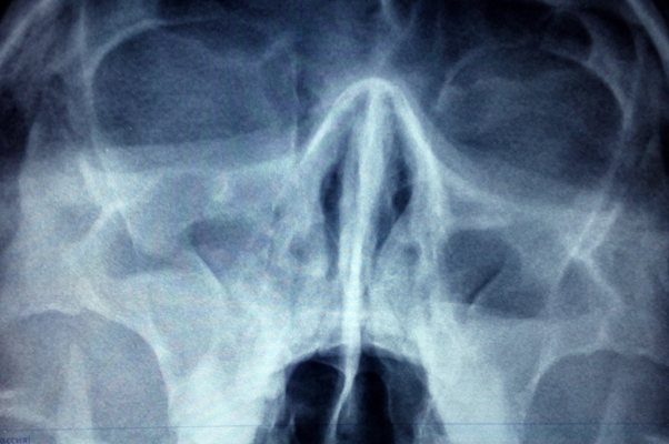
X-rays of the paranasal sinuses are carried out in specially equipped diagnostic rooms using an X-ray unit. Before the procedure, you will need to remove metal objects and outer clothing, and put on a protective apron with a lead coating. If it is necessary to take a picture in the nasomental projection, the patient presses his nose and chin to the device, opening his mouth. In the chin projection, the chin is pressed against the installation, and the nose is located 2 cm from it. Patient position for x-ray of the paranasal sinuses
corrected by the doctor. The patient is then asked to hold his breath and not move while a photo is taken.
X-ray with contrast
The contrast agent blocks x-rays and thus makes the image clearer. It is inserted into the paranasal sinus before the x-ray. The administration of contrast is an unpleasant procedure, so the doctor first administers anesthesia. However, recently, contrast X-rays of the paranasal sinuses have been used less frequently, as MRIs
And .
Treatment of frontal sinusitis
After diagnosis, the doctor prescribes appropriate treatment. The treatment regimen depends on the severity of the disease and the cause that caused it. Treatment can take place at home. Only those with severe bilateral frontal sinusitis are sent to the hospital.
The main goal of treatment is to provide drainage, i.e. outflow of inflammatory contents from the frontal sinuses. Additionally, measures are taken to restore normal breathing and prevent complications. At the first stage of treatment, medications are used:
- antibiotics: Amoxiclav, Sumamed, Doxycycline, Ceftriaxone. A specific drug is prescribed taking into account the causative agent of frontal sinusitis;
- local antibiotics: Bioparox, Polydexa. They are produced in the form of a spray, and are used to act on bacteria directly at the place of their reproduction - in the nasal cavity;
- local antiseptics, for example, Miramistin. It can be used to rinse the nose. The drug will help wash away bacteria from the mucous membrane and remove the contents of the nasal cavity out;
- antihistamines: Diphenhydramine, Suprastin, Tavegil. Prescribed for allergic nature of the disease;
- for nasal rinsing: Aqua Maris, Afrin, Quicks. They cleanse the nasal cavity of purulent contents, moisturize the mucous membrane and facilitate breathing;
- vasoconstrictor drops: Galazolin, Naphthyzin, Nazol. They are necessary to restore normal nasal breathing;
- antipyretics: Panadol, Paracetamol, Nurofen. Used when the temperature rises above 38.5°C;
- homeopathic remedies: Sinupret, Cetirizine, Tavegil. They help relieve swelling and inflammation, ensure the opening of the sinuses and normal breathing.
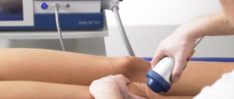
Physiotherapy methods are used in combination with drug treatment. Several procedures show good results for frontal sinusitis:
- laser therapy;
- UHF therapy;
- quartzization of the nasal cavity;
- irradiation with a Sollux lamp.
Surgery
If the disease cannot be controlled with the help of medications, then doctors perform surgery. There are several types of operations performed.
1. Puncture of the frontal sinuses. The procedure is also called a puncture. It takes place in several stages:
- the patient is given local anesthesia, after which a special needle is inserted into the nose;
- along its end, which remains outside, purulent contents flow out of the nose;
- when all the liquid comes out, the specialist applies an antiseptic inside to rinse the cavities;
- if there is a lot of discharge or it is purulent, then the doctor can insert a catheter through the needle, which will remain there for a while. Through the device, secretions can come out, and washing is also carried out through it.
2. Sinoplasty. An endoscope is inserted into the nasal cavity, and a special balloon is inserted through it. The latter is inflated to widen the anastomosis. Through the wide opening, secretions can easily flow out.
3. Open. Radical interventions are rarely performed, since they severely injure the tissue. Open surgery is performed through an incision above the bridge of the nose, a hole in the bone at the bottom of the frontal sinus, and sometimes through the nose.
The operation is also performed when frontal sinusitis was caused by a deviated nasal septum, adenoids or polyps. Intervention helps to get rid of these defects.
Indications for frontal sinusitis
To speed up recovery, with frontal sinusitis the patient should adhere to some recommendations:
- regularly ventilate the room in which the patient is located;
- humidify the air daily;
- observe bed or semi-bed rest;
- provide plenty of warm drinks and easily digestible food.
During frontal sinusitis, it is very important to regularly rinse your nose so that you can breathe normally and the nasal cavity is not filled with inflammatory contents. A regular syringe without a needle is suitable for rinsing. A solution is drawn into it, which is then poured into one nostril.
It is better to carry out the procedure over a sink. The head must be turned to the side. The solution is poured into the nostril, which, when tilted, turns out to be the top. The fluid should come out of the other nostril. A sea salt solution is considered a classic solution for washing. For a glass of boiled water, 1/3 teaspoon of salt is enough.
It is important that its crystals completely dissolve, otherwise the mucous membrane can be injured. You can rinse your nose up to 5-6 times a day. If the nose is stuffy, then before the procedure it is necessary to instill vasoconstrictor drops, otherwise the solution will not be able to pass through the nasal passages.
Contraindications for frontal sinusitis
Warming up during frontal sinusitis is not only not useful, but in some cases even dangerous. Heat exposure can only increase swelling. Warming up also creates favorable conditions for further growth of bacteria. Also, the patient may accidentally warm up the lymph nodes, which will also increase inflammation and even lead to complications.
Thermal procedures for frontal sinusitis are allowed only if the patient does not have a fever or purulent nasal discharge. But even here it is important that the treatment occurs under the supervision of a doctor. Only he will be able to determine whether warming up will do any harm at a specific stage of the disease.
What else should not be done if you have frontal sinusitis:
- continue to go to work and maintain your usual lifestyle. In the acute form, it will be difficult for the patient to do this due to severe weakness, but in the chronic form, patients often neglect bed rest. This often leads to complications;
- smoke. It is necessary to give up cigarettes at least for the duration of treatment of the disease. Cigarette smoke irritates the mucous membrane, which only increases its swelling and delays recovery;
- use vasoconstrictor drops for a long time. They can be used for no more than 7-10 days. It is advisable to instill only before bedtime in order to get a good night's sleep. With the constant use of such drops, addiction develops, and the nasal mucosa also grows, which only worsens the course of frontal sinusitis.
Prevention of frontal sinusitis
To avoid diseases of the paranasal sinuses, including sinusitis, it is necessary to follow the following preventive measures:
- wash your hands thoroughly after visiting public places, before eating, before touching your face;
- avoid exposure to chemical air pollution, dust, allergens and tobacco smoke;
- drink more fluids and eat healthy, vitamin-rich foods, strengthen the immune system;
- do not be exposed to hypothermia, avoid contact with people with acute respiratory viral infections, and get vaccinated against influenza.
If frontal sinusitis nevertheless develops, our clinic invites you to the paid services department for treatment of this disease. The most qualified doctors work here, who consider the characteristics of each individual case. All necessary procedures and interventions can be completed here. This relieves the patient of discomfort and speeds up recovery.
Advantages of treatment at the clinic of JSC "Medicine"
Everyone who comes to the clinic of JSC "Medicine" undergoes an examination in accordance with medical standards. This helps to accurately identify the cause of the disease and prescribe effective treatment. By contacting JSC “Medicine”, you can be confident in the provision of quality services. Each patient here receives:
- Personal access to medical history via the Internet.
- A personal curator who will accompany the patient throughout the entire therapy, even if other specialists take part in it.
- An accompanying person for quick movement around the clinic.
- Explanation in clear language about treatment and prescribed medications.
- Possibility of receiving all necessary services on the day of treatment.
- The wait for an appointment is no more than 20 minutes, subject to prior registration.
- Providing examination results on a CD.
The Clinic of JSC "Medicine" is always ready to discuss in case of justified claims. If you were not provided with certain services properly, you can receive financial compensation.
The administration considers any requests, since JSC “Medicine” works on the principle that only the client evaluates the quality of the services provided. This is what became the key to the clinic’s success in providing medical care. Each patient leaves us not only healthy, but also completely satisfied with the services provided.
Progress of the MRI procedure of the paranasal sinuses
The scan is carried out according to a standard plan and takes from 15 minutes (in normal mode) to 50 minutes (using a contrast protocol). Full instructions on the MRI of the sinuses are provided by the staff of the diagnostic center.
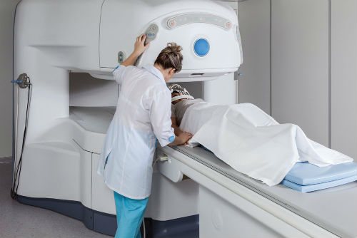
The main steps of the procedure include:
- Registration and survey. At this stage, the necessary documentation is created in the medical institution, the radiologist on duty gets acquainted with the client’s medical history, determines the area of study, selects the necessary mode and equipment settings. The MRI protocol for a cyst is somewhat different from diagnosing sinusitis. The patient is given detailed instructions on what to expect from the procedure and how to behave.
- Preparatory manipulations. The patient is escorted to the room with the equipment, placed on the mobile part of the installation, sensors are attached to the area being studied, and, if necessary, fixed in a stationary state with special belts. The examination should be carried out in a calm state, so it is important to know that there will be no pain or discomfort during the session.
- MRI scanning of the nose. At this stage, the conveyor with the patient is placed inside the unit, the machine can slowly move along the body and produce an audible mechanical noise. At this time, diagnosticians are in an adjacent room and monitor what the screening shows. If discomfort or complaints arise during the MRI process, it is easy to contact the staff via loudspeaker.
- Waiting for results to be interpreted. Based on the results of the examination, diagnosticians prepare a final conclusion; this requires time. The patient is asked to wait in the lobby or a specially equipped waiting room. If an MRI was performed on a cyst, diagnosticians need to determine its size, the degree of growth of the cyst, the nature of the contents and the need for surgical removal of the formation. Software helps in diagnosing, which, through comparative analysis with the norm, determines the nature of the pathology.