Colposcopy is an instrumental diagnostic technique that is used in gynecology to identify a number of pathological processes. The essence of the study is a visual examination of the mucous membrane of the walls of the vagina and cervix using a colposcope (a diagnostic optical device that looks like a tube). There is a regular (examination only) and extended colposcopy. During extended colposcopy, the gynecologist can also perform a biopsy - taking a section of tissue for subsequent histological examination under a microscope and identifying the possible development of a tumor process. Extended colposcopy with biopsy is a fairly traumatic procedure, and therefore requires certain restrictions after its implementation.
Our clinics in St. Petersburg
Medical center on Pionerskaya Polikarpov Alley 6k2 Primorsky district
- Pionerskaya
- Specific
- Commandant's
Medical center South-West Marshal Zhukov Ave. 28k2 Kirovsky district
- Avtovo
- Avenue of Veterans
- Leninsky Prospekt
Medical center in Devyatkino Okhtinskaya alley 18 Vsevolozhsk district
- Devyatkino
- Civil Prospect
- Academic
For detailed information and to make an appointment, you can call +7 (812) 640-55-25
Make an appointment
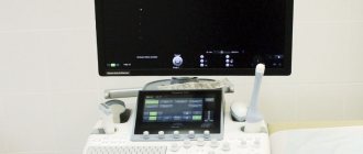
The examination is carried out using a special device - a video colposcope, thanks to which an image of the examined organ and tissues is displayed on the computer screen in real time. Images of problematic areas of the internal genital organs can be enlarged, saved digitally and printed.
This diagnostic method is especially effective during asymptomatic gynecological diseases, and also makes it possible to detect the disease even with slight changes in the color and structure of the cervix and vagina, and invisible tissue defects.
At Medicenter, video colposcopy is performed by highly qualified gynecologists using modern equipment that allows them to see the smallest pathological lesions on the mucous membrane of the vagina and cervix.
What is included in the concept of “gynecological examination”
A gynecological examination means a set of medical procedures, including consultation, examination by a gynecologist and various studies prescribed by a doctor, depending on the patient’s health status, her age and goals.
The following methods of examining gynecological patients are used at MedicCity:
- taking anamnesis;
- gynecological examination;
- ultrasound diagnostics;
- laboratory research;
- hysteroscopy, colposcopy, etc.
History taking
This is the primary diagnostic method, its purpose is to obtain complete information about the state of general and gynecological health of the patient. The doctor may be interested not only in the characteristics of a woman’s menstrual cycle and sexual life, but also in her heredity, past illnesses and operations, the woman’s living conditions and the health status of her husband.
Particular attention is paid to the age of the patient. After all, certain features of her health are considered in some cases as a pathology, and in others as the norm. For example, the absence of menstruation during reproductive age indicates the presence of endocrine or gynecological disorders, and in childhood and old age it is an indicator of health. Another example is intermenstrual bleeding. During reproductive age, this may be considered a complication after an abortion or a symptom of pelvic inflammatory disease. Vaginal bleeding in an elderly woman can signal tumor processes; in a teenage girl, it can indicate ovarian dysfunction.
The patient’s lifestyle can also affect the state of women’s health. Working in a “harmful” industry, constant overheating or hypothermia of the body, hard physical labor, professional sports - all this affects the reproductive system. The same goes for nutrition. Addiction to various diets can lead to delayed development of the genital organs, menstrual irregularities and infertility. (If you want to solve the problem of excess weight without harming your body, contact our highly qualified nutritionist).
1 Gynecological examination

2 Gynecological examination

3 Gynecological examination
Gynecological examination
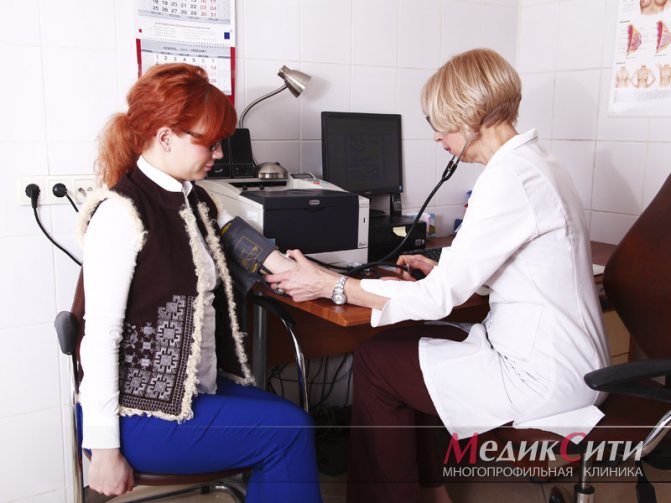
A gynecological examination is an examination in an examination chair. This indispensable examination allows you to detect such “asymptomatic” diseases as ovarian cysts, some infectious and inflammatory diseases of the pelvic organs.
Preparation for a gynecological examination is as follows: the night before you need to empty your bowels, and in the morning, immediately before visiting the doctor, you need to empty your bladder. Otherwise, palpation and inspection will be difficult. Before visiting the doctor, you should take a shower, but you should not douche - in this case, the smear will be uninformative.
It is necessary to tell your doctor the date of your last menstruation, since the condition of the internal genital organs depends on the day of the cycle.
A gynecological examination on a chair (manual and using mirrors) helps the doctor assess the condition of the vulva, vaginal mucosa, cervix and at the same time take biomaterial for examination. During a bimanual examination, pain, size of the uterus, and the condition of the uterine appendages are assessed. Thanks to this method, the doctor may suspect the presence of a uterine or ectopic pregnancy, ovarian cysts, inflammation of the appendages, uterine fibroids, and endometriosis. Ultrasound and laboratory tests are used to clarify the diagnosis.
The first days after the end of menstruation are best for a gynecological examination. However, in case of acute pain or menstrual irregularities, you should not postpone the visit.
Ultrasound method
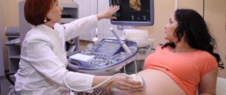
An ultrasound examination is necessary if you need to see the condition and size of the ovaries, uterus and fallopian tubes, determine the presence of mature follicles or the corpus luteum in the ovaries, and also identify the structure and thickness of the endometrium.
Using ultrasound monitoring (or folliculometry), you can monitor the function of the ovaries and monitor the maturation of the egg over time.
Ultrasound during pregnancy helps to analyze the development of the fetus and detect intrauterine defects.
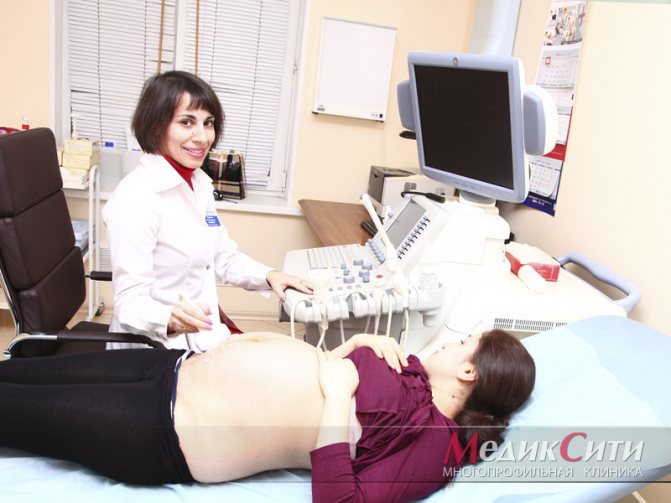
1 Ultrasound examination
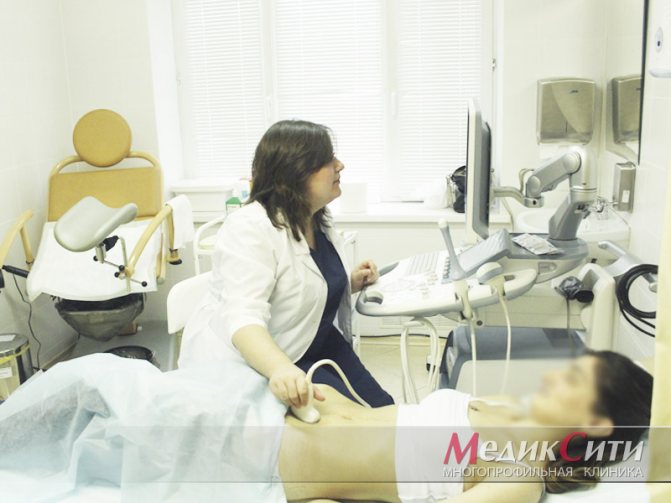
2 Ultrasound examination
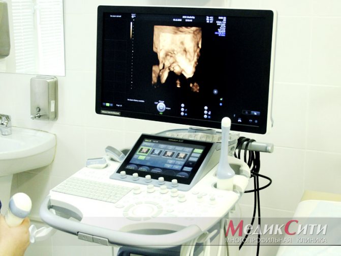
3 Ultrasound examination
Endoscopic diagnostics - colposcopy, hysteroscopy
Video colposcopy procedure
Video colposcopy feels a little different from a regular examination. The doctor inserts a speculum into the woman and applies Lugol's solution, iodine or acetic acid to the cervix, which allows identifying the affected tissue. When the solution takes effect, the specialist, using a video colposcope (a binocular microscope with a video camera attached to it), begins to conduct an examination. During video colposcopy, the gynecologist can at any time save the image to a personal computer and demonstrate it to the patient on the monitor. In difficult situations, the process of video colposcopy can be observed on the monitor by several doctors. On average, the procedure takes 10-20 minutes. After video colposcopy, a woman should limit sexual activity for several days.
What happens after a colposcopy?
You will probably have no problems after the colposcopy and biopsy. If your results show any problems, your doctor may suggest additional tests or treatment.
What should I do after the colposcopy procedure?
After a colposcopy, your vagina may feel slightly sore for a few days. If you have had a biopsy, you may also have a dark-colored vaginal discharge. Use a sanitary pad, it is not recommended to take a bath for 2 weeks (only hygienic shower).
Here are some other things to keep in mind after a colposcopy and biopsy:
- You can take a shower whenever you want.
- If you have not had a biopsy, you can have vaginal sex at any time.
- If you have had a biopsy, wait about 3 days before having vaginal sex. This allows the cervix to heal.
- If you are taking any medications, continue to take them as usual, including birth control.
Advantages of the method:
- Possibility of diagnosing cancer at early stages and timely treatment.
- Video colposcopy is an absolutely painless procedure that lasts no more than half an hour.
- The image is broadcast on the screen, which makes it possible for several doctors to participate in the examination, and for patients to clearly see the nature of changes in the organ.
- Thanks to the image magnification by 30 times, the device allows you to diagnose the smallest structural and functional changes in the walls of the vagina and cervix and determine the nature of the pathology.
- The resulting image is saved on a digital medium, which in the future will make it possible to track the dynamics of the development of the disease focus and evaluate the results of treatment.
Doctors at the Medical Center recommend that every woman undergo video colposcopy at least once a year.
What does video colposcopy show and is it painful?
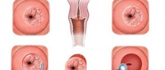
How does this procedure work? In order to assess the condition of the tissues of the cervix and vagina, the doctor uses a special device that is immersed inside through the vagina. The device has a special backlight and a camera, which makes it possible to magnify the image up to thirty times or more.
Thus, on the surface of the tissues of the uterus and its cervix, one can see various inflammatory processes, cervical erosion, chlamydia, polyps, and mechanical damage. And, what is extremely important, video colposcopy in our medical center in St. Petersburg can be done to identify both benign and oncological tumors at an early stage.
This diagnostic technique has a number of advantages over others:
- absolute safety, which allows you to use it as often as necessary;
- According to our patients, this study is absolutely painless;
- the ability to thoroughly study tissues when scaling;
- the ability to record examination results on external media to track dynamics when the procedure is repeated;
- compatibility with other studies, for example, with biopsy;
- the price for video colposcopy is affordable for any patient.
Thus, this procedure can be called the most effective modern existing means for diagnosing diseases of the female reproductive system.