Ultrasound examination of the uterus is an indispensable diagnostic procedure for monitoring the health of this organ. Thanks to ultrasound, you will receive all the necessary information about the correct position of the uterus, the characteristics of the blood vessels and other important parameters. Ultrasound of the uterus gives excellent results for diagnosing pathologies of this organ, so it is optimal to make it planned and regular - about once a year, but not less often.
The total time of such an ultrasound examination is about half an hour. Qualified specialists performing ultrasound in our medical center have the most modern equipment, and therefore receive the most accurate results. This will allow you to obtain complete and reliable information about the condition of the uterus, track changes over time, or obtain indications for subsequent treatment of pathologies.
- What is uterine ultrasound
- Preparation
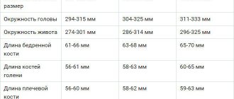
- Normal indicators
- Indications for ultrasound examination of the uterus
- Advantages and disadvantages of the procedure
- When is the best time to perform the procedure?
What is uterine ultrasound?
Ultrasound examination of the uterus is the simplest and most effective way to find out about the condition of this organ, to diagnose possible diseases and pathologies at the earliest stages, when they are positively amenable to treatment. The study is carried out on the direction of a gynecologist after a consultation appointment. To save time, in our medical center you can combine a visit to a gynecologist and an ultrasound scan of the uterus.
Ultrasound examination of the uterus is completely safe for the body, as evidenced by the fact that it is prescribed even during pregnancy (to verify the presence of a fetus in the uterus, to exclude ectopic pregnancy, etc.). Ultrasound waves allow a clear image of the organ to be transferred to the monitor, and a specialist can thoroughly examine and evaluate its condition.
There are several types of ultrasound examination. Let's look at the main ones.
Indications for examination
It is not recommended to neglect the examination during pregnancy planning and pregnancy, after childbirth and surgical interventions, as well as when reaching menopause. It is necessary to make an appointment for an unscheduled consultation if the following symptoms appear:
- cycle disruption;
- bloody or purulent discharge;
- heavy and prolonged periods;
- pain during sexual intercourse;
- if neoplasms are suspected;
- spasmodic pain in the lower quadrant of the abdomen.
Once a year, preventive examinations by a gynecologist and an ultrasound of the pelvic organs are recommended for all representatives of the fair sex, regardless of age. Timely identification of problems can significantly reduce pregnancy complications and the risks of cancer.
How to properly prepare for an ultrasound examination of the uterus?
The first rule of almost any ultrasound is the absence of intestinal gases, which interfere with the correct passage of ultrasound waves and, accordingly, can significantly distort the results of the study. It is recommended to stop consuming foods that increase gas formation - in particular, carbonated drinks and foods rich in fiber - at least two days before the procedure. To eliminate excess gases in the body, a day before the ultrasound, take the recommended dose of activated carbon (or another drug aimed at eliminating excess gas formation). Other preparation features depend on the specific method (transvaginal or transabdominal), as well as the time of day for which the study is scheduled.
How to properly prepare for a transvaginal ultrasound procedure?
It is very important to sign up for an ultrasound scan at a certain time - given the characteristics of the female body, you will get the most complete picture with this method of examination for prevention at the very beginning of the menstrual cycle. Therefore, in this case, you need to sign up for the first day without menstruation. If you are going to do an ultrasound because you suspect endometriosis, it is optimal to choose the second half of the cycle. For pregnant women, transvaginal ultrasound is prescribed exclusively in the first trimester, if there are direct indications for this. The only reason why you should undergo this procedure without delay and as soon as possible is bleeding of an unknown nature.
When is an ultrasound with Doppler ultrasound performed?
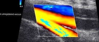
Most often, ultrasound with Doppler and 3D is performed in the third trimester, but in the presence of pregnancy complications (intrauterine growth retardation, placental insufficiency, etc.) and earlier - in the second trimester.
This procedure is harmless and painless. Its implementation is necessary, since in many cases it is thanks to Doppler measurements that it is possible to identify problems in a timely manner and avoid accidents. This sensor shows the entanglement of the umbilical cord and allows you to see what it is: one-, two-, or three-fold; it allows you to diagnose fetal hypoxia and insufficiency of placental function. As a result, obstetricians promptly select the necessary treatment for the pregnant woman and correctly determine the tactics of labor management and the timing of delivery.
How to properly prepare for a transabdominal ultrasound procedure?
The main point in preparing for any ultrasound examination is to minimize gas-forming processes in the body, for which the patient takes activated carbon tablets on the eve of the procedure, and at least two days before excludes from the diet products that stimulate the formation of gases (carbonated water, legumes, fiber, etc. .). Among the specific recommendations is that you need to drink water (at least half a liter) an hour before the procedure and refrain from visiting the restroom.
Types of ultrasound examination
The doctor will refer you for an ultrasound if inflammatory processes are detected or if you complain of pain and discomfort in the lower abdomen. The reason for the examination is the absence of menstruation or, on the contrary, heavy blood loss. Ultrasound of the female reproductive system is carried out using two complementary methods:
- Transvaginal method, in which the examination of the pelvic organs is carried out with a vaginal sensor on certain days of the cycle. The method is highly informative and allows you to most accurately diagnose pregnancy in the early stages, pathologies of the cervix and organs of the reproductive system. The procedure does not require special preparation, other than standard hygiene measures. Also, a few days before the test, foods that cause increased gas formation are excluded from the diet.
- Transabdominal examination is carried out through the anterior abdominal wall with a special portable sensor. An important condition for the procedure is a full bladder, for which it is recommended to drink at least four glasses of water an hour before the procedure. The examination is indicated for women of all age categories, including adolescents and pregnant women in the second and third trimesters.
Depending on the presumptive diagnosis and the goals of diagnostic measures, the gynecologist individually determines the approximate dates and selects the ultrasound technique. In some cases, for the most accurate assessment of the condition of the pelvic organs, a combination of transvaginal and transabdominal ultrasound is used.
Indications for ultrasound examination of the uterus
You should sign up for an ultrasound of the uterus if you suspect:
- Ovarian cysts.
- Uterine fibroids.
- Tumors.
- Internal endometriosis.
- Endometrial polyps.
- Internal inflammatory processes.
- Ectopic pregnancy, etc.
Ultrasound of the uterus is also done to diagnose hematuria, during biopsy manipulations, to diagnose the size of the ovaries, with painful urination, with vaginal non-menstrual bleeding, with abdominal pain of unknown origin, etc.
Our advantages
By contacting our center for an ultrasound scan of the uterus and appendages, you can get:
- Accurate results To conduct ultrasound of the ovaries and uterus, modern equipment from General Electric, one of the leading manufacturers of medical equipment, is used. Thanks to this scanner, the image of the uterus and ovaries is very clear, and the professionalism of doctors allows you to accurately decipher the result;
- Quick examination. The patient undergoes the procedure strictly according to the appointment time. You do not have to wait in line and return to the clinic for the result - all data on the condition of the uterus and ovaries will be entered into the medical record within 10-15 minutes after the examination;
- Budget savings. "Alfa Health Center" is a large network of private clinics. We always offer affordable prices for ultrasound of the appendages and uterus and other procedures. Our clinic regularly holds promotions. This makes prices for ultrasound of the uterus and appendages even more profitable;
- Comprehensive services. After receiving the results of the procedure, you can immediately make an appointment and undergo examination by specialized doctors. All manipulations for the diagnosis and treatment of diseases of the uterus and ovaries can be carried out in our clinic.
Transvaginal ultrasound examination of the uterus
Transvaginal ultrasound is a procedure that shows the most accurate picture, so it can be called the most effective and efficient gynecological examination. The only disadvantage of the procedure is that, due to the peculiarities of its implementation, it can only be prescribed to women who are already sexually active.
To carry out this procedure, a woman, having first undressed as for a regular appointment with a gynecologist, sits on a gynecological chair or a special couch, then places her legs in a position favorable for examination (spread to the sides and bent at the knees). The prepared ultrasound device (with a medical condom on and a special gel applied) is inserted a minimum distance inside the vagina. Thanks to this method, an accurate picture is obtained that conveys all the features of the organ being studied. An ultrasound of the uterus should not feel painful, so if you feel discomfort or pain, be sure to point this out to the specialist performing the procedure.
In addition to the uterus, such a study will allow us to study other organs - the ovaries, fallopian tubes, bladder and more. In addition, in some cases, it is possible to conduct an exclusively transvaginal examination - for example, in case of obesity, problems with conceiving and bearing a child, excessive gas formation, and inability to keep the bladder full. Before the procedure, you should not drink water or other drinks for at least four hours before the test - your bladder must remain empty to obtain accurate data.
How is an ultrasound scan of the uterus performed?
- The most informative method is transvaginal, in which the sensor is inserted into the vagina. It does not require preparation and allows the device to be as close as possible to the area being examined. However, this method has contraindications: changes in the structure of the genital organs (congenital, resulting from injury or surgery) and virginity.
- In these situations, the examination can be performed transrectally (through the rectum). This technique gives the same accurate results as the transvaginal one. Its only inconvenience is that you need to prepare for it: do an enema several hours in advance to cleanse the intestines.
- Abdominal ultrasound (through the surface of the abdomen) can only provide general information about the state of blood flow in the uterus. It is considered ineffective in the presence of pathologies that require detailed study. You need to prepare for it: 1 hour before the procedure you should drink a liter, or better yet more, of liquid. Typically, an abdominal examination is prescribed to women in the last weeks of pregnancy to ensure that the condition of the vessels of the uterus and fetus is normal. At the slightest deviation, the expectant mother is referred for a transvaginal examination.
The medical department has created all the conditions to make the procedure as comfortable as possible for you. Our doctors will conduct it very delicately and will certainly answer all your questions. We provide everything necessary for the examination.
| № | Name of service | price, rub. |
| 1 | Color duplex scanning of the veins of the lower extremities/upper extremities. | 2 100 |
| 2 | Color duplex scanning of the arteries of the lower extremities / upper extremities. | 2 100 |
| 3 | Ultrasound color duplex scanning of the vessels of the lower extremities (veins and arteries). (UZDG, UZDS, UZAS, DS of vessels...) | 3 200 |
| 4 | Triplex scanning of the vessels of the upper extremities (veins and arteries). | 3 200 |
| 5 | Duplex scanning of the brachiocephalic vessels of the neck for children over 1 year old and adults. (BCA of veins and arteries, USDG of extracranial, extracranial parts of the brain). | 2 100 |
| 6 | Transcranial color duplex scanning of brain vessels. TCD for children over 1 year old and adults. Intracranial examination of the vessels of the head. | 2 500 |
| 7 | Duplex scanning of the vessels of the brain and neck with functional tests for children from 1 year and adults. | 3 200 |
| 8 | Renal vessels and abdominal aorta (duplex scanning of veins and arteries). | 1 800 |
| 9 | Triplex scanning of the abdominal aorta and inferior vena cava | 1 500 |
| 10 | Ultrasound of the pelvic vessels (Doppler study of blood flow in the uterus and appendages). Transvaginally. | 1 700 |
| 11 | Neurosonography with Doppler analysis. NSG or ultrasound of the brain for infants. | 1 500 |
| 12 | Ultrasound video recording on DVD. | 500 |
| 13 | You can order duplex scanning of blood vessels and other studies at home, see the page “Ultrasound at home” |
Transabdominal ultrasound examination of the uterus
The simplest method of ultrasound of the uterus - transabdominal - is similar to most other standard ultrasound examinations. The patient lies down on the couch and removes clothes from her stomach. The doctor applies a special gel and examines the organ using an ultrasound device. Naturally, this method of research is not at all uncomfortable and does not cause any unpleasant sensations.
Unfortunately, transabdominal ultrasound of the uterus has some disadvantages. For example, a qualitative study may be hindered if the wall of the abdominal cavity is somewhat thicker than normal, or the sensor used for the procedure does not meet modern standards. In addition, the image is not very detailed, which is sometimes very important for clarifying diagnostic data.
When exactly is it best to perform an ultrasound of the uterus and ovaries?
Try to sign up for the procedure in the first half of the menstrual cycle - on the fifth to seventh day. If you choose a different part of the cycle, the study may show an incorrect result and will have to be redone. The only exception is the diagnosis of endometriosis, which, according to indications, should be done in the second half of the cycle.
If you have previously had an allergic reaction to latex products, consult your doctor first - latex condoms are used during the procedure, which can cause negative consequences.