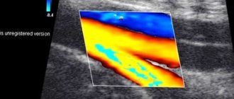
Ultrasound based on the Doppler effect (USDG or Doppler ultrasound) allows you to identify blood clots, inflammations of various types and thoroughly examine the structure of blood vessels, as well as the veins of the lower extremities. Diagnostics is non-invasive, has no contraindications, can be carried out frequently, and is completely safe for health. The examination results are interpreted by a phlebologist.

Indications for the use of Doppler Doppler Ultrasound (USD) of the veins and vessels of the lower extremities
A phlebologist can refer you for an ultrasound scan after examining the patient, especially if there are complaints of cramps, pain in the legs, a feeling of numbness, or muscle weakness. The procedure is also indicated when swelling occurs, changes in the color and structure of the skin, and when noticeable veins appear. Ultrasound examination of the veins and vessels of the lower extremities makes it possible to establish a diagnosis even in the early stages of the development of pathologies. The procedure helps to find the cause of the disease, as well as evaluate the effectiveness of the treatment received.
Diseases and conditions for which ultrasound examination is necessary:
- Violation of vascular patency.
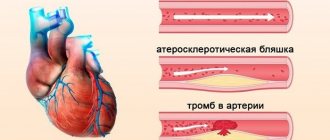
- Atherosclerosis of the vessels of the lower extremities.
- Varicose veins
- Inflammatory processes in blood vessels and veins.
- Thrombosis.
- Other disorders associated with venous circulation.
Sometimes a doctor may prescribe Doppler ultrasound for ailments that are not related to the veins of the extremities. Such conditions include excess weight, asymmetrical pulse, high blood pressure, previous heart attack, systematic dizziness, arm weakness and others. It is recommended to periodically undergo ultrasound examination in case of thrombophelitis, diabetes and a possible hereditary predisposition to the development of vascular diseases. You should not postpone the examination if you feel pain of any kind when walking, since the advanced stages of many diseases cannot be cured.
Doctors in this field
What is examined during Doppler Doppler Doppler (US) of the veins and vessels of the lower extremities
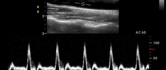
During the procedure, the doctor evaluates the speed of blood flow, the patency and lumen of blood vessels, the functioning of the valves, the location and size of blood clots. The narrowing of the arteries and the thickness of the vascular walls are also examined. The arteries are examined in detail; with the help of ultrasound, the doctor can identify many diseases: Raynaud's disease, arterial insufficiency, obliterating atherosclerosis and endarteritis, as well as aneurysms.
If atherosclerotic plaques are detected during diagnostics using ultrasound, their size and structure must be assessed. In case of thromboembolism (blood rupture), an ultrasound scan of the blood vessels is performed urgently to find the detached blood clot and the site of blockage. This condition requires urgent intervention, as it is very dangerous to human health.
The advantages of vascular Doppler ultrasound include the ability to examine the extremities of bedridden patients, since portable devices can be used and there is no need to transport or disturb patients.
How to properly prepare for an ultrasound examination of the veins and vessels of the lower extremities
Ultrasound examination does not require preparation, except for hygiene procedures before visiting a doctor. The patient does not need to limit himself in food or water. An ultrasound examination lasts about 20-25 minutes.
How does an ultrasound scan of the veins and vessels of the lower extremities work?
Before starting the diagnosis, the patient must remove clothing covering his legs. The uzologist applies a special conductor gel to the examined areas of the limbs. Ultrasound examination can be performed from different angles, so the patient may be asked to lie on a couch or take an upright position. The examination begins with the lower veins, then the outer parts and structures of the arteries are examined. Be sure to check the structure of the popliteal, femoral, small, and posterior tibial veins. Data from the examination of the vessels of the extremities are displayed in the form of a graphic image (black and white picture) on the monitor. The doctor describes the characteristics obtained and draws up a conclusion.
How is the ultrasound procedure performed?
Before the examination begins, the doctor examines the patient, palpates (feeling) the vessels and determines those that need special attention during the scan. Diagnostics is performed both in a sitting and in a lying position of the patient. This nuance is caused by the property of superficial veins to change their lumen depending on the position of the body.
During an ultrasound scan of the veins of the lower extremities, special tests are used to assess the function of their valves: scanning is carried out before and after the subject performs certain actions (squats, a series of deep breaths and exhalations).
Service cost*
| NAME | PRICE (Kolomenskaya) | PRICE (Vidnoe) |
| Duplex scanning of the veins of the lower extremities (on the device Voluson, Mindray, Logiq) | 4100 rubles | 4100 rubles |
| Duplex scanning of vessels (arteries and veins) of the lower extremities (on the device Voluson, Mindray, Logiq) | 6300 rubles | 6300 rubles |
| Ultrasound Dopplerography of the arteries of the lower extremities (on the device Voluson, Mindray, Logiq) | 2900 rubles | 2900 rubles |
| Duplex scanning of the arteries of the lower extremities (on the device Voluson, Mindray, Logiq) | 2900 rubles | 2900 rubles |
| Ultrasound Dopplerography of vessels (arteries and veins) of the lower extremities (on the device Voluson, Mindray, Logiq) | 3800 rubles | 3800 rubles |
| Ultrasound Dopplerography of the veins of the lower extremities (on the device Voluson, Mindray, Logiq) | 3500 rubles | 3500 rubles |
We accept payment:
*Attention! The indicated prices are provided as reference information and do not constitute a public offer. Check current prices by phone and directly in clinics.
Ultrasound of veins
Ultrasound examination of veins is a method for studying the vascular wall of veins and arteries, which allows one to simultaneously obtain reliable information about the state of blood flow in the vessels. The indisputable advantages of the technique include:
- absolute safety – allows you to carry out the procedure an unlimited number of times, which is ideal for preoperative and postoperative diagnostics, tracking the dynamics of treatment;
- simplicity of execution - does not cause any discomfort to the patient;
- accessibility – the price of the service is low, acceptable for all segments of the population; efficiency – it takes little time, the result is provided within a few hours.
Ultrasound is based on the ability of mobile blood cells to reflect beams of ultrasonic waves with a certain change in their characteristics. A special sensor picks up these reflected signals, and the computer calculates the speed of blood movement, which makes it possible to judge the presence or absence of blood flow disturbances.
The value of ultrasound of the veins of the lower extremities, upper extremities and arteries lies in the possibility of detecting the early stages of vascular diseases, when their clinical manifestations are minor or completely absent.
Preparation for ultrasound examination of veins and arteries
No special preparation is required for this study. However, before performing the procedures, it is recommended to remove hair from the skin of the lower extremities. The patient is prohibited from smoking, drinking strong coffee, tea, or energy drinks immediately before the examination.
Algorithm for performing ultrasound of the lower extremities
Before the procedure, the patient is asked to remove clothing from the lower extremities. According to the decision of the functional diagnostics doctor, the patient is examined lying on the couch or standing.
Diagnosis is carried out as follows
the patient is placed on his stomach on the surface of the skin, the gel for research is evenly applied, then the sensor moves gradually, following the course of the great vessels, then the patient is placed on his back, the gel is applied, the study of arteries and veins is continued during a dynamic study, compression tests are performed, the gel is washed off at the end of the procedure.
After completing the session, the doctor describes and analyzes the data obtained from studying the vessels.
Doppler ultrasound of the veins of the lower extremities: indications
Examination of the leg veins is prescribed when the following symptoms appear:
- swelling of the legs;
- cramps of the calf muscles;
- leg pain;
- redness, inflammation along the veins;
- unnatural skin tone on the legs - red, purple, bluish;
- visible dilated veins or spider veins;
- decreased temperature of the extremities, sensation of goosebumps;
- pain when walking;
- pallor of the skin on the legs.
All these manifestations may indicate disturbances in the proper functioning of the venous system. When planning treatment, the phlebologist prescribes an ultrasound scan. This simple informative study is necessary for an accurate diagnosis and assessment of the condition of the vessels of the lower extremities. Such an examination is also required if atherosclerotic changes in the arteries of the legs are suspected. For patients with diabetes, it is recommended to do Doppler ultrasound of the legs once or twice a year. Preventive examinations are also indicated:
- pregnant women;
- people over 45 years old;
- smokers (especially those who have been using tobacco products for more than 15 years);
- persons with a family history.
Indications
In each specific case, the area of localization of the vessels for which ultrasound scanning is required is determined by the doctor. Among the most common indications are:
- thrombosis, phlebitis or thrombophlebitis;
- phlebeurysm;
- congenital anomalies of vascular development;
- diagnostics before and after vascular operations;
- injuries with possible damage to blood vessels;
- swelling, numbness of the limbs, change in skin color;
- pain along deep veins, etc.
Ultrasound diagnostics of the veins of the lower extremities is mandatory for diabetics, people suffering from excess weight and problems with blood pressure. As a preventative measure, it is advisable for smokers to undergo testing.
Ultrasound Dopplerography: price and features of Dopplerography
Doppler ultrasound (USDG) is a modern diagnostic technique that is used to assess the condition and identify pathologies of blood vessels. Like regular ultrasound, Doppler ultrasound is completely safe and has no contraindications; it cannot be performed unless there are open wounds on the skin of the extremities. This is a non-invasive, painless procedure that can be performed on patients of any age.
Using Doppler ultrasound, you can identify various pathologies of arteries and veins, including those that are asymptomatic or accompanied by uncharacteristic symptoms. Atherosclerotic plaques are detected; the degree of vascular stenosis and the presence of thrombosis are determined; The condition of the valvular apparatus of the veins and the effectiveness of collateral blood flow are assessed. The study allows the doctor to obtain information about the condition of the blood vessels. In particular, thanks to Doppler sonography the following parameters become known:
- Blood flow speed.
- The thickness of the vascular walls.
- Dimensions of the lumen of blood vessels.
- Pulsation index.
- Resistance index.
- Systole-diastolic indicators.
This information helps the doctor make the correct diagnosis and prescribe adequate therapy. Doppler ultrasound is also needed to monitor the effectiveness of treatment and assess the condition of blood vessels after the necessary procedures.
Ultrasound of the veins of the lower extremities
Types of ultrasound of arteries and veins of the lower extremities
Doctors at the Scandinavian Health Center assess the condition of the blood vessels in the legs using one or more types of Doppler ultrasound (USD) or duplex ultrasound scanning (USA) simultaneously. The center's equipment allows you to do:
- Duplex ultrasound of the legs is supplemented with functional tests: holding the breath, changing body position, etc. They make it possible to learn about the operation of the valves of the veins and collateral (additional) arteries in case of obstruction of the great vessels.
- Triplex examination: a modern method of ultrasound examination of blood vessels, combining visual examination of the vessel with mapping in color or power Doppler mode. The main advantage of the technique is its high information content and accuracy.
Of all the techniques, triplex scanning is the most advanced method for diagnosing diseases.
Indications for ultrasound of leg vessels
- swelling, tingling, aching, heaviness in the legs after walking or at rest;
- pain syndromes in the lower extremities;
- feeling of numbness;
- changes in skin color;
- visible dilated veins;
- spider veins, asterisks;
- muscle cramps;
- skin thickening along large vessels;
- trophic ulcers that do not heal for more than a month.
Doctors recommend undergoing an ultrasound of the vessels of the lower extremities as a preventative measure for people who have:
- metabolic disorders;
- diabetes mellitus type I or II;
- sharp fluctuations in blood pressure;
- angina pectoris or previous myocardial infarction;
- arrhythmia, other diseases of the heart and blood vessels;
- numbness, weakness in the legs, pain when walking or at rest;
- elevated cholesterol levels in a biochemical blood test;
- feeling that your feet are constantly freezing;
- bad habits.
There are no contraindications to ultrasonography of the veins and arteries of the lower extremities. For ultrasound examination of the veins and arteries of the lower extremities, special preparation for the study is not required (except for ultrasound examination of the vessels of the pelvic organs).
Features of ultrasound examination
Ultrasound of the vessels of the lower extremities lasts from 15 to 25 minutes, depending on the complexity of the diagnosed vascular problems. The procedure is performed in a vertical or horizontal position. During the examination, the doctor evaluates:
- vascular patency—no signs of thrombosis;
- quality and condition of the vessel wall, diameter and uniformity of the lumen;
- work of the valve apparatus of veins;
- speed indicators of blood flow.
Having assessed the data obtained, the doctor makes a conclusion and immediately gives it to the patient.
The Phlebology Department of the Scandinavian Health Center uses modern methods for diagnosing and treating venous diseases.