Types of ultrasound: Doppler ultrasound, duplex scanning and color Doppler mapping of the veins of the lower extremities
You can often hear the question, what is an ultrasound examination called? The question arises due to the fact that there is more than one ultrasound technique: triplex angioscanning, duplex ultrasound (USDS or USAS), Doppler ultrasound (Doppler ultrasound or USDG) of the veins of the lower extremities. The complexity of the terminology causes some confusion, but only for the uninitiated person. Regardless of how an ultrasound examination is done, it is always an ultrasound. Only the hardware method for diagnosing the veins of the lower extremities is changing. Let's look at both the terminology presented above and the differences between these research methods:
Doppler ultrasound or Doppler ultrasound of veins
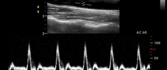
The ultrasound system of veins and arteries of the lower extremities is based on the Doppler effect - the reflection of ultra-high frequency sound waves from red blood cells moving in the blood vessels. Based on this, the specialist receives information about the speed of blood flow, the lumen of blood vessels, the presence and absence of varicose veins, weakness of the vascular walls, etc.
!
Doppler is the name of an Austrian physicist, but now it has become a household name.
You can often hear that an ultrasound examination with Doppler is prescribed. This means that a procedure called Doppler sonography is ahead. At its core, it is a conventional ultrasound examination. To obtain the most informative data, Doppler ultrasound can be performed in duplex or triplex modes. Doppler ultrasound is considered one of the simplest ways to diagnose venous valve pathologies and assess blood flow.
Duplex scanning or ultrasound scanning of veins
What does duplex scanning do? What is the difference with Doppler ultrasound? This ultrasound examination of the veins of the lower extremities is considered the most complex and combines several research techniques.
Expert opinion
Doppler helps to study blood vessels, blood flow and their characteristics. A scanner operating in B-mode visualizes the received information on the monitor in on-line mode as two-dimensional gray scale images of anatomical structures. Ultrasound scanning makes it possible to differentiate pathological conditions of blood vessels that affect the decrease in blood flow, including cholesterol plaques and blood clots.
Vascular surgeon, phlebologist
Osipova Ekaterina Yakovlevna
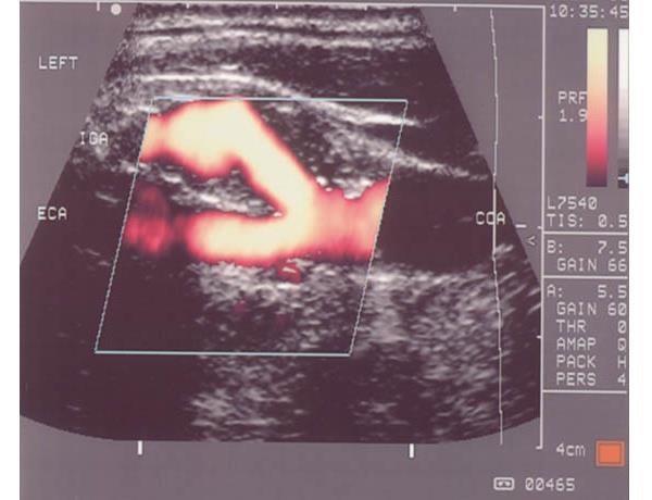
Ultrasound ultrasound with color mapping / color Doppler mapping / triplex angioscanning
How is ultrasound performed using this technique? Triplex angioscanning allows you to maximally examine the blood flow of not only superficial and deep veins and arteries, but even the smallest connecting capillaries of the lower extremities. This is possible due to the combination of all the methods described above and the visualization of all the vessels of the legs with blood flow in color. This type of study is the most highly accurate in terms of determining not only the patency of blood vessels, but also their stenotic lesions.
Indications for ultrasound examination of the veins of the lower extremities
The difference between duplex scanning and triplex scanning of blood vessels
Duplex ultrasound scanning of blood vessels
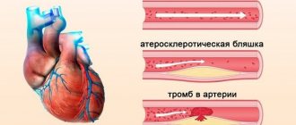
Ultrasound duplex scanning, firstly, allows you to visually assess the patency of the vessel and identify the reasons for the violation of its patency - tortuosity of the course, thickening of the walls (stenosis), the presence of blood clots, plaques, developmental anomalies, installed stents, postoperative joints of vessels, etc. Secondly, the speed and direction of blood flow. Thus, two functions are examined, i.e. duplex.
In a duplex study, an ultrasound scanner using the echolocation method can construct a flat two-dimensional image of the lumen and wall of the vessel in real time. It should be noted that the lumen of the vessel itself can be shown both along and across.
Triplex ultrasound scanning of blood vessels
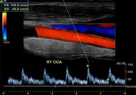
Ultrasound triplex scanning expands the capabilities of duplex scanning by adding color Doppler modes, i.e. color image of the movement of blood in the vessels, from which one can more clearly judge the direction of blood flow and its speed, as well as allowing a more accurate assessment of the patency of the vessels and the degree of stenosis. Thus, three goals are achieved, which determined the name of the method - triplex:
- the anatomy of blood vessels is studied
- blood flow is assessed
- an accurate assessment of vascular patency is made in color mode
Duplex and triplex scanning make it possible to accurately assess the diameter and course of blood vessels, identify the structure of the walls of arteries and veins, and promptly and quickly identify atherosclerotic plaques or abnormalities in vascular development. Reception is conducted by:
Anna Igorevna Zimovskaya , ultrasound doctor, Doppler ultrasound doctor, cardiovascular surgeon Polukhina Tamara Andreevna Ultrasound doctor, Doppler ultrasound doctor Lyudmila Vasilievna Boyko - ultrasound doctor, Doppler ultrasound doctor
Ultrasound diagnostics are carried out at the following addresses:
6th Sovetskaya street, 24-26, 3rd floor, Yesenina street, 2, building 3.
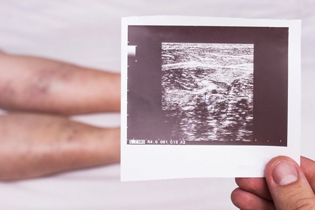
Ultrasound of the veins of the lower extremities: indications and contraindications
Ultrasound of the leg veins is informative and, most importantly, a safe diagnostic method, as more than 60 years of medical practice has shown. In this regard, there is more than one indication for which an ultrasound of the legs is performed to check the veins and arteries:
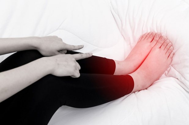
- constant feeling of fatigue and heaviness in the limbs;
- pain syndrome;
- increasing or constant swelling;
- constant or sudden redness along the veins, accompanied by a burning sensation;
- manifestation of the vascular pattern – asterisks;
- the formation of trophic ulcers localized in the lower legs and feet;
- paroxysmal contractions of the leg muscles;
- changes in skin color;
- tingling feeling;
- cold feet.
All these signs may be symptoms of vascular disease. If they are detected in oneself, a person should consult a specialist for advice. He will examine the limbs and conduct a survey.
!
If necessary, if pathology is suspected, the doctor will certainly prescribe an ultrasound scan of the veins and vessels of the lower extremities.
To be able to monitor the state of health and the dynamics of diseases, a phlebologist prescribes an ultrasound scan of the veins of the lower extremities for:
- varicose veins;
- thrombophlebitis;
- diabetes mellitus;
- atherosclerosis;
- lymphatic edema;
- chronic venous insufficiency;
- thromboangiitis;
- postthrombotic disease;
- postoperative period, etc.
As mentioned earlier, ultrasound is a safe diagnostic method. But does this mean a complete absence of contraindications? Despite the safety of the method, it is important to note that there are absolute contraindications to its implementation. Therefore, it is impossible to find out the condition of the arteries and veins of the lower extremities using ultrasound if:
- infectious diseases and skin lesions;
- myocardial infarction;
- exacerbation of heart failure and arrhythmia;
- asthmatic attacks;
- cerebrovascular accidents in the acute stage;
- mental disorders during their decompensation;
- burn disease.
Carrying out an ultrasound examination in any of these conditions can provoke complications of the disease. However, it is worth noting that ultrasound of the veins of the lower extremities is not prohibited during pregnancy. As you know, during this period of a woman’s life, safety comes first.
Main similarities of ultrasound research techniques
Ultrasound scanning and ultrasound scanning have many positive common similarities, which are as follows:
- both studies are non-invasive methods, which makes them completely safe and painless; During the procedure, the patient does not feel any discomfort or inconvenience;
- Ultrasonic waves are completely safe for the human body, so these diagnostic methods can be used in pregnant women and young children;
- high information content.
May be of interest: What is duplex scanning of the veins of the lower extremities? Decoding the results
Preparation for ultrasound of the veins of the lower extremities
In order for the result to be reliable, most laboratory and instrumental diagnostic methods require certain preliminary preparation.
How to prepare for an ultrasound examination? There is no advance and complex preparation for venous ultrasound. There is no need, for example, for a diet. But it is important to exclude negative factors that affect the condition of the vessels of the lower extremities. This includes smoking and alcohol – at least 24 hours before. These 2 factors provoke dilation of veins and arteries, which can indirectly affect the result obtained.
Expert opinion
Before giving a referral for this diagnostic procedure, the patient must receive preparation recommendations from the doctor. If the patient is taking medications that have a vasoconstrictor or vasodilator effect, the doctor will most likely stop them a day before the test. This is necessary in order to eliminate diagnostic inaccuracy.
Vascular surgeon, phlebologist
Osipova Ekaterina Yakovlevna
On the day of the study, some time before it, the patient performs hygienic measures. Takes a shower or washes lower limbs. If you wish, for your own convenience, you can choose comfortable clothes that will allow you to free the part of the body being examined to the maximum and without temporary delays.
How is ultrasound diagnostics of leg veins performed?
In what cases is ultrasound examination performed?
Ultrasound diagnostics with the Doppler effect are more often used to diagnose large vessels (carotid, vertebral and cerebral arteries, as well as leg veins).
The main indications for ultrasound scanning are:
- constant migraines combined with dizziness;
- frequent fainting;
- noise in ears;
- used in patients who have had a stroke;
- diseases of the spinal column;
- persistent increase in blood pressure;
- decreased visual function;
- diabetes;
- neurological diseases;
- swelling of the legs, accompanied by a feeling of heaviness and pain;
- the appearance of a vascular network, change in skin color on the legs;
- excess weight.
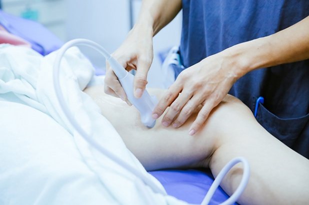
How is ultrasound examination of the veins of the lower extremities performed?
If an ultrasound scan of the veins of the lower extremities is prescribed, the question may arise as to how it is performed? Most likely, each of us has undergone ultrasound diagnostics at least once, and even in general terms, we have an idea of how this process is organized.
When you need to check the condition of the blood vessels in your legs using ultrasound, it will take from 20 minutes to an hour of your time. The first thing the doctor will ask the patient to do is to remove clothing covering the lower limbs. This is necessary for unhindered access to the area requiring research. After which the patient takes one of the positions required to begin the diagnosis - lying on his back and stomach, as well as a standing position. The doctor will tell the patient the order of positions.
When the patient has taken the required position, the specialist applies a contact medium – a universal gel – to the skin of the legs. It is on top of this product that the doctor will advance the ultrasound sensor.
In some cases, accuracy will require the application of special cuffs that allow blood pressure to be measured in the diagnostic area. Testing may also be required. What are they and how are they made? These are actions with the help of which an ultrasound diagnostic specialist will be able to establish the absence or presence of vascular damage.
!
Some of the most common ultrasound examinations of leg veins are: Valsalva stress and the Hackenbruch-Sicard test (cough test).
For varicose veins on the legs, these tests allow for effective analysis and identification of the disease even at its earliest stages. Valsalva stress is a specific breathing technique, and the Hackenbruch-Sicard test is a series of cough movements.
If the specialist considers that these manipulations are not enough for a complete diagnosis, then additional studies may be required.
Interpretation of ultrasound results of leg veins
Features of ultrasound examination
Doppler ultrasound is considered a mandatory test if the patient needs to check vascular patency, the characteristics of blood flow through them and the condition of the venous valves. The basis of this method was the Doppler effect. It lies in the fact that ultrasonic waves are reflected from moving blood cells.
The method has a number of features:
- During the diagnostic process, the doctor does not see any veins or arteries.
- It is quite difficult to identify the causes of the found pathologies, especially if a patency disorder is diagnosed.
- The method does not make it possible to see the integrity of the walls of blood vessels, their possible changes that affect the speed of blood flow.
- This method is used in diagnosing hypertension, varicose veins or identifying the cause of headaches.
This method allows you to see only three parameters, but the ultrasonic scanning method is more informative.