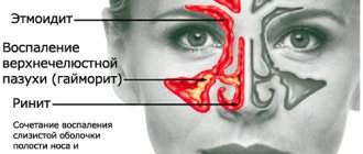
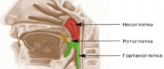
A person has four pairs of sinuses, which have names associated with their location: frontal, maxillary, ethmoid, sphenoid. They perform the functions of humidifying and warming the air, resonating, lightening the overall mass of the skull, etc.
Computed tomography of the nose and paranasal sinuses is a non-invasive, painless and quick diagnostic method. CT scans of the nose and paranasal sinuses are classified as x-ray methods, so you need to know that the body is exposed to some kind of radiation exposure.
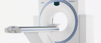
The computer tomograph of the Yusupov Hospital is a modern device of the latest generation, which makes it possible to obtain hundreds of layer-by-layer images in a short time, recording them both on film and on media.
You can sign up for an examination by phone or online. An individual consultation with a specialist from the Yusupov Hospital will help you understand the pathology and clarify all your questions regarding diagnosis and treatment. Yusupov hospital is open
The essence of the method
The essence of computed tomography of the paranasal sinuses is the mathematical modeling of the internal structure of the maxillary, frontal sinuses and surrounding structures based on the results of the passage of x-rays.
On a CT scan you can see the structure of the paranasal sinuses themselves, the level of fluid in them, and space-occupying formations. During a computed tomography scan, radiologists determine the condition of the walls and mucous membrane of the maxillary and other paranasal sinuses, the presence of secretions and excess tissue in their lumen. Using CT, doctors detect underdevelopment of the sinuses, bullous or paradoxical middle concha, an invented uncinate process, and hyperpneumatization of the ethmoidal crest cells. If there is a deformation of the nasal septum, computed tomography can reveal the severity, location, and shape. Using CT, the condition of structures closely associated with the nasal cavity and paranasal sinuses is determined - the bottom of the anterior cranial fossa, the walls of the orbit.
Computed tomography is 50 times more sensitive than classical radiography. When performing a CT scan of the maxillary sinuses, they are rarely limited to obtaining an image of one “slice” several millimeters thick. Modern tomographs that the Yusupov Hospital is equipped with make at least 10 sections about 1 mm thick. They are performed in different increments (usually several millimeters).
In order to navigate the location of the resulting layers relative to the human body, the radiologist immediately takes an overview digital image of the entire studied area (x-ray topogram), on which the various layers are displayed. CT scan of the maxillary sinuses using contrast allows you to increase the contrast of the image, highlight vascular formations, cysts, benign and malignant tumors, and their metastases.
What will a CT scan of the maxillary sinuses show?
Based on the results of a CT scan of the upper jaw and maxillary sinuses, the doctor determines:
- the location of the nasal septum and bones forming the maxillary cavities;
- sinus drainage pathways;
- symmetry of the left and right sides of the nose and sinuses;
- degree of pneumatization of the sinuses;
- condition of the tear ducts.
CT scan of the upper jaw reveals:
- inflammatory processes, including the presence of exudate in the sinuses;
- tumors of the mucous membranes;
- polyps;
- granulomas and cysts;
- abscesses;
- foreign objects in the nasal sinuses (filling materials, root fragments after tooth extraction, dental implants);
- damage or deformation of the nasal bones;
- additional voids (ostia);
- pathological communication of the maxillary sinus with the oral cavity (oroantral fistulas).
Thanks to the high resolution of the computed tomography apparatus, it is possible to detect the smallest pathological formations, assess their size, location and relationship with surrounding tissues. Based on the interpretation of CT data and comparison with the results of other types of examination, the dentist or ENT makes a diagnosis.
How is a CT scan of the upper jaw performed?
CT scan of the upper jaw and maxillary sinuses looks like this:
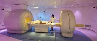
- the patient lies down on a retractable couch, the neck is fixed using a special stand;
- the head end of the couch smoothly slides into a ring-shaped device where sensors and an X-ray emitter are located;
- moving in a spiral, the scanning complex takes a series of layer-by-layer images of the studied area;
- if necessary, a contrast agent is injected into the vein and the examination continues;
- Upon completion of the diagnostics, the device turns off.
Any movement of the patient during a CT scan of the upper jaw can degrade the quality of the images, so it is necessary to maintain a static position throughout the entire scanning period.
Indications
Indications for CT scanning of the nose and paranasal sinuses are quite numerous. It is also necessary to take into account that in some pathologies it is not advisable to administer a contrast agent, and sometimes performing a CT scan without contrast reduces the information content of the method to zero. Indications for CT scanning of the nose and paranasal sinuses are:
- diseases of inflammatory origin of the nasal cavity and paranasal sinuses;
- cysts in the nasal cavity and paranasal sinuses;
- tumors in the nasal cavity and paranasal sinuses (benign and malignant);
- polyps in the nasal cavity and paranasal sinuses;
- osteomyelitis;
- developmental anomalies of the nasal cavity and paranasal sinuses;
- traumatic injuries to the face;
- as one of the stages of preoperative preparation;
- sinusitis;
- frontal sinusitis;
- sphenoiditis;
- ethmoiditis;
- constantly recurring nosebleeds;
- toothache and headache of unknown etiology;
- purulent process of the lacrimal sac, etc.
If any of the above diseases are present, it is necessary to conduct a CT scan of the nasal sinuses in Moscow at the Yusupov Hospital.
Registration is carried out by phone and online. Make an appointment
CT scan of the paranasal sinuses: what is it?
CT scan of the paranasal sinuses is a type of X-ray tomography. During the examination, X-rays pass through different areas and tissues of the body. The X-ray tube takes many pictures in different planes, then the computer analyzes the information received and creates a layer-by-layer 3D image of the desired area.
A computed tomograph is widely used in ENT practice and helps the otorhinolaryngologist to see in detail and clearly the condition of the paranasal sinuses and adjacent areas. CT scan of the nose shows abnormalities in the structure of the nasal cavity; helps to recognize the beginnings of the inflammatory process or determine the extent of its spread. A computed tomogram of the sinuses allows the ENT doctor to draw correct conclusions about the patient’s health status, correctly diagnose the disease and prescribe appropriate treatment.
Contraindications
Contraindications to computed tomography of the paranasal sinuses and nasal cavity differ depending on the need for administration of a contrast agent. If a patient has an allergic reaction to an iodine-containing medication, then a CT scan becomes impossible. General contraindications to computed tomography, both with and without contrast:
- pregnancy regardless of term;
- lactation period;
- decompensated diabetes mellitus;
- uncontrolled bronchial asthma;
- renal and liver failure;
- the patient's condition requiring artificial life support;
- acute cardiovascular pathology;
- thyroid diseases.
If the patient’s body weight exceeds 140 kg, then this is considered a relative contraindication, since the computed tomograph is not designed for such a weight.
In patients with mental illness, CT scanning is also a relative contraindication. In cases where CT is a vital procedure, it is performed under general anesthesia.
Some experts consider the lactation period to be a relative contraindication. Considering the risks, doctors recommend performing a computed tomography scan with contrast, but avoiding breastfeeding for two days. The final decision on restrictions for the study is made by the attending physician.
Indications for MRI of the sinuses
This type of diagnosis is prescribed, as a rule, in cases where it is not possible to identify the cause of the disease for quite a long time. These include:
- systematic pain in the nasal sinuses, in which there are signs of a pathological course on x-rays;
- periodic headaches for no apparent reason, which even painkillers do not always help relieve;
- frequent nosebleeds;
- copious nasal discharge, uncharacteristic of a cold;
- any significant facial injuries;
- a sharp decrease in the sense of smell, a drop in visual acuity;
- systematic congestion of the nasal passages;
- in the presence of symptoms of various diseases, which include polyposis, sinusitis, sinusitis, cyst proliferation.
Most often, specialists prescribe MRI for sinus cysts and sinusitis, and the procedure is performed on patients multiple times. In addition, MRI for cysts allows in many cases to avoid the need for surgical intervention if treatment is started in a timely manner.
Preparation
Computed tomography of the paranasal sinuses does not require special preparation. Before a diagnostic examination, you must remove all metal objects from yourself, including jewelry, glasses, hairpins, dentures and other objects that may interfere with the interpretation of the results.
A few hours before the manipulation, it is forbidden to consume food, and in some cases, water.
During a CT scan, the patient must lie still throughout the examination to ensure clear images. For this purpose, special rollers are often placed.
The main advantage of computed tomography is the minimal radiation exposure to the human body. This procedure does not take much time (about 30 minutes), so it is suitable even for people suffering from severe pain and patients with mild claustrophobia.
How the research is carried out
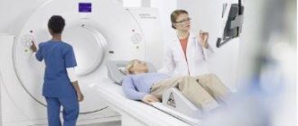
No special preparation is required for the procedure. You must remove all metal objects or clothing with metal elements. The patient is placed on the couch and his body is secured with belts, and his head is placed on a special headrest. You should not move during the entire procedure, as movement may affect the accuracy of the result. If the patient cannot lie still (for example, children or people with mental disabilities), the doctor may use anesthesia.
If the CT procedure requires the injection of a contrast agent, such as iodine, into the patient’s blood, you should refrain from eating for several hours before the procedure.
Then the couch on which the patient lies is pushed into the tomograph. After this, the doctor retires to another room where a computer is installed that records the tomograph signals. All this time, with the help of sound equipment, the doctor and the patient are in touch. If the patient feels any discomfort, he reports it, and the doctor hears it. The study itself lasts no more than ten minutes.
Then the specialist deciphers the tomogram and gives the patient an x-ray and a disk recording the results of the study, as well as a medical report.
CT, as a method of radiation diagnostics, involves radiation exposure to the body. Therefore, it is recommended to do CT scans no more than three times a year. The cost of this study varies from 3,000 to 5,000 rubles.
Progress of the study
Computed tomography of the sinuses is performed with the patient in a horizontal position - he lies on his back on the table, and the tomograph ring moves around his head. There is no staff in the equipment room, the patient is alone, but the equipment allows communication if necessary. The intervals between sections are assigned individually. The patient may feel discomfort during a CT scan due to noise and feel hot, but this goes away on its own after some time. After completion of the study, photographs and digital media are provided. It is necessary to understand that the conclusion is not a diagnosis. The final diagnosis is made by the attending physician.
CT with contrast
To clarify the nature of detected pathological changes or determine the extent of tumor cells, CT PPN can be supplemented with intravenous administration of a contrast agent.
Computed tomography of the paranasal sinuses with contrast is contraindicated:
- patients with renal failure;
- patients with severe diabetes mellitus;
- during pregnancy and lactation;
- patients with an allergic reaction to an iodine-containing contrast agent.
Advantages
Computed tomography of the paranasal sinuses in Moscow is one of the safest methods for studying all ENT pathologies. This is a reliable way to visualize all the sinuses and sinuses of the nose. The main advantage of computed tomography is the simultaneous acquisition of images of bones, soft tissues, blood vessels and lymph nodes.
Unlike magnetic resonance imaging, computed tomography can be performed if the patient has any implanted medical device.
The diagnosis, which is established after a computed tomography scan, may eliminate the need for additional diagnostic methods or even surgery.
For many years, the Yusupov Hospital has been famous for its specialists and innovative equipment that meets all standards. The accuracy and speed of CT scanning allows patients to immediately receive results and begin treatment. CT scanning of the paranasal sinuses in Moscow is popular due to its enormous capabilities and the absence of side effects.
Benefits of the procedure
The main advantages of CT are:
- painlessness and safety of the study;
- high research speed;
- allows for timely diagnosis of pathological processes in the paranasal sinuses;
- the ability to check the skull bones, soft tissues and blood vessels;
- the resulting image is of high quality, on which all changes will be visible;
- use in diagnosing patients who have had medical devices installed;
- the use of CT in an emergency allows you to diagnose disorders and begin immediate treatment.