3D radiography (or computed tomography) is a modern diagnostic method that is used in many medical fields. Dentistry cannot do without it either. Three-dimensional photographs of the dental system are performed when preparing patients for orthopedic, orthodontic manipulations or dental implantation. With their help, dentists and surgeons monitor the quality of treatment. We tell you what the features of dental computed tomography are and in which Moscow clinics it can be done.
How is a 3D dental image performed?
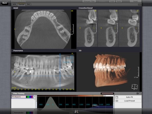
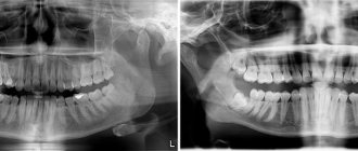
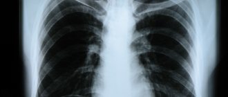
The procedure is based on scanning the jaw with an X-ray beam. Only the radiation does not spread like a fan, as with ordinary x-rays, but in a cone. During one revolution around the head, a cone-beam tomograph takes from 150 to 200 images of the patient’s teeth in different projections. The images are immediately transferred to a computer, which builds a 3D model of the dental system based on them.
Including data processing, the study takes no more than 10 minutes. The results are saved to a CD and, if necessary, printed onto digital film.
The uniqueness of tomographic images lies in the fact that with their help the doctor can examine any tooth from all sides and in all conceivable sections. On a computer, a 3D model can be rotated, individual fragments can be selected from it, enlarged and studied layer by layer. Compared to two-dimensional radiography, dental computed tomography provides many times more information and reduces the risk of diagnostic errors.
Contraindications
There are not many contraindications to the study:
- Pregnancy;
- Diabetes;
- Some liver and kidney problems;
When performing computed tomography with the introduction of a contrast agent, an allergic reaction to iodine is a contraindication, since iodine is contained in the contrast.
Patients with hyperkinesis are allowed to be diagnosed with special caution, since it is important to maintain a motionless position during the procedure. Movement during the examination may blur the images.
Main advantages of CT
- Speed of research. The study itself takes 15-20 seconds. Its results are immediately reproduced on a computer. The radiologist spends several minutes transcribing the data, burning a CD, and printing the images. The patient does not need to wait for the doctor to “develop” the images.
- Convenient storage and transfer of data. Film photographs fade and lose clarity over time. Images on a CD can be stored indefinitely. Over the Internet, data from the disk can be sent to the attending physician or other specialists for third-party consultation.
- Safety. The radiation dose of CT is lower than that of traditional panoramic examination (orthopantomography).
- High reliability and accuracy. 3D computer photographs of teeth are clear and detailed. This makes it possible to get an accurate picture of the location, size and shape of all structures of the patient’s dental system. The doctor does not have to do repeated studies to clarify the diagnosis.
Advantages of an intraoral 3D scanner
The modern technique has many advantages:
- highest accuracy - the error of data from intraoral 3D scanners is only 12 microns versus 100 microns in the case of making an impression using silicone materials;
- maximum convenience for the doctor and the patient - the manipulations are simple and do not cause discomfort;
- the device scans each print in just 24 seconds;
- excellent information content - the three-dimensional model visualizes all defects and shortcomings of the maxillofacial system.
Digital impressions are sent to the laboratory or other dental clinic without delay. They show as accurately as possible the condition of the teeth and the jaw as a whole. The dentist can study the impression in detail and suggest the most suitable treatment option, taking into account all the features of the case.
Review of Moscow clinics where you can get a CT scan
Many clinics offer dental x-rays in Moscow. For review, we selected five of the most reputable ones with high-quality equipment and a staff of qualified radiologists.
Unident
The Unident network includes 16 clinics located in different areas of Moscow. The clinics are staffed by pediatric and adult dentists in five areas: orthodontics, orthopedics, therapy, periodontology and implantation. The Unident brand has a good reputation and extensive experience: the first clinic of the network was opened in 1999. Prices for most services are average market prices.
You can get a 3D dental x-ray at 4 locations: in clinics on Yugo-Zapadnaya, Arbat, Taganka and Bobrovy Lane. The procedure is carried out using French Carestream tomographs, capable of taking images of both individual segments of the oral cavity (3 teeth), and both jaws with the paranasal sinuses and the temporomandibular joint. The tomograph in Bobrovy Lane is equipped with a special head holder (cephalostat), which allows you to take pictures for the purposes of orthodontics.
Cost of the service: The website shows only the lower price threshold - 1870 rubles. (for 3 teeth). The cost of decoding and printing the data is not indicated. An initial consultation with a specialist at the clinic will cost 900-1200 rubles.
More information about the clinic - unident.ru.
SM clinic
A network of multidisciplinary clinics operating on the market for more than 10 years. Dentistry departments with tomographic diagnostics are presented in clinics near the Voykovskaya, Tekstilshchiki and VDNKh metro stations. Modern German Siemens tomographs are used here, the advantage of which is low radiation exposure. A CT scan is done immediately for both jaws and paranasal sinuses. It is not possible to take photographs of individual teeth in the dental dental clinic.
Cost of the service: Together with data decoding and recording on a CD, a CT scan of both jaws will cost 6,300 rubles. An appointment with any dentist costs 1,500 rubles.
More information about the clinic - smclinic.ru.
All yours
An extensive network of dentistry, including 27 clinics in Moscow, St. Petersburg and Nizhny Novgorod. The chain strives to implement international dental standards and often runs promotions and discounts. Discount cards are offered to regular customers.
Computed tomography of teeth is carried out in the diagnostic center on Taganka using a Finnish tomograph Planmeka Pro Max. Patients can have both jaws photographed for periodontal or orthodontic treatment. The clinic does not mark missing teeth (that is, patients planning implantation will not be able to take a picture here).
Cost of the service: The cost of a CT scan is 3,750 rubles, interpretation of images is 1,000 rubles. The initial consultation with a specialist is free.
Read more about the clinic vse-svoi.ru.
ROOTT
A network of clinics founded by the international Open Association of Dentists. Many of the doctors undergo internships in Switzerland. The clinics represent all dental areas: implantation, orthopedics, orthodontics, therapy and surgery. The work is carried out using expensive modern equipment, but at the same time affordable prices are maintained.
In the clinic, you can take both general and targeted 3D photographs of your teeth. For patients planning for implantation, the radiologist will take a radiograph with markings. Patients are given a CD and, if desired, film images.
Cost of the service: Cost of CT scan of 3 teeth - 1300 rubles, both jaws - 5000 rubles, both jaws + paranasal sinuses + temporomandibular joint - also 5000 rubles.
Burning to CD is included in the cost of the service. Marking for implantation, depending on the size of the area being studied, will cost 1000-4500 rubles. When receiving treatment on the ROOTT network, data decoding and marking of missing teeth are provided free of charge. Initial consultations with blade specialists are also free. More information about the clinic. To get a consultation
Indications for CT scan of the jaw
Preparation for implantation
— to assess the condition of the jaw bone and determine the optimal locations for placing implants.
- Bone grafting, sinus lifting - to determine the volume of bone in the alveolar processes, the location of the maxillary sinuses.
- Surgical intervention - to study dental roots, impacted/dystopic teeth, identify neoplasms, diagnose inflammation in the root area.
- Orthodontic treatment – to correct complex malocclusions.
- Orthopedic treatment - to create the most accurate dentures.
- Endodontic treatment - to diagnose the condition of complex dental canals.
Possible harm
Cone beam dental computed tomography is the safest and fastest diagnostic method. Thanks to the use of a conical X-ray beam, the radiation dose received during the study is 10 times less than when using spiral CT. And the pulsating mode of the X-ray beam further reduces the radiation dose. The three-dimensional computed tomograph SOREDEX Scanora 3D is one of the safest devices in terms of X-ray radiation dose - only 0.035 m3v.
However, despite the safety of the study, CT also has contraindications. If we just talk about dental tomography, it is not performed during pregnancy (in the 1st trimester). 3D dental x-rays with contrast are prohibited for pregnant and lactating women, patients with endocrine disorders (diabetes mellitus, thyroid pathologies), renal failure and intolerance to iodine-containing drugs.
The process of creating inlays, veneers and crowns with the 3shape TRIOS scanner
Below we will look at how the process of creating a prosthesis is carried out using a 3shapetrios laser scanner. It includes the following sequential steps:
1. Preparatory stage. Before scanning your teeth using digital technology, the specialist will find out which specific teeth he will be working with, and will also clarify what materials the dentures will be made of. Next, he will start the computer, open the necessary program and prepare the equipment for operation.
2. After this, a 3D scanning of the teeth is performed directly , during which the doctor receives a highly accurate digital image. The entire process of creating a virtual impression takes 10-40 minutes. The scanner sensor is compact in size and equipped with a built-in miniature camera, so the entire procedure does not cause any discomfort to the patient, allows you to obtain a clear computer image, and is performed in a short time.
3. Next, the specialist studies the resulting image and evaluates the structural features of the teeth. If no problems are found, the virtual impression file will be sent to the dental laboratory for subsequent creation of a crown or veneer. During your second visit to the dental clinic, you can already count on the installation of a dental structure.
Thus, a 3D dental scan performed in our clinic will allow you to avoid long waits and erroneous results. We value your time, so we strive to reduce the process of prosthetics as much as possible, namely, to 2 visits: conducting a virtual scan using the 3shape TRIOS device and installing aesthetically attractive and high-precision orthopedic structures.
[]
Our clinic's orthopedic dentists:
Vasilyeva Anastasia Alexandrovna
Orthopedic dentist
Clinical experience over 10 years
Inessa Khautievna Fitsezh
Dentist-therapist, orthopedist
Clinical experience over 15 years
All doctors of our clinics »
An effective way to fix dental implants
An important point is that 3D scanning of teeth can also be used when installing crowns on implants. In this case, it is possible to obtain a digital abutment, which is fixed to the implant in a special computer program. However, it should be remembered that the virtual version is not applicable to all implantation systems. Strauman dental implants are among the most universal designs that have all the necessary virtual copies. In this situation, intraoral examination allows us to determine the shade and shape of the future prosthesis, which will subsequently be installed on the adapter.
Also considered unique is the fact that possible inaccuracies can be avoided and stress factors can be eliminated at the planning stage, when it is necessary to clearly determine the location of the future structure on the bone tissue. This is facilitated by the use of an intraoral 3D scanner, which allows you to take an accurate picture of the jaw and digitally design the area of fixation of the dental implant. Next, with the help of special computer technology, information is supplied to the appropriate device, allowing you to create a template, thanks to which the specialist has the opportunity to fix the implant in the bone tissue as clearly as possible. Based on the above facts, we can conclude that the 3shape Trios scanner plays a vital role in the implantation procedure: it eliminates the risk of any inaccuracies at the planning stage, and also helps prevent errors in the manufacture of the implant prosthesis. As a result, the patient receives a full guarantee of a successful result and gains confidence in the perfection of the dental restoration procedures performed.