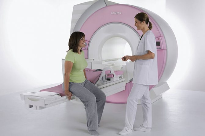
MRI or magnetic resonance imaging is the most accurate diagnostic test that provides greater detail of the internal structures of the body without the use of radiation. MRI uses a magnetic field, radio waves, and a computer to create clear, exceptionally detailed “images” of the area being examined. The MRI itself is a painless procedure.
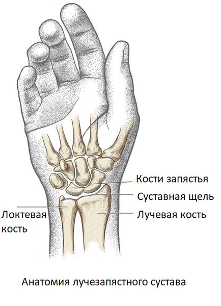
Anatomy of the wrist joint
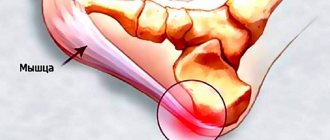
The wrist joint is one of the most mobile and complex articulations in the human body, formed by three small carpal bones (scaphoid, lunate and triquetrum), the articular surface of the radius and a cartilage disc. Normal wrist joint function is essential for the precise, intricate hand movements needed in many areas of human activity. This is a rather fragile joint, easily damaged by injuries, which is due to the not very powerful ligaments and complex structure.
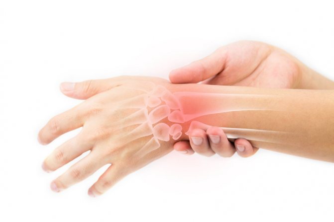
Diseases of the wrist joint
The most common injuries to the wrist joint are fractures of the head of the radius and ulna, subluxations and dislocations. The most common mechanism of fracture is a fall on outstretched arms. Dislocations of the wrist joint most often occur in children under 5 years of age, which is due to the weakness and high extensibility of the ligaments that strengthen the joint. The most common mechanism for dislocation of the wrist joint in children is a tug on the hand with simultaneous rotation along the longitudinal axis of the joint, which occurs when an accompanying adult tries to keep the child from falling.
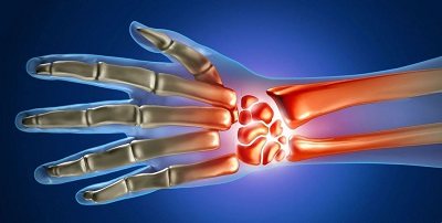
The wrist joint is susceptible to inflammatory processes, for example in rheumatoid or other arthritis. The cause of the disease can be specific infections (gonorrhea, chlamydia, tuberculosis), autoimmune diseases (rheumatism, rheumatoid arthritis), metabolic disorders (gout). Less commonly, the cause of inflammation of the wrist joint is a direct hit of a nonspecific infection during injury or through the bloodstream (purulent arthritis). The inflammatory process may involve the synovial bursa of the joint and tendon.
With age, the risk of degenerative-dystrophic phenomena increases. Osteoarthritis of the wrist joint occurs as a result of metabolic disorders in the articular cartilage. Professional activity may be a predisposing factor - most often PC operators, artists, workers who use jackhammers, rotary hammers and drills, and musicians encounter osteoarthritis.
Space-occupying formations of the wrist joint are a large group of benign (synovial cysts, hygromas, fibromas, chondromas) and malignant neoplasms. MRI of the wrist joint is ideal for diagnosing all possible diseases in this area, due to the ability of the method to “see” soft tissue, unlike radiography or computed tomography. The latter methods are good for diagnosing fractures, but not other diseases.
Indications and contraindications for MRI of the wrist joint
Magnetic resonance imaging of the wrist joint is indicated in the presence of the following complaints:
- pain;
- limited mobility, inability to bend or straighten the hand;
- swelling, redness of the skin in the wrist area;
- deformity of the wrist joint;
- nodes, swelling, tumor-like formations in the area of the wrist joint.
MR imaging of the wrist joint is contraindicated:
- children under 5 years of age (the study is carried out only in departments specially created for this, since MRI in children under 5 years of age requires sedation and medicated sleep due to the inability of small patients to remain still during the examination);
- patients with defibrillators, pacemakers, neurostimulators, cochlear implants and other electronic devices implanted into the body (magnetic fields disable implants);
- patients with metal foreign bodies (bullets, shot, shrapnel, metal shavings in the eyes) or medical devices (vascular clips, stents, prostheses) in the body (risk of injury or bleeding due to the displacement of a metal object under the influence of a magnetic field);
- patients with excess body weight, weighing more than 130 kg and torso girth more than 150 cm (physically do not fit into the tomograph);
- pregnant women during the 1st trimester (in the 2nd and 3rd trimester without restrictions).
MRI of the hands
Duration of the examination
: 30 min min.
Preparing for the examination
: No
Contraindications
: Yes
Preparation of the conclusion
: 40-60 min min.
Restrictions
: No
Magnetic resonance imaging is an ultra-precise way to diagnose various body systems. MRI of the hand is practically the only method for determining pathologies of the wrist joints. The technology of the procedure allows you to obtain high quality images and detailed visualization of hand tissues. Thanks to the capabilities of tomography, the most effective and adequate treatment is developed.
The essence of the method
MRI of the hand works on the basis of nuclear magnetic resonance. The tomograph uses a magnetic field to capture the energy coming from the nuclei of atoms. The information from the device is transmitted to the computer screen in the form of a high-resolution image. A feature of the diagnosis is the display of soft tissues without bone structure. This facilitates the process of diagnosing the hand of the upper limb.
MRI of the hand, in its technological features, is second only to arthroscopy. The latter method is a surgical procedure in which a camera is inserted into the joint of the upper limb. The technology is effective, but expensive. In addition, the operation damages the skin, so it is better to do an MRI first.
Standard diagnostics can be performed with a contrast agent. The patient is injected intravenously with a special drug that is absolutely safe for the body. The substance indicates neoplasms in the area being examined. The use of contrast is effective when it comes to surgical intervention and monitoring the surgical process.
An MRI of the hand has a number of advantages:
- obtaining images of pathology in different planes;
- the only diagnostic method for nerve damage;
- no radiation exposure;
- information content during surgical intervention;
- the effectiveness of contrast, which rarely causes allergic reactions;
- speed of results.
The manipulation is planned at a convenient time. After the procedure, the patient can return to their normal lifestyle.
For what diseases is it performed?
Pathologies of the hands are usually accompanied by pain, redness and swelling of the tissues. The causes of diseases can be genetic and acquired. These disorders lead to dysfunction of the wrist joint. Therefore, it is important to carry out tomography as early as possible to prevent the development of pathology.
Hands are involved in almost all areas of life. If the wrist joint hurts, the patient is like “without hands.” The main diseases in this area:
- Osteoporosis of the hands is the destruction and thinning of the joint.
- Dupuytren's contracture is a pathology caused by the flexion of the fingers towards the palm while extension is impossible.
- Arthritis, osteoarthritis - inflammation of the joint due to infection.
- Kienböck's disease is an aseptic necrosis of the lunate bone resulting from heavy physical exertion.
- Gout is the accumulation of uric acid in the hands.
- Osteonecrosis is tissue death as a result of circulatory problems.
- Carpal tunnel syndrome is a neurological pathology caused by pain and numbness in the fingers.
Magnetic resonance imaging technology can detect tumors, infections, fractures, injuries, ligament damage, and damage to the nerve fibers of the wrist joint.
Indications for diagnosis
Tomography is a safe and painless way to diagnose pathologies. MRI of the hand is performed if there are indications:
- persistent pain in the joints;
- swelling of soft tissues;
- redness of the hands;
- joint stiffness;
- insufficient finger mobility;
- pinched nerves;
- clicking when moving a limb;
- suspected tumors;
- lack of treatment effectiveness;
- inflammatory process of soft tissues.
Magnetic resonance imaging is also performed to assess the effectiveness of therapy.
Preparation for the procedure
Carrying out an MRI of the hand requires preparation in compliance with certain rules. First, you need to remove all your jewelry. Secondly, the clothes in which you will undergo the procedure should not have zippers, rivets, or metal buttons. It is better to use disposable clothing that will be offered to you at the clinic. There is no need to follow any special diets.
If the tomography involves the introduction of a contrast agent, you must refuse food ten hours before. Sometimes the drug causes nausea and vomiting. In addition, the contrast is not always perceived by the patient, and allergic reactions occur. The specialist first injects a small amount of the product. If there is no reaction to the substance, then you can begin the procedure. If there are signs of allergies, you should refuse tomography.
If the patient has diseases of the genitourinary system, then it is worth warning the attending physician about this. The fact is that the contrast agent is difficult to remove from the body. Pathologies of the kidneys and genitourinary tract can aggravate the patient's condition. In this case, the decision to carry out the procedure is made by a qualified specialist.
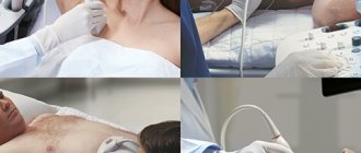
How is the procedure performed?
The patient lies down on the medical table in a comfortable position. The patient's arms and legs are secured with straps to keep him stationary. Then the patient is put on headphones and the table is pushed into a special flask. If you're scared, don't hesitate to ask for a sedative. The duration of MRI of the hand is 30 minutes, with contrast - 50 minutes. The procedure occurs with noise and tapping, but the patient has two-way communication with the doctor. If something goes wrong, tell the doctor, he will pause the session. But don't worry, the tomography process is completely safe.
results
Based on the data obtained from the tomograph, the doctor draws up a description of the pathology. The result depends on the clinic, but in most cases it takes from 30 minutes to an hour. The specialist gives the patient a disc with a recording, a photograph, or a printed document. The signal that the conclusion is ready will come in the form of an SMS message or another option according to the rules of the clinic. You do not need to interpret the result yourself, but go straight to the attending physician who has given you a referral for magnetic resonance imaging.
Contraindications
Carefully read the contraindications before agreeing to an MRI procedure of the hand:
- the presence of a pacemaker, insulin pump, iron crowns and other metal objects in the patient’s body;
- claustrophobia - fear of closed spaces;
- mental illnesses such as epilepsy;
- inability to fit into a tomograph due to increased body weight;
- hyperkinesis - involuntary movements of individual parts of the body;
- children's age up to 7 years.
There are restrictions on MRI of the hand for patients with acute heart failure, pathologies of the kidneys and genitourinary system, and pregnancy. Expectant mothers should not undergo the procedure unless there is an emergency. In any case, an MRI of the hand is performed if the attending physician gives permission. If you decide to undergo magnetic resonance imaging of your hand, you can go to any clinic in Moscow.
Return to list
Preparing for MRI of the wrist joint
MRI of the wrist joint is performed without prior preparation. The procedure does not require a special diet or fasting, pre-medication or other measures. However, due to the magnetic field, magnetic resonance imaging requires compliance with safety regulations. The presence of foreign objects in pockets or on the body, such as jewelry, body jewelry, piercings, cell phones, and other wearable electronics, can cause burns or injury. Magnetic fields cause heating of metal products, disable and lead to fire of devices and gadgets.
Additional information about the procedure
Before starting an MRI scan, you need to make sure that there are no iron objects in your body (removable dentures, cochlear implants, artificial heart valves, etc.). You also need to take off all iron items (earrings, watches, hairpins, etc.), and also put out your credit card and mobile phone. Their presence can greatly distort the image. Any movement can reduce the quality of the image, so it is important to remain motionless throughout the entire examination.
After the tomograph finishes its work, you will be given images on X-ray film or disk. If desired, you can reset the information to any electronic medium. Next, the specialist draws up his medical opinion. If you don’t have time, just leave your email to the clinic staff, and within 24 hours the results will be emailed to you.
Prices for MRI of joints
Prices for MRI of joints from 2600 rubles. Prices for MRI of the hip joint from 2600 rubles. Prices for knee MRI from 2600 rub. Prices for MRI of the ankle joint from 2600 rubles. Prices for MRI of the foot start from RUB 3,700. Prices for MRI of the shoulder joint from 2600 rubles. Prices for MRI of the elbow joint from 2600 rubles. Prices for MRI of the wrist joint from 2900 rubles. Prices for MRI of the hand from RUB 3,990. Prices for MRI of the temporomandibular joints from RUB 3,290.
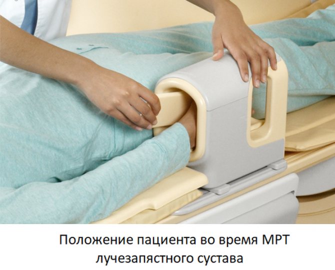
How to do an MRI of the wrist joint
MRI examination of the wrist is performed using an MRI scanner. This is a large cylindrical device with a hole in the center, forming a tunnel 60 cm long and wide. This is the working part of the installation; the patient lies in the tunnel during the examination. Before the procedure begins, the patient is positioned, with the arms either extended above the head or folded along the body, depending on which additional coil is used.
Before starting the examination, the patient needs to wear headphones, since the tomograph makes quite loud sounds during operation. Headphones are also needed to communicate with the operator and listen to music - it helps you relax and make the procedure more comfortable. The duration of an MRI examination of the wrist joint is 30 minutes. In some cases, with inflammatory processes or neoplasms, contrast is used to increase diagnostic accuracy. Contrast drugs are administered intravenously in the middle of the study - the contrast is evenly distributed throughout the body, accumulating exclusively where there are signs of disease. When using contrast enhancement, the duration of the procedure increases to 45 minutes.
After completing the MRI scan, the radiologist begins interpreting the images. This process lasts no more than 30 minutes. After decoding, the patient is given the images on a CD and a specialist’s report. They must be handed over to the attending physician. In addition, the radiologist at our center will definitely tell you about the results of the study and show the problems found in the images, and recommend a doctor to whom you can contact if the patient comes without a referral.
How is an MRI of the thumb performed?
The study of the phalanges does not require specific preparation. The procedure takes place in a separate room where the MRI scanner is located. Before sitting on the retractable table of the device, the patient must take off his clothes with metal objects, leave behind accessories, gadgets, etc., that could affect the examination process. A magnetic coil is fixed around the patient's hand. During an MRI, cross-sectional images of the thumb and all phalanges of the hand are taken.
As a result of the influence of the magnetic field, a simulated image of the area of interest is displayed on the monitor. The duration of the diagnosis does not exceed 15 minutes. If during the scanning process it is necessary to introduce a contrast agent to improve the visualization of pathologies (often required for tumors), the total examination time increases to half an hour. MRI is completely harmless and safe. The drug used for highlighting has a minimum of side effects and is well tolerated by patients. The photo clearly determines the size of the tumor, the involvement of surrounding tissues, and the presence of metastases.
It is prohibited to do an MRI if there are non-removable metal objects (fixing plates, pins, etc.) and devices in the body (cardiac and neurostimulators, hearing aids, insulin pumps, etc.) for women in the first trimester of pregnancy. The problem of fear of closed spaces should be discussed with a doctor, who will suggest ways to solve it.
What will an MRI of the wrist joint show?
MRI images are a series of 1 mm thick sections taken through the examination area in several planes. They clearly show the bones that form the wrist joint, the articular cavity and articular cartilage, the membrane of the articulation, ligaments, muscles and their tendons.
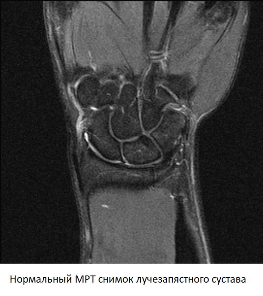
When interpreting the images, the doctor pays attention to the width of the joint space, the condition of the cartilage and articular membrane, and the integrity of the ligamentous apparatus. In the presence of space-occupying formations, MRI allows one to determine with high accuracy whether it is a cyst or a tumor. By studying the dynamics of contrast accumulation, early signs of inflammation or a malignant process can be identified.