Computed tomography of the throat is one of the new ways to examine bone structures and soft tissues in this area. Diagnostics provides an opportunity to identify existing pathological conditions and structural features of the tissues of the larynx. Through scanning, you can obtain highly accurate images of the part of the body being examined, and therefore making the correct diagnosis will not cause difficulties for a specialist.
What does a CT scan of the larynx reveal?
Using CT images, specialists have the opportunity to visualize a number of diseases and pathological conditions:
- neoplasms of soft and bone tissues of the larynx with the ability to assess their size and location;
- presence of esophageal diverticula;
- places of destruction of cartilage in the larynx;
- observe changes in the structure of the walls of the larynx, which may be a sign of a developing tumor;
- identify the presence of foreign bodies in the area being examined;
- determine the severity of injuries received;
- assess the condition of the lymph nodes;
- identify existing inflammatory processes in the cavity being examined;
- diagnose various developmental pathologies;
- examine the vascular network of the larynx.
What does the study provide?
Using computed tomography diagnostics, we study:
- Upper respiratory canals.
- Soft and hard fabrics.
- Region of the thyroid and parathyroid glands.
- Blood and lymphatic vessels.
A CT scan can be used to study the condition of the thyroid and parathyroid glands.
Performing a CT scan of the trachea and larynx helps to identify various disorders and abnormalities in this area of the human body. Among them:
- neoplasms and formation of metastases;
- cysts, adenomas;
- presence of inflammation;
- polyps;
- presence of foreign bodies;
- change in blood flow in the vessels of the neck.
In addition, a CT scan of the larynx shows the existence of the following diseases:
- lymphadenitis;
- bone formations in the nuchal ligament;
- destruction of cartilage tissue;
- atherosclerosis;
- stenosis;
- abscess;
- diverticula of the larynx, as well as areas of the esophagus;
- thyroid diseases;
- laryngitis;
- narrowing of the larynx.
In this video you can see how a CT scan of the larynx and trachea is performed:
CT scan of the throat and larynx using a contrast agent
A CT scan of the larynx, performed without contrast enhancement, allows one to obtain information about the condition of the bone tissue and the existing pathological process in the hyoid bone and in the cervical spine. Using this diagnostic method, the cartilage of the larynx and the soft tissues of the neck are practically not distinguished, which is a significant disadvantage of this scanning method.
Therefore, in order to improve the quality of images when performing a CT scan of the throat, the doctor may prescribe a CT scan of the throat and larynx using a contrast agent.
Contrast is most often injected into the patient's body intravenously, after which it immediately spreads along with the bloodstream to all tissues and organs. Through the use of a contrast agent, it is possible to examine anatomical formations and organs in great detail. They acquire detail and the clearest outlines.
Contrast can also be introduced into the patient gradually throughout the entire scan. Exactly how the contrast will be administered is always decided on an individual basis, based on the purposes of the examination and the intended disease.
Regardless of how the contrast enters the body, the following discomfort cannot be ruled out:
- the appearance of a metallic taste in the mouth;
- feeling of warmth in the body;
- feeling of rising heat.
Such symptoms are considered not dangerous. They represent a normal reaction of the body to the injected drug, so there is no need to stop the procedure.
Dangerous signs that may indicate the appearance of intolerance to contrast during a CT scan of the throat are:
- the appearance of severe swelling of the face;
- sore throat;
- dizziness;
- nausea, vomiting;
- a skin rash and severe itching;
- decrease in blood pressure.
The presence of at least one such symptom becomes a compelling reason to stop administering the drug and provide first aid to the patient. Although such cases are extremely rare. Ideally, each patient should undergo a sensitivity test before using contrast. If, based on the test results, the doctor has no doubts about the safety of the contrast agent for the patient, then you can safely proceed to the next stage of the examination.
How much does a laryngeal x-ray cost?
The cost of the examination depends on the location - a private diagnostic center or a public clinic. The price is also affected by the equipment used for the study - classic X-ray or digital. On average, the cost varies from 1000 to 1900 rubles.
The attending physician, usually an otolaryngologist, can refer you for an X-ray examination. You should not decide on your own about the need for the procedure; after all, diagnosis requires a certain dose of radiation.
X-ray of the larynx is an accessible method for identifying pathology of the respiratory tract, however, it is not the most informative. In some cases, an additional CT or MRI may be necessary.
Indications for CT scanning
Each patient can receive a referral for a CT scan of the throat and larynx from a doctor in the following cases:
- in case of previous neck injuries, as a result of which there is a high probability of injury to internal organs;
- when foreign bodies get into the throat;
- with existing pathologies of the development of the throat and larynx;
- with progressive inflammatory diseases;
- for pathologies of the vascular network of the neck;
- for the purpose of precise location of the pathological process before surgery;
- to assess the effectiveness of treatment after surgery.
Doctors prescribe a CT scan of the larynx only in cases where another diagnostic method carried out in the future will not provide sufficient information and will complicate diagnostic measures.
As a rule, the patient is initially referred to undergo ultrasound diagnostics or x-rays, and only if these examination methods do not lead to results, the patient will be sent to a CT scan of the throat and larynx.
Symptoms
Most often, neoplasms of this type are observed in men 40-60 years old.
Throat cancer is difficult to determine at an early stage, since the symptoms are very similar to a common cold. The patient may feel:
- aching sore throat;
- swelling in the neck area;
- difficulty swallowing food;
- voice change.
If you ignore the symptoms of cancer at the initial stage, then it can manifest itself in the form of:
- sharp pain in the throat that even strong painkillers cannot relieve;
- enlarged lymph nodes;
- cough with bloody discharge;
- weakness, high temperature;
- strong bad breath.
At the first symptoms of a malignant tumor of the throat, you should immediately contact an appropriate specialist.
Contraindications for diagnostics
CT scan of the throat and larynx has some contraindications for the following patients:
- women expecting the birth of a baby;
- patients with excess body weight;
- patients with mental illness.
CT scans are performed with extreme caution:
- children under 12 years of age;
- women breastfeeding babies;
- patients with kidney problems;
- patients with multiple myeloma.
Carrying out computed tomography using contrast is unacceptable:
- during pregnancy and breastfeeding;
- patients with disorders of the liver and kidneys;
- diabetics;
- patients with an allergic reaction to contrast.
Preparation and performance of ultrasound
Special preparation in the form of diet or taking certain medications is not provided. The patient is required to come for examination in comfortable clothes, with the throat area exposed and without neck jewelry (chains, beads, etc.). If the study is carried out specifically to identify a malignant tumor, you need to stop taking antitumor medications a couple of days in order to obtain objective results.
The procedure itself is performed with the patient in a horizontal position. The area under study and the ultrasound sensor are treated with a medical gel that conducts ultrasound waves. The doctor moves the sensor quietly along the neck. Ultrasound waves are reflected by a return echo signal, which a computer program converts and displays an image of the organs on the monitor. The time interval of the procedure ranges from a quarter of an hour to 30 minutes.
The results are recorded by the ultrasound specialist in a protocol, which the patient presents to the otolaryngologist who referred for the ultrasound. He also determines the treatment tactics for identified pathologies.
How to properly prepare for a CT scan of the larynx
For those patients for whom CT of the throat and larynx is planned to be performed in the native mode, there is no need to observe any strict restrictions in their usual lifestyle or follow a special diet.
However, those patients who are indicated to undergo a scan using a contrast agent will have to comply with some restrictions. So, before undergoing a CT scan of the throat, it is prohibited to eat any food 6 hours before the start of the scan. However, drinking water in small volumes is allowed.
All patients, regardless of how the examination will be carried out: with or without contrast, must remove absolutely all jewelry and objects with metal parts before entering the office. You will also have to leave your mobile phone, watch, bank cards outside the office - all these items can cause poor-quality photographs.
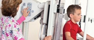
How is the examination carried out?
X-rays of the larynx are performed in two projections - lateral and direct. Direct projection can be anterior or posterior. Depending on the projection in which the picture needs to be taken, the patient will be asked to lie on his stomach or side.
The X-ray tube creates a beam of rays that is directed at the area being examined. The tissues of the human body have different densities. The softer the fabric, the better it transmits the beam. The bones prevent light from reaching the film. The resulting image will be a negative, so the lighter a certain area is, the darker it actually is. The hollow structures will appear black in the image, while the bones will appear white or light gray. If you need to see in more detail what an x-ray of the larynx will show, a procedure with contrast may be performed. To do this, a contrast agent is sprayed into the laryngeal cavity.
Progress of CT scan of the larynx
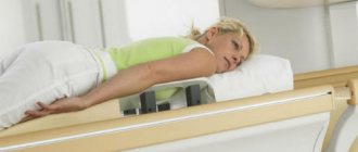
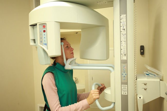
To conduct a CT scan of the throat and larynx, the patient is asked to take a horizontal position on a sliding table, where he will remain for the duration of the study.
His body is fastened with belts for secure fixation, after which the table slides inside the device. All that is required of the patient during a CT scan of the throat is to remain completely calm and not make any movements. The tomograph scans, resulting in a series of images. As a rule, the entire examination process takes no more than 15-20 minutes.
During the scan, the patient is alone in the room. Medical staff monitors the examination progress through a special window from an adjacent office. The patient is not left for a minute without the supervision of doctors. Both the patient and the doctors have the opportunity to communicate through the use of a microphone, so if there are any changes in well-being, the patient will always be able to inform the staff and not be afraid that he is isolated from the outside world.
The latest generation of tomographs meets all safety requirements, so they do not pose any significant harm to the health of patients. Although CT is based on X-ray radiation, it has a very low intensity and, when compared with a classic X-ray examination, it is quite insignificant.
However, due to the fact that X-rays are used during a CT scan of the larynx, this procedure cannot be called absolutely harmless, and it has certain time restrictions for repeated scanning.
Thus, it is unacceptable for women who are expecting the birth of a baby to undergo a CT scan, regardless of the stage of pregnancy, as well as for patients who have already undergone this type of examination in the recent past.
A CT scan of the throat cannot be performed an unlimited number of times, since the radiation dose cannot be exceeded. And only in exceptional cases, if there is strong evidence for this, specialists can prescribe an additional CT examination.
What preparation is required
Preparing a patient for a CT scan does not involve performing special actions for the examination. The only thing that can interfere with the correct diagnostic process is the presence on the patient’s body of any objects with the presence of metals, including jewelry. Therefore, before the procedure, it is recommended to get rid of existing similar things.
How to perform the procedure
The examination session involves the patient lying down on a mobile table. The patient's head should be slightly elevated with the help of a pillow and secured with special straps. The person must remain motionless until the end of the procedure. In this position, it is sent inside the tomographic ring, while the X-ray unit moves evenly in a spiral around the larynx, scanning and sending the corresponding image to the device monitor. The test usually lasts approximately 15 minutes.
The procedure itself is very simple and does not take much time.
Alternative diagnostic methods
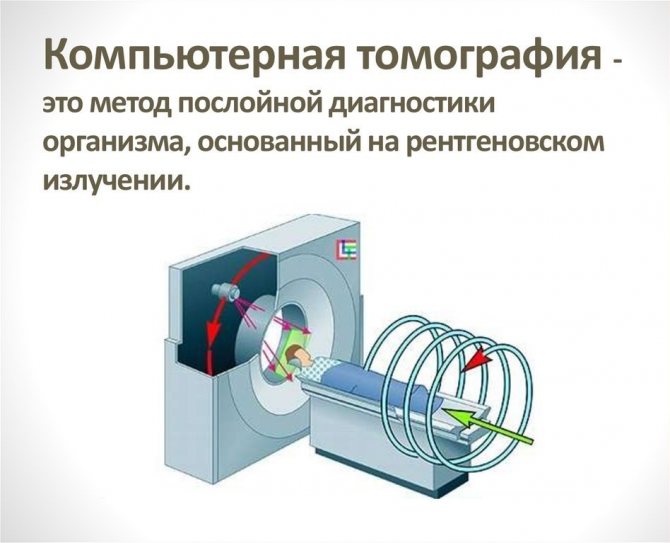
Alternative diagnostic methods include ultrasound and magnetic resonance imaging. However, CT of the throat has a clear advantage - this method is the most informative and fastest. In addition, it allows you to identify even minor pathological processes and evaluate the structures of internal organs.
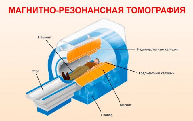
If we talk about which type of diagnosis is better: MRI or CT, then it will be quite difficult to answer this question unambiguously. Both research methods serve completely different purposes, although many of the data obtained may be duplicated.
A CT scan of the throat is performed using X-rays, so it allows you to examine dense formations and hollow organs. Magnetic resonance imaging is the best way to identify soft tissue conditions and is widely used to evaluate injuries to the neck and ligaments.
If there is a need to scan patients in an emergency situation, the choice of one or more examination methods is at the discretion of the doctors, based on the nature of the pathological condition.
How can a x-ray of the larynx be replaced: research analogues
X-ray of such a section of the respiratory tract is an accessible method for diagnosing pathologies of the larynx, however, in terms of information content it is inferior to other studies. Anatomical structures overlap each other, making it difficult to identify certain objects.
For this reason, they often resort to computed tomography - the most informative diagnostic method. CT helps to obtain a layer-by-layer image of an organ, compare sizes and shapes. However, the radiation exposure from CT is many times greater than that received from x-rays. Therefore, computed tomography is performed when absolutely necessary.
The safest diagnostic method
If radiation exposure is extremely undesirable for the patient, magnetic resonance imaging is used. The method is based on the use of a magnetic field and the absence of radiation. MRI can be performed several times in a row without adverse effects on the body. If necessary, you can take a contrast agent for a detailed picture.
Where to get a CT scan of the throat and larynx
Regardless of the exact reasons for which the patient was prescribed a CT scan of the throat, a fundamentally important point is to find a suitable clinic. She must not only satisfy the patient with her pricing policy, but also perform the examination at the highest level.
Unfortunately, sometimes it is not so easy to make the right choice: some medical centers do not have modern equipment, and tomography performed on old scanners does not provide a complete picture of the existing pathological process. In other clinics, the medical staff does not have sufficient experience, so there is a high probability of making an incorrect diagnosis.
We are pleased to offer our services to patients: on our website, every visitor can see for free a large list of medical clinics, as well as detailed information on each of them.
After selecting the required medical center, you can call the phone number listed on the website to make a preliminary appointment. We provide a discount on CT scanning to everyone who turns to us for help.
By contacting us for help, you can easily decide on the choice of the necessary medical center from a huge list, and also receive a guarantee of the lowest price.
Additionally
When examining the larynx, a qualified ophthalmologist will not ignore possible changes in the thyroid gland. The reason to check the functionality and condition of the body’s endocrine system is the pathologies detected on an ultrasound of the larynx:
- hyper- and hypothyroidism (impaired synthesis of hormones in the thyroid gland);
- purulent abscess;
- the formation of one or more cysts or nodes in the thyroid gland;
- postoperative complications (if surgery was performed on the thyroid gland);
- the presence of tumor formations;
- nodular goiter;
- abnormal increase in gland volume (the norm for men is 2.5 cm, for women – 1.8 cm).
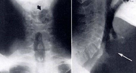
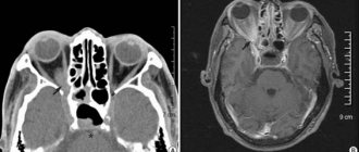
Diagnosed with laryngeal cancer. Photo from ultrasound scan
In the study protocol, these indicators will be reflected as information for the endocrinologist. To obtain more extensive data, your doctor should prescribe an additional blood test for thyroid hormones. Despite the progressive development of medical technologies, ultrasound remains one of the most popular diagnostic methods. A timely examination will help to identify oncological diseases of the hypopharynx at the initial period of their development.
How is tomography performed?
A CT scan of the larynx is performed with three samples:
- The inhalation test makes a CT image of the vocal folds in the position of their greatest stretch, reveals the degree and uniformity of their mobility, as well as the width of the glottis.
- The phonation of the sound “I” brings the vocal cords to the position of their closest approach.
- The Valsalva maneuver best allows you to assess the condition of the sinuses and walls.
Examination of the larynx is usually performed with a slice thickness of 3-5 mm. The screening area is from the hyoid bone to the lower edge of the thyroid cartilage plate. CT scan of the trachea covers the level of the tracheal bifurcation to the pyriform sinuses. The study is carried out with breath holding while exhaling and inhaling.
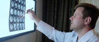
The scanning itself takes 2-3 minutes. It will take another 30-40 minutes to decipher the results. Based on the results of the diagnosis, the patient will receive a report from a radiologist and tomograms printed on film or recorded on a disk or flash drive.
Leading doctors of St. Petersburg

Pashkova Anna Alexandrovna
specialization: MRI doctor, radiologist of the highest category, candidate of medical sciences. Appointment - at the AMI Medical Center, Standard MRI on Ladozhskaya
medical experience - 21 years
Online registration
Khalikov Aziz Jalanovich
specialization: MRI doctor, radiologist of the highest category, candidate of medical sciences. Registration - at MC Scandinavia
medical experience - 20 years
Online registration
Trufanov Gennady Evgenievich
specialization MRI doctor, MD, professor. Recording - Federal State Budgetary Institution "National Medical Research Center named after. V. A. Almazova"
medical experience - 35 years
Online registration
Kholin Alexander Vasilievich
specialization MRI Doctor Professor of the Department of Radiation Diagnostics Northwestern State Medical University Appointment - CMRT Staraya Derevnya, CMRT Petrogradskaya
medical experience - 35 years
Online registration