In the presence of various diseases and pathologies of the larynx and throat, sooner or later the need for detailed diagnosis arises. Due to its complex structure, examination of these areas requires the use of modern diagnostic methods, which can be magnetic resonance imaging.
This examination method is one of the most highly accurate and safe, due to which it is very popular in our time among both doctors and patients. And, if 10 years ago it was not possible to get an MRI of the throat in every clinic, even in the capital, now everyone can undergo a scan either upon a referral from a doctor or on their own initiative. The price of an MRI of the larynx is quite reasonable.
MRI of the throat and larynx: what diagnostics show
MRI of the larynx includes a detailed examination of the larynx, salivary glands and thyroid gland. An MRI of the throat can be done if there are suspicions of inflammatory diseases, neoplasms, or infectious processes. To undergo an examination, one or more of the following symptoms is sufficient:
- the appearance of laryngeal edema;
- breathing problems;
- change in the size of the thyroid gland (increase in size);
- copious mucous and purulent discharge from the respiratory tract;
- a feeling of sore throat occurring simultaneously with an increase in temperature.
Regardless of what an MRI of the throat and larynx shows, a scan is performed either to confirm or exclude the suspected diagnosis. In some cases, diagnostics are prescribed to monitor the effectiveness of treatment for diseases of the ENT organs.
MRI of the larynx is prescribed for:
- pathologies of the thyroid gland;
- injuries to the throat and larynx;
- the presence of neoplasms and metastases;
- diseases of the salivary glands, lymph nodes;
- osteochondrosis, intervertebral hernia.
MRI of the throat and larynx shows a huge number of different diseases and pathological conditions. It is also important to remember that if the patient has a high risk of developing certain diseases, an MRI of the throat should be done for preventive purposes at least once a year.
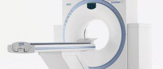
How is an MRI of the neck performed?
If contrast enhancement is not used for diagnosis, then special preparation for the study is not required. At the doctor's office, the patient will be asked to remove all metal-containing items. Do not forget even about such small details of clothing as buttons or zippers; you must remove keys and plastic cards from your pockets - the presence of these items may interfere with the operation of the device.
The patient will not experience any discomfort or pain, but will hear noise made by the device.
The neck MRI diagnostic procedure will take about 30 minutes. If injection of a contrast agent is required, the diagnostic time will increase by approximately 15 minutes.
During the entire examination, the patient is given headphones to muffle the sounds of the operating tomograph, as well as a signal bulb in case the patient wants to stop the scan.
Contraindications for MRI of the larynx

Despite its high accuracy and safety, MRI of the throat has some contraindications:
- Patient's age. Thus, it is not possible to do an MRI of the throat for children under the age of 7 years, since they cannot comply with the main condition - to ensure complete immobility during the examination.
- Obesity. Magnetic resonance imaging is not performed on patients whose weight exceeds 120 kg for the reason that the diagnostic table is not designed for large body weights.
- Patients with metal objects in the body. These can be any implants and stimulators that may begin to move or cause a burn during an MRI of the trachea.
- Patients with uncontrollable tremors of the limbs. There is no point in having tomography performed on such patients because the images will be inaccurate.
- Patients with claustrophobia. Finding a patient inside a scanner can cause a panic attack, so for safety reasons, it is advisable for such people to undergo magnetic resonance imaging only in a state of medicated sleep.
In exceptional cases, an MRI of the larynx can be performed on a pregnant woman if there are compelling indications. However, scanning with contrast will be strictly contraindicated, since contrast has a toxic effect on the body of the developing fetus, which can lead to irreparable consequences.
How to properly prepare for magnetic resonance imaging
MRI of the throat and larynx shows a variety of diseases, even though the larynx is one of the most difficult areas to examine. The patient does not need to follow any strict preparatory measures before the scan, which is a huge plus for patients. It is only important to adopt some recommendations that may be useful during an MRI of the trachea:
- For 3-5 days, you should eliminate fatty and spicy foods from your diet, and also give up any foods that can cause increased gas formation and discomfort in the intestines. This recommendation should be given special attention if the diagnosis will be carried out using a contrast agent.
- 5 hours before the start of the examination, you should refuse any food, as food can cause discomfort at the most inopportune moment.
- Before scanning, you need to remove all metal objects, including dentures and hearing aids. You can also change into hospital clothes, as they are more comfortable. During the examination, the patient should experience maximum comfort.
- If you have a tendency to have panic attacks, it is better to drink any sedative. You can ask your doctor to prescribe the most effective and appropriate one.
How to scan and where to get an MRI of the throat
If you have decided to undergo an MRI of the throat for a child or yourself, the first thing when choosing a center is to ask whether they perform MRI of the neck on a low-field or high-field machine. If the purpose of tomography is to search for an oncological process or differentiate a space-occupying lesion, then only a closed device should be used; if the purpose of the examination is routine (non-life-threatening) pathology, then the power of an open scanner is sufficient. Also, open devices are preferable for scanning children, since the profile of such devices allows the parent to be nearby and be in the child’s field of view.
No preparation is required for the native examination; if contrast is administered, it is not recommended to eat three hours before the procedure. During scanning, a radiofrequency coil in the form of a half ring is placed on the neck area. The scanning time takes about 30-40 minutes, and the patient must remain completely still; swallowing is also prohibited; therefore, children most often undergo MRI under the influence of anesthesia.
Tomography results are usually given to the patient within 20-40 minutes (in private institutions), but in some cases the results are ready only after 72 hours (in government agencies).
Progress of the survey
Before undergoing an MRI of the throat, the patient must submit all documents to the radiologist (results of previous studies, an extract from the outpatient card, personal medical record), and also inform about potential contraindications and the presence of chronic diseases.
Even seemingly insignificant diseases can act as strict contraindications to undergoing magnetic resonance imaging. The radiologist must be aware of everything. The examination will be carried out in several stages:
- If an MRI of the larynx is performed using a contrast agent, then before the examination begins, a special drug will be administered intravenously to the patient. As a rule, an allergy test is performed before its administration. If its results did not reveal the presence of intolerance, then there will be no contraindications to this method of examination.
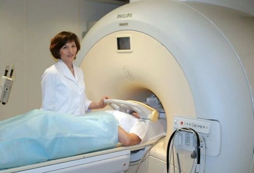
- The patient will be asked to go to a special room where the tomograph is located. To ensure complete immobility and eliminate any involuntary movements of the limbs, the body will be secured with special straps. The patient should not be frightened by such a need, because this is done in his interests.
- After this, the retractable table is rolled into the tomograph and the device begins its work. Medical personnel leave the room and monitor the progress of the MRI of the trachea from the next room through a special window.
- The examination continues for 20-30 minutes. During operation of the scanner, specific noises are produced. This is normal and you should not be afraid of these sounds.
As soon as the scanning is completed, the patient can immediately leave the office, change into personal clothes and go to wait to receive a report from the doctor. If a person does not have enough time, the survey results can be sent to him by email. In any case, as soon as the conclusion is received, it should be taken to the specialist who has written a referral for an MRI of the throat and larynx.
Use of contrast in MRI of the neck
Contrast enhancement in magnetic resonance imaging of the soft tissues of the neck is mainly used in cases of suspected benign or malignant neoplasm, metastases to the organs of the neck, as well as after medical and surgical treatment of tumors.
Contrast is carried out using intravenous administration of gadolinium-based drugs (most often gadodiamide), which, accumulating in the affected area, increase the contrast of pathologically altered tissues in the images, which makes it possible to clearly determine their boundaries and localization in relation to healthy tissues.
The use of contrast agent is harmless. However, contraindications to its use may include pregnancy, breastfeeding and severe renal failure.
Decoding the results
The images obtained by MRI of the larynx can be printed or recorded on any digital media. This opportunity allows the patient to show the results of the examination to absolutely any specialist, regardless of where exactly he goes.
Interpretation of the results obtained should be carried out exclusively by a competent specialist with extensive experience, because only in this case can the patient fully rely on the professionalism of the doctor.
The radiologist will compare the obtained images with samples without pathology and determine the presence of abnormalities, if any. After this, the specialist prepares a conclusion, on the basis of which further therapeutic tactics are made.
If necessary, the scan can be rescheduled, and even frequent scanning will not have a negative impact on the patient’s health due to the absolute harmlessness of the diagnosis.
Advantages of MRI of the larynx over other diagnostic methods
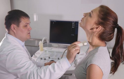
Various methods can be used to identify diseases of the pharynx. Most often, patients are referred for a computed tomography scan, ultrasound diagnostics or endoscopy. However, MRI of the trachea has a number of advantages over other diagnostic methods.
First of all, this list includes complete harmlessness to the patient’s body. To date, no harmful effects of the magnetic field on the body of the subject have been identified. The only exception is pregnant women. The magnetic field can have a negative effect on the developing fetus, so tomography is not prescribed to expectant mothers in the early stages of pregnancy.
Another undoubted advantage is non-invasiveness, which is why magnetic resonance imaging is very popular among patients. It is impossible not to mention the high information content - tomography allows you to obtain highly accurate images, and if necessary, the finished images can be viewed in a volumetric projection.
MRI of the throat and larynx also provides the opportunity to detect the presence of even the most minor neoplasms, regardless of their nature: benign or malignant.
Despite the many advantages, tomography also has some disadvantages. For example, if during an MRI of the trachea one can learn about disturbances in the functioning of the thyroid gland, then a more specific study of this organ should be carried out separately.
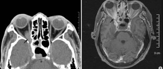
What does an MRI of the soft tissues of the neck show?
Magnetic resonance imaging is a safe technique for scanning the body using magnetic waves, which, unlike X-rays, are safe for humans.
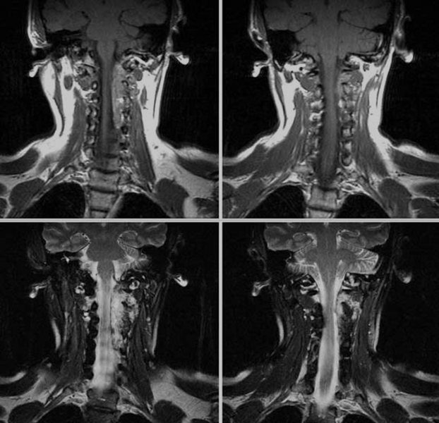
MRI images of the neck in coronal projection
An MRI of the neck area may be prescribed if the following symptoms occur:
- pain in the neck that does not go away for a long period of time;
- feeling of a lump in the throat, feeling of fullness or difficulty swallowing;
- the appearance of swellings of various sizes and shapes;
- headaches of unknown origin;
- violation of voice functions.
The appearance of the above symptoms requires an early examination by a doctor, based on the results of which magnetic resonance imaging of the soft tissues of the neck may be prescribed.
MRI diagnostics of the neck area can be carried out either as an independent study or in conjunction with scanning of other areas to identify oncological and hematological diseases.