Indications for the study
The specialist will recommend a CT scan of the eye orbits and optic nerves when he suspects:
- Traumatic damage to the eye sockets (orbits) of the optic nerve, eye muscles;
- Presence of a foreign body;
- Tumor process in this area;
- Inflammatory changes.
There are symptoms for which you should definitely consult a doctor and have your orbits scanned. These include:
- Significant decrease in vision over a short period of time.
- Pain in the eye area that occurs for no apparent reason.
Features of CT scan of the orbit
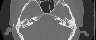
The operating principle of diagnostics is based on the use of X-ray radiation. The rays are directed strictly to the upper part of the head and pass through the area being studied. Thanks to this, a visualization is gradually formed, which includes layer-by-layer images of the eye sockets themselves.
Content:
- Features of CT scan of the orbit
- Indications and contraindications
- Preparation rules
- How is the procedure done?
- The norm and deviations from it
This approach guarantees a high-quality image of not only the structure of the optic nerve, but also the vessels of the retina, as well as the immediate eyeball, the muscles that hold it, and even the condition of the lacrimal glands. Moreover, tumor growth is not always the cause of a sharp deterioration in well-being in a given area.
Sometimes the result of the analysis provides information about an extensive inflammatory process of a completely different etiology, or even degeneration, the consequences of a recent severe injury.
Computed tomography is a more advanced method of studying the health of the orbit than a classic x-ray.
Experts highly value this method of medical progress for its reduced percentage of harmful radiation during manipulation and high information content compared to a simple X-ray image.
Another undoubted advantage is non-invasiveness. Since the human eye is one of the most sensitive organs, any interference with its normal functioning is fraught with serious complications and long-term pain.
Classic examination options involve the use of various instruments, which in fact, at the slightest contact with the membrane of the eye, guarantee, if not injury, then discomfort. There will be no unpleasant sensations here.
Another significant victory for the authors of the procedure was the period of its implementation. Unlike an MRI, which takes about half an hour, a computed tomography scan lasts several times less.
This is suitable for patients who simultaneously suffer from claustrophobia, or who complain of regular pain in the visual area.
Possibilities of the method in studying orbits
Computed tomography is a diagnostic procedure based on the action of x-rays. Our medical center is equipped with an expert-class Siemens Somatom tomograph, which allows us to improve the quality of diagnostics.
After the scan, the doctor analyzes the resulting images, which visualize:
- Bone structures that form the basis of the orbits.
- Soft tissue lining the orbital cavity.
- Lacrimal glands.
- Eyeballs.
- Part of the optic nerve.
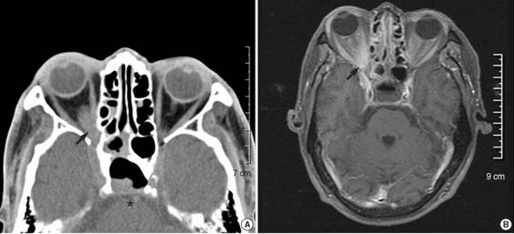
Thanks to the high accuracy of the images and the qualifications of the specialist, CT of the eye orbits shows:
- Neoplasms of this area with determination of the stages of the process (including metastatic lesions).
- Bone fractures caused by trauma.
- Foreign body, its location and affected area.
- Various inflammatory processes with determination of their prevalence and degree of tissue destruction.
If the question of the need for a CT scan of the eye orbits has not been resolved, our call center staff will help you. A telephone consultation is also possible.
What is retinal tomography?
Computed tomography of the retina (also known as optical coherence tomography) is popular and trustworthy among ophthalmologists. As you know, medicine does not stand still, and these days we have the opportunity to undergo a retinal examination using such a non-contact and painless method as computed tomography.
A tomograph uses X-rays to scan the top of the patient's head. Ultimately, the specialist displays images of the eye sockets layer by layer on the screen, which allows him to assess the condition of the retina and optic nerve, identify the initial stages of diseases, and therefore prescribe timely treatment to the patient.
Contraindications to CT of the eye orbits
Regardless of the examination area, contraindications to computed tomography are always the same. Among them are:
- Pregnancy, since X-ray radiation has a detrimental effect on the actively developing fetus.
- Behavioral disorders accompanied by increased excitability, when the patient cannot remain motionless in the tomograph.
- The technical weight limit applies to patients with high degrees of obesity (more than 150 kg).
Children can undergo this study only if:
- when other reliable diagnostic methods cannot be used;
- mandatory referral from a doctor.
If after a standard CT scan of the eye orbits the eyes should be examined using a contrast agent, then additional contraindications must be taken into account. These include:
- Allergic reactions to iodine and iodine-containing drugs. In this case, allergies of any severity are taken into account: from skin rashes to swelling of the larynx.
- Presence of renal failure. The removal of the contrast agent from the body occurs primarily through the kidneys, which increases the physiological load on them.
- The presence of other diseases that affect the kidneys. These include severe diabetes mellitus, long-term malignant hypertension and atherosclerosis of the renal arteries.
The period of breastfeeding is not an obstacle to CT. However, before the procedure you should pump or prepare formula, as it is not recommended to feed your baby breast milk for 24 hours after the scan.
The decision on the possibility and necessity of performing a CT scan of the eye orbits should be left to the doctor. Only a specialist will be able to correctly assess the situation of each individual patient.
When is optical coherence tomography of the eye prescribed?
Indications for the procedure
Optical coherence tomography of the eye is a common diagnostic method, so ophthalmologists quite often resort to this procedure. The main indications for this study are the following:
- presence of foreign bodies or suspicion of them;
- sharp decrease in vision;
- pain in the eyes;
- tumors of the walls of the orbits (benign or malignant);
- injuries to the eye sockets or orbits;
- protrusion of the eyeball (in medical terms, exophthalmos);
- inflammatory processes;
- lesions of the lacrimal glands caused by autoimmune diseases.
Preparing for the study
There is no need to carry out special preparatory measures for standard tomography of the orbits.
If a contrast study is planned, it is necessary to check the blood creatinine level. Analysis done in advance will be valid for 2 weeks. If it was not possible to determine blood creatinine in advance, some centers provide the opportunity to be tested immediately before scanning using a test strip.
You should not go to a contrast examination completely hungry, as this may lead to nausea and dizziness. Some time before the scan you need to eat lightly.
When planning a CT scan, you need to tell your child in detail about the study in advance. You can also watch a video of the procedure together or play “we are in the CT room.” After this, the child will feel more confident.
To provide complete information about the patient’s health and the rationale for the prescribed procedure, you must bring:
- medical referral for CT scan of the eye orbits;
- conclusions from previous studies in this area (not just CT).
In the absence of medical documentation, research is also possible.
Examination at the Miracle Doctor clinic
The medical center uses a modern coherence tomograph OPTOVUE RTVue100, manufactured in the USA, which performs eye scanning at high speed and with maximum resolution. The technical data of medical equipment ensures diagnostic accuracy for a wide variety of eye diseases.
The clinic employs professional ophthalmologists with extensive experience. They make smart decisions in any situation and provide timely assistance. Specialists work with all ophthalmological problems and find a way out of a seemingly hopeless situation.
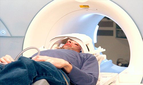
Methodology for CT scanning of the eye orbits
After all the documents have been completed, the nurse will tell you and show you what to do next.
The room with the tomograph and the medical staff's office are two adjacent rooms. Such separation is necessary due to the effect of x-ray radiation.
The nurse will guide the patient to the machine and help him lie down on a special table. It drives into the annular part of the tomograph to the level of the orbits.
Next, the nurse returns to the nursing office, where the doctor and other staff can observe the patient through the window. A special device is provided for voice communication.
A couple of minutes after turning on the device, the painless procedure will be completed. When contrast is administered, the examination time increases by 15 minutes.
You should remain still during the scanning process, as any movement will significantly degrade the image quality.
CT diagnostic services at CELT
The administration of CELT JSC regularly updates the price list posted on the clinic’s website. However, in order to avoid possible misunderstandings, we ask you to clarify the cost of services by phone: +7
| Service name | Price in rubles |
| CT ear | 7 500 |
| CT scan of the temporomandibular joint | 7 000 |
| CT scan of facial bones with three-dimensional reconstructions | 9 000 |
All services
Make an appointment through the application or by calling +7 +7 We work every day:
- Monday—Friday: 8.00—20.00
- Saturday: 8.00–18.00
- Sunday is a day off
The nearest metro and MCC stations to the clinic:
- Highway of Enthusiasts or Perovo
- Partisan
- Enthusiast Highway
Driving directions
Diagnostic result
At the end of the scan, the doctor receives many cross-sectional images. After analyzing them, the specialist makes a conclusion and recommendations. Waiting times vary by center but can usually be spent in the medical center or on a walk-in basis. If you don’t have time to wait, you can receive the result by email or arrive on any other day.
Depending on the purpose for which the CT scan was done, the result may be as follows:
- medical center paper form;
- printouts of the most revealing photographs;
- writes all images to disk.
Electronic media in the form of a disk is provided free of charge in most clinics.
Also, after diagnosis, the radiologist will answer all questions regarding this study. This consultation will help the patient more accurately understand the CT scan result.
Preparatory measures before CT eye
No special preparation is required from the patient before performing a CT scan. All you need to know is that when scheduling a health check with a booster, you will need to stop eating a few hours before your session (approximately 6-8 hours). There are no restrictions before the standard examination. You can eat regular foods, drink any liquid, take medications, etc.
More detailed information about diagnostics can be obtained on the official website 36go.ru. Our company was created specifically to help people find the most profitable and suitable option for a medical organization engaged in various testing methods. Using the services of the portal, you will receive information about available places for examinations for several days in advance, the current cost of the examination, and will also be able to read real reviews from clients who have visited different clinics.
CT scan
The principle of the method is based on the interaction of body tissues with x-ray radiation. Some tissues absorb Rg - the rays are better than others (bone tissue), and fluid accumulations, on the contrary, do not interact with them.
However, in tomographs it is possible to regulate the “hardness” of the radiation, thanks to which any structures of the human body can be examined.
CT has many advantages, including:
- Non-invasive or minimally invasive with intravenous contrast.
- High accuracy of the results obtained.
- Credibility. Often they are sent for testing to clarify the diagnosis.
- The procedure takes little time, despite its complexity. This is especially important when you need to quickly choose a treatment strategy.
- Possibility of re-examination with image intensification (if necessary).
- Availability.
Magnetic resonance imaging
MRI is based on recording the “excitation” of protons in the body after exposure to an electromagnetic pulse. In this case, the person is in a magnetic field.
Using this diagnostic method, soft tissue pathology is better visualized.
The scanning time for MR diagnostics exceeds that for CT. This factor causes some inconvenience, especially in patients with difficulties in maintaining immobility.
One of the contraindications to MRI is the presence of implants that contain ferromagnets (metals that can be magnetized).
Advantages of the method
First, the main advantage of retinal computed tomography
is non-contact, since the eyes are highly sensitive to any touch and interference. Secondly, the procedure takes no more than a minute (provided that no contrast is used). Thirdly, the diagnosis is absolutely painless (due to the fact that there is no physical intervention). Retinal OCT allows doctors to obtain detailed and clear information about the patient's eye condition, which is a definite advantage. Finally, this diagnostic method is quite inexpensive, its cost can reach 3000-4500 rubles.