Previous Next Magnetic resonance imaging of the knee joint is one of the preferred methods for diagnosing most articular pathologies of the knee area. During MR scanning, it is possible to successfully identify various types of degenerative changes and inflammatory processes at an early stage, including bone fractures, damage to cartilage tissue and soft structures of the muscular and ligamentous apparatus. A tomograph scan of the knee is non-invasive. The patient does not experience any discomfort, the examination is not dangerous to his health and can be carried out repeatedly if necessary. An MRI of the knee joint can be done either upon the doctor’s direction or on the patient’s personal initiative.
- MRI
- Ultrasound
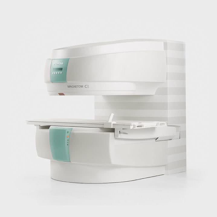
MRI tomograph:
Siemens Magnetom C
Type:
Open (expert class)
What's included in the price:
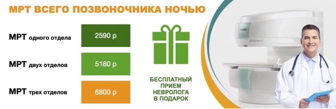
Diagnostics, interpretation of images, written report from a radiologist, recording of tomograms on CD + free consultation with a neurologist or orthopedist after an MRI of the spine or joint
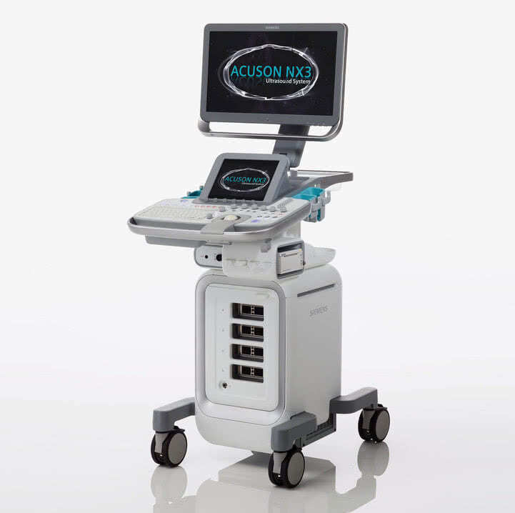
Ultrasound machine
HITACHI HI VISION Avius
Class:
Expert (installation year 2019)
What's included in the price:
Diagnostics, interpretation of images, written diagnostic report
What is magnetic resonance imaging of the knee joint
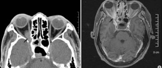
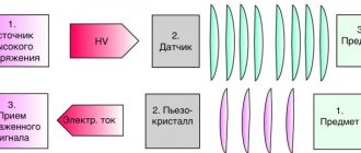
Magnetic resonance imaging of the knee is a scanning technique in which images are obtained using the physics of the phenomenon of nuclear magnetic resonance. During the examination, the patient's knee is placed inside a tomograph, where a strong magnetic field is created and radio frequency pulse sensors work. Under this influence, hydrogen atoms in tissue cells begin to perform oscillatory movements. This resonance is recorded by the installation computer, digitizes the data and converts it into three-dimensional images of a black and white spectrum. The more water is contained in the tissues of the examined area, the more contrast and clarity the scans will be. Muscles, tendons, and cartilage contain enough water, so these tissues are very well visualized on tomograms. Bones have little water in their structure, so they are poorly detailed in photographs. A more effective method for diagnosing bone pathologies of the tibia and femur will be computed tomography.
Are there any contraindications?
Since a strong magnetic field is used during the study, it is contraindicated for people who have any metal implants or foreign bodies in their bodies. Most often, wearing a pacemaker, defibrillator-cardioverter, or metal joint prostheses is a contraindication.
No evidence has been found that a magnetic field can cause harm to an unborn child during pregnancy, however, in expectant mothers this diagnostic method is used with caution and only in cases where it is absolutely necessary. Magnetic resonance imaging is especially not recommended in the first trimester of pregnancy, when the baby's internal organs are forming.
The patient must remain motionless during the examination. Otherwise, the pictures will turn out to be of poor quality. MRI is difficult to perform in people with mental disorders in a state of agitation or persistent cough. During the procedure, the patient is inside the device, in a confined space, so claustrophobia is another contraindication. Most devices are designed for people weighing no more than 150 kg.
A separate group of contraindications is associated with the use of contrast. In rare cases, allergic reactions occur. In people with severe renal impairment, contrast can lead to a complication called nephrogenic systemic fibrosis. New mothers should not breastfeed for 24 to 48 hours after receiving gadolinium.
Get a consultation with a doctor
Indications
The following symptoms may make you think about the need to do a magnetic resonance imaging scan of the knee joint:
- persistent pain in the knee;
- swelling of the skin and swelling;
- accumulation of blood and suppuration in the joint capsule;
- change in the shape of the knee;
- problems bending or straightening your leg;
Patients who are planning knee surgery are often asked to do an MRI. Tomography data allows us to evaluate the anatomical features of the patient’s knee joint and build the correct surgical plan.
Initial appointment with an ORTHOPEDIST
ONLY 1800 rubles!
(more about prices below)
What will an MRI of the knee show?
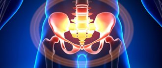
The structure of the knee joint includes very diverse tissues: ligaments, tendons, bones, fatty tissues, cartilage. All of them are subject to inflammatory or degenerative changes. MRI of the knee joint allows for a comprehensive assessment of changes in the ligamentous apparatus, menisci, and bone structures that form the joint. Most often the knee suffers from:
- bone-traumatic injuries;
- degenerative changes in cartilage tissue (menisci, articular cartilage);
- ligamentous injuries;
- osteochondropathy;
- necrosis, arthrosis and arthritis;
- tendinitis.
Tomogram data will help to make a differential diagnosis of these diseases, assess the degree of damage to the articular apparatus and build a prognosis for possible treatment and recovery of the patient. If the patient has undergone active therapy, using a series of MRI images, the doctor can conduct a comparative analysis and assess the degree of progress or regression.
MRI after knee injury
MRI of the knee is one of the most effective ways to diagnose knee injuries. According to the World Health Organization, 80% of people in the world suffer from this or other diseases of the musculoskeletal system. Some scientists believe that diseases of the joints and spine are humanity’s retribution for upright walking. Whether this is true or not is difficult to say. But the fact remains that half of the people on the planet will sooner or later encounter pathologies of the knee joint, especially if you are a fan of active sports. Up to 50% of all sports injuries are related to knee damage.
If a knee injury has occurred in your life, an orthopedist will most likely give you a referral for magnetic resonance imaging, since it shows much better the soft tissues, ligaments and tendons, which are much more often and more easily injured than the bone component of the joint. An MRI scan is a safe test due to the absence of x-rays, unlike computed tomography (CT) and radiography. MRI is the only method that provides comprehensive information about a knee injury such as a subchondral (hidden) fracture. This type of fracture is called occult because its signs remain invisible on x-rays and arthroscopy. On an x-ray, this joint will appear undamaged. Meanwhile, this knee injury requires prompt treatment to avoid secondary degenerative changes in the cartilage.
Previous Next
MRI for meniscal damage
Damage to the meniscus (meniscopathy) is one of the most common knee injuries. Most meniscus damage happens, as they say, out of the blue. For example, an unsuccessful leg movement or squatting can be associated with this grass. When the meniscus is pinched or torn, the patient experiences sharp, stabbing pain in the knee. After such an injury, the leg usually swells. With such symptoms, the patient should come to see an orthopedist and have an MRI of the knee joint. Based on its results, the doctor will be able to assess the extent of the damage and answer the question of whether the damaged meniscus requires surgical intervention.
MRI for Baker's cyst
In addition to damage to the meniscus, a person may suffer from a phenomenon called Baker's cyst. It forms when the stretched capsule of cartilage in the knee bulges back and fills with synovial fluid. This cyst does not necessarily cause pain, but always limits movement. The patient feels that the knee cannot be fully bent.
The Baker's cyst can be independently felt by the patient under the knee in a bent position. Usually this tumor does not cause severe pain and is benign. But at some point, the popliteal cyst can fester and lead to purulent arthritis. This protrusion can also cause venous thrombosis. Therefore, it is important that a Baker's cyst is diagnosed and removed. Magnetic resonance imaging will again help in diagnosing the exact location of the popliteal wrist.
Advantages of undergoing MRI diagnostics at NEARMEDIC
Modern technical base
Diagnostics are carried out using unique equipment made in Japan HITACHI APERTO with the latest software.
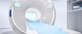
An open-type tomograph with a 0.4 T permanent magnet allows you to take clear, high-quality images with high image quality due to the high uniformity of the constant vertical magnetic field. Reduced noise level (0.3 dB) increases comfort. Highly qualified MRI doctors
NEARMEDIC's specialized specialists have the necessary basic knowledge in pathophysiology, anatomy, pathological anatomy, as well as in the field of computer and electronic technologies.
Experienced doctors will competently decipher the results of the study and write a professional conclusion. Affordable cost
The company's reasonable pricing policy allows patients to have an MRI of the knee joints at reasonable, affordable prices that are affordable to the majority of the population.
How often should you have an MRI of your knee?
Typically, magnetic resonance imaging of the knee joint is prescribed only if the patient has suffered an injury or has pain. However, there is a group of people for whom knee imaging is recommended on a regular basis. Sport is not only about medals and victories, but also about grueling hours of training and daily testing of the body’s strength. With increased stress on the body and sports-related injuries, it becomes extremely important to monitor any changes in the condition of the athlete’s musculoskeletal system. This is where MRI comes to the rescue. The general recommendation of doctors for people leading an active sports lifestyle is to conduct an MRI of the entire spine and an MRI of joints that are subject to heavy load due to sports at least once a year.
MRI of the knee at NEARMEDIC
Using MRI of the knee, you can obtain information not only about bone, but also about soft periarticular tissues.
MRI makes it possible to see the condition of the tendons of the knee joint, especially the patella and quadriceps muscles, and obtain information about internal damage or tissue rupture.
Tomography allows you to determine the degree of damage:
- Fluid has accumulated near the ligament;
- the fibers of the ligament are partially destroyed.
- The ligament is completely torn.
Advantages of the method
- Tomography is based not on X-rays, but on magnetic resonance (a physical phenomenon). Therefore, the procedure is completely safe and has no contraindications, including for pregnant women and children.
- Highly informative and clear images ensure accurate diagnosis, and the ability to create a three-dimensional computer model of the knee joint makes it possible to study in detail the condition of its structures and, if necessary, develop a plan for surgical intervention.
- The procedure is completely painless and does not require special preparation.
How to do a tomography of the knee
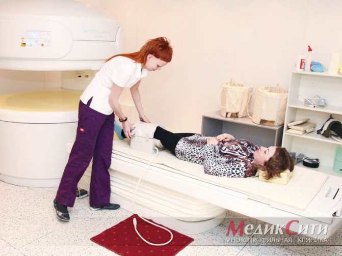
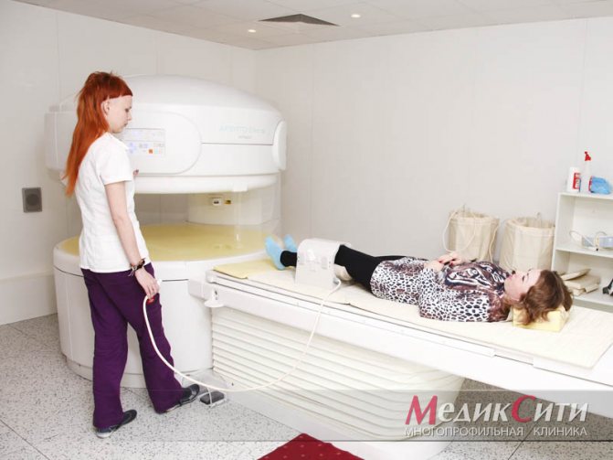
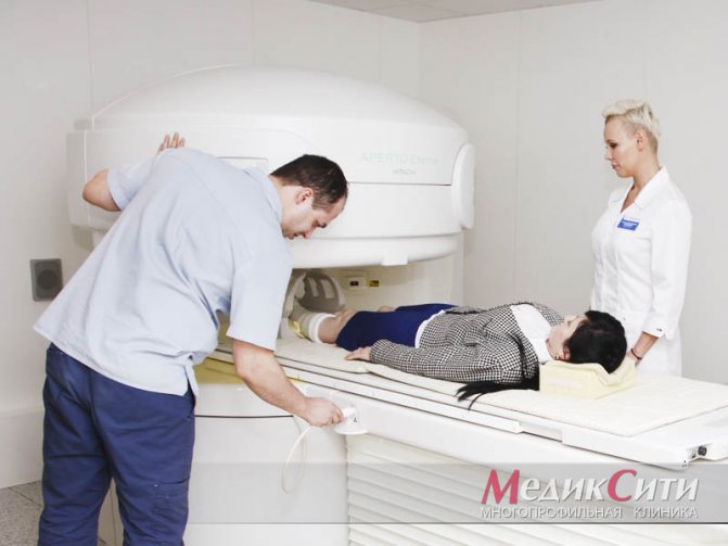
The magnetic resonance imaging procedure takes place in a comfortable environment for the patient. In most diagnostic centers in St. Petersburg, it is carried out by appointment. Before entering the MRI room, the subject will be asked to remove all metal objects from the body and remove all electronic devices from their pockets. A tomograph is a big magnet. Having metal objects near him may cause him to attract them towards him. In addition, the presence of metal in the scanning area reduces the quality of the images.
The second factor that affects the quality of magnetic resonance imaging is the immobility of the patient. In the diagnostic room, he will be asked to lie down on a mobile table, a special intensifying coil will be put on his knee and brought inside the scanning part of the tomograph. The patient will be asked to remain still throughout the examination. The average screening duration is 30 minutes. Usually the patient does not feel anything other than slight heating in the examination area. The only thing that can be annoying is the noise of a working tomograph. If it is too strong, a person can always ask to be given noise-canceling headphones.
Questions about diagnostics
Dress code
You can enter the MRI room in any clothing that does not contain metal. When going to the clinic, it is best to wear loose, non-restrictive clothing without metal elements (zippers, rivets, hooks), in which you can lie comfortably. For women, we recommend bringing a T-shirt or not wearing a bra with metal wires or hooks.
Preparation
This tomography does not require any preparatory steps from the patient.
Is MRI harmful to health?
MRI is a completely harmless diagnostic method for the human body. This method of examination can be carried out at any age and for any disease an unlimited number of times, unless you have contraindications.
Contraindications
Some pacemakers and foreign objects in the body may pose serious limitations to tomography. In particular, cochlear implants, vascular clips, stents, heart valves and insulin pumps, pacemakers, neurostimulators, steel screws, staples, pins, plates, joint endoprostheses may be a contraindication to diagnosis. The patient must notify the radiologist about all implanted objects in the body. The diagnostician will be able, based on information about the composition and model of the implant, to assess the possibility of conducting diagnostics.
If you are having an MRI with contrast, be sure to report any allergies to medications or kidney problems. It is also necessary to inform the radiologist about a possible pregnancy.
Is it possible to do an MRI with braces and dental implants?
Dental implants and crowns are not a contraindication to magnetic resonance imaging. The magnetic field does not have any negative effect on them. Fixed brace systems can produce artifacts on tomograms during MRI of the head. If the light effect is too strong, the doctor will stop the study and offer the patient alternative diagnostic methods.
Is the device noisy?
Any MRI machine in working condition makes noises reminiscent of tapping. The open tomograph is one of the quietest installations. The noise from its operation is significantly lower compared to closed tomographs. If the sounds of the operating unit cause you anxiety, you will definitely be offered special noise-canceling headphones.
What should I do if I have claustrophobia?
An open tomograph is the optimal solution for patients suffering from panic attacks in a closed space. It is open on the sides on three sides and does not create a claustrophobic feeling.
Can I take sedatives before an MRI?
If you are a little nervous, before the tomography you can take mild sedatives, for example, valerian, motherwort infusion or afobazole. Taking sedatives does not have a negative impact on the quality of MRI.
Why is it important not to move during the test?
Any movement during the examination reduces the quality of the resulting images. Multiple motion artifacts may appear on the images, and the MRI results will be uninformative.
Can I do the research with an accompanying person?
Absolutely yes. You can invite any accompanying person from among your family and friends to the MRI room. It is important that your companion does not have metal implants or artificial pacemakers in his body.
Decoding
The results of the tomography will be black and white images in three-dimensional format. They are deciphered by a radiologist. He carefully evaluates the anatomical features of the patient’s knee structure and notes all the anomalies found in his conclusion. The conclusion, together with MRI images printed on film or recorded on electronic media, is handed over to the person being examined. The decryption procedure takes on average 40-60 minutes. In complex and controversial cases, doctors are sometimes forced to resort to a second independent opinion. This is a situation where the patient’s images are additionally assessed by a radiologist of the highest category and expresses his independent opinion on the accuracy of the diagnosis. With the tomography data, the subject must make an appointment with his or her attending physician. It is he who, summing up all the data on the medical history, the results of analyzes and tests, makes the final diagnosis and proposes a treatment strategy.
An example of MRI decoding of the knee joint
On a series of MRIs of the right knee joint, the relationships in the joint are not disturbed.
In the area of the upper inversion and lateral pockets, an accumulation of effusion is determined in the joint cavity. The synovial membrane is hypertrophied. In the anterolateral sections, at the pre- and infrapatellar level, areas of edema of the subcutaneous fatty tissue are determined.
The patella is in the middle position. MR signal from the subchondral parts of the patella without pathological changes. The ligaments holding the patella are not changed. Hoff's fat body has a homogeneous structure. The patellar ligament has a homogeneous structure, the area of the tibial tuberosity is not changed.
The cartilaginous covering of the condyles of the femur and tibia is unevenly thinned, the articular cavity is moderately narrowed, and is more pronounced in the medial part of the joint. The bone contour of the condyles of the femur and tibia is not deformed. In the postero-central internal sections of the medial condyle of the tibia, a local area of cyst-like restructuring is identified subcortically, without pathological changes from adjacent trabecular sections. Small antero- and posterolateral marginal bone growths of the condyles of the femur and tibia are identified.
The anterior cruciate ligament is defined throughout its entire length, its structure is heterogeneous, the contour of the ligament is clear, and its overall integrity is preserved. Extrusion of the medial meniscus and an increase in the intensity of the MR signal from the posterior horn of the meniscus, with signs of damage, are determined. The intensity of the MR signal from the lateral meniscus, posterior cruciate and collateral ligaments is not changed, their integrity is not compromised.
In the posteromedial thigh, in the area of the intermuscular space of the medial head of the gastrocnemius and semimembranosus muscles, a cystic expansion of the mucous bursa is noted.
Conclusion:
MRI signs of grade 2 deforming arthrosis of the right knee joint; damage to the posterior horn of the medial meniscus; degenerative changes of the anterior cruciate ligament. Synovitis. Baker's cyst.
Contraindications
Since MRI examinations are performed using a magnetic field, you need to remember that metal objects in a magnetic field can move and heat up. Therefore, the presence of metal in the body is a limitation to MRI scanning.
A pacemaker will also be a contraindication for diagnosis. In a strong magnetic field, any electronic devices may fail or their operation may be impaired. If there is an electronic device in the body, you need to consult a doctor about the possibility of performing a tomography.
When patients come for an MRI, the question is often asked: are crowns and dental implants a contraindication? And the answer in most cases will be no. Modern dentures are mostly made of non-magnetizable material and will not be a limitation in tomography.
Author: Belik Ekaterina Mikhailovna
Radiologist with 19 years of experience
What does the price of magnetic resonance imaging of the knee depend on?
The cost of an MRI of the knee joint is determined by several basic factors:
the power of the tomograph, which is measured in Tesla;
- the need to use contrast enhancement;
- personnel qualifications;
- availability of discount promotions.
The most inexpensive way to do a tomography of the knee is to use open machines. The most accurate and most expensive scanning will be an examination using an ultra-high-field tomograph with an induction power of 3 Tesla.
| Service | Price according to Price | Discount Price at Night | Discount Price During the Day |
| from 23.00 to 8.00 | from 8.00 to 23.00 | ||
| MRI of the knee joint | 4000 rub. | 2990 rub. | 3690 rub. |
| Appointment with an orthopedist | 1800 rub. | free after MRI | free after MRI |
| First aid program for joints (8 studies + appointment with an orthopedist + MRI of the joint) | 13000 rub. | 7500 rub. | 7500 rub. |
What determines the cost of tomography?
![]()
Tomograph power
![]()
Applying Contrast
Personnel qualifications
![]()
Promotions and discounts
MRI, X-ray, ultrasound or CT: similarities, differences, advantages
The attending physician (orthopedist, traumatologist, rheumatologist) recommends one or another diagnostic method, based on the type and form of the disease, as well as the individual characteristics of the patient.
Radiation diagnostics of joints, which includes radiography and computed tomography, is indispensable when you need to find out the state of the bone structure and the relationship of the bones, to see changes in the shape and size of the joint space.
X-rays will also indicate sprained ligaments, various dislocations and bone injuries.
When diagnosing changes in soft tissues, X-rays and CT scans are not very informative. In this case, MRI or ultrasound of the joints is preferable.
Given the various strengths and weaknesses of each method, specialists often prescribe all possible diagnostics to obtain the most accurate picture.
It should be noted that radiological diagnostics are by no means harmless and are not suitable for pregnant patients or people who need repeated examinations (which are necessary, for example, when monitoring the progress of treatment).
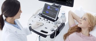
Ultrasound of joints is used to determine the condition of cartilage structures, identify inflammatory processes and cracks.
Ultrasound diagnostics is used in cases of chronic and acute inflammatory diseases, tendon and ligament ruptures and other injuries.
Compared to MRI of joints, ultrasound is much more affordable, but magnetic resonance imaging is much more informative. Using this method, you can obtain a 3D image of any joint and soft tissues in any section and any projection.
MRI relieves the patient of such painful research methods as puncture and biopsy. This is one of the best ways to diagnose chronic arthritis and degenerative joint diseases.
A unique feature of MRI is that it can detect pathological changes at a stage when there are no symptoms. Such early detection of the disease, provided immediate and adequate measures are taken, gives the patient hope for healing.
1 MRI of joints
2 MRI joints
3 MRI joints