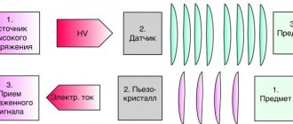
How is an ultrasound of the uterus and appendages performed?
Ultrasound of the uterus and appendages does not cause pain or inconvenience to the patient, since the examination is done in most cases through the anterior abdominal wall. Before the procedure, the patient undresses to the waist or frees access to the abdominal area. During an abdominal ultrasound of the uterus and appendages, the woman lies on her back, the doctor applies a gel to her stomach and moves a sensor over it.
With other forms of examination, the position of the patient, as well as the location of the sensor, may vary. In some cases, the sensor is inserted into the rectum or vagina.
It is important to note that the decision about when and in what form is best to do an ultrasound of the uterus is made by a specialist based on a number of symptoms or pathologies of the reproductive system noticed during examination.
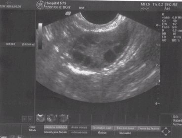
Normal uterus
Rice. 1. Normal uterus. Second phase of the cycle. The myometrium is homogeneous. The thickness of the M-ECHO corresponds to the day of the cycle.
When assessing the condition of the uterus with ultrasound, you can determine:
- Position of the uterus. Normally, the uterus is either deviated towards the bladder, that is, anteriorly (this position of the uterus is called anteflexio), or deviated towards the rectum, that is, posteriorly, (retroflexio).
- Dimensions of the uterus (longitudinal, anteroposterior and transverse). The average dimensions of a normal uterus are from 4.0 to 6.0 cm in length, anterior-posterior from 2.7 to 4.9 mm. The dimensions of the uterine body vary depending on the woman’s age, constitution and obstetric-gynecological history.
- The condition of the endometrium (its thickness varies depending on the day of the menstrual cycle). Immediately after the end of menstruation, the endometrium is visualized in the form of a strip 1-2 mm thick. In the second phase of the cycle, the thickness of the endometrium (M-ECHO) can range from 10 to 14 mm on average.
- Condition of the myometrium. Normally, the myometrium should be homogeneous and not have pathological formations in its structure (fibroids, adenomyosis, etc.)
How to prepare for an ultrasound of the uterus?
Since the examination can be carried out in various ways, preparation for an ultrasound of the uterus will depend on the method.
Transvaginal diagnosis
On the day of the examination, it is recommended not to eat foods that cause increased gas formation. Before the procedure, you must empty your bladder.
Transrectal examination
Since the examination is performed rectally, it is necessary to prepare for an ultrasound of the uterus more thoroughly. 6-8 hours before the procedure, the patient needs to empty the rectal ampulla using an enema or laxative.
Abdominal diagnostics
Abdominal ultrasound of the uterus and appendages is performed through the anterior abdominal wall. Therefore, increased gas formation in the intestines may affect the examination results. To eliminate distortions, it is recommended to remove foods that cause increased gas formation from the diet the day before the procedure: brown bread, cabbage, legumes, carbonated drinks.
Since abdominal ultrasound of the uterus and appendages is performed with a full bladder, you need to drink a liter of water an hour before the procedure or not urinate for 2-3 hours before the examination.
How to prepare for the procedure
Features of preparation for ultrasound examination depend on the method in which it will be carried out.
Preparing for the abdominal method
The day before the procedure, you need to change your diet to reduce gas exchange as much as possible: cabbage, beans, carbonated drinks, nuts, black bread and other gas-forming foods should be excluded.
The bladder should be full. Therefore, 1 hour before the ultrasound, a woman should drink 1 - 1.5 liters of non-carbonated liquid and not urinate.
Preparation for the invaginal method
The main requirement is an empty bladder, so immediately before the procedure the woman must urinate. Also, to improve the quality of the examination, it is recommended to empty the intestines of gases the day before using special medications (smecta, espumizan, activated carbon, etc.).
How is the procedure performed?
Abdominal ultrasound of the uterus and appendages is performed through the anterior abdominal wall. The patient undresses to the waist and takes a horizontal position face up. The sensor is placed on the stomach.
During a cervical examination or transvaginal ultrasound
a special sensor in a disposable condom is inserted into the vagina. Before the procedure, the patient removes clothing from the lower part of the body, including underwear. After this, the woman lies on her back and bends her knees.
For transrectal examination, the thinnest probe is used, which is inserted into the rectum. Before the procedure, the patient removes all clothing below the waist and lies on her left side. The sensor is inserted into the rectum in a special condom. A gel is used to improve the conductivity of ultrasound.
Why is it necessary to monitor the length of the cervix during pregnancy?
The cervix is a very important part of a woman’s reproductive system, the condition of which determines the successful outcome of pregnancy. The organ is similar to a tube, it has a length of approximately 4 cm and a diameter of 2-2.5 cm. The cervix connects the vagina to the uterus and is divided into two parts - the upper, supravaginal, and lower, vaginal.
The cervix of a non-pregnant woman is lined with a muscle layer, but during gestation, normally starting from 20-23 weeks, it is transformed into connective tissue, due to which the cervix becomes very elastic (see also: short cervix at 20 weeks of pregnancy). Changes in the cervix cause the uterus to stretch, so the connecting tube itself becomes shorter.
Starting from about 12 weeks, the connecting canal between the uterus and vagina gradually shortens. Just before childbirth, the cervix normally changes greatly, shrinking to a length of two centimeters.
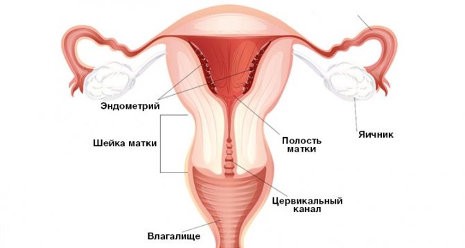
These changes are physiological and indicate a normal rate of cervical ripening. The norms of size indicators for primiparous and multiparous women may be different. In the case when the connecting canal does not change in size or rapidly shortens, doctors note a pathology that is dangerous to the health and life of the expectant mother and her baby.
Decoding the results
On the ultrasound transcript, the uterus is pear-shaped with a smooth, clearly defined contour, the ovaries are clearly visible. Fuzzy boundaries of the uterus may indicate inflammation. An uneven contour also occurs in other pathological conditions, such as tumors.
If after an ultrasound of the uterus the interpretation shows a deviation from the norm, the specialist will prescribe additional examinations or make a diagnosis based on the available data.
Normal sizes of the uterus and ovaries on ultrasound
The size of the uterus according to ultrasound may vary, the norm depends on the age of the patient, pregnancy and the onset of menopause.
For patients aged 15-40 years, the normal sizes of the uterus and ovaries according to ultrasound data will be: uterus - 4.5-6.7 cm in length, 3-4 cm in thickness and 4.6-6.4 cm in width, each ovary – 3-4.1 cm in length, 2.0-3.1 cm in width and 14-22 mm in thickness. 20 years after menopause, the uterus becomes significantly smaller.
An increase in the size of the uterus occurs during pregnancy or due to a tumor. A deviation from the norm to a lesser extent indicates an infantile uterus.
Normal ovaries
Rice. 2. Normal ovary with follicular apparatus. There is no dominant follicle, since the study was carried out on the 3rd day of the menstrual cycle.
When assessing the condition of the ovaries using ultrasound, it is determined:
- Position of the ovaries. Normally located on the sides of the uterus, most often asymmetrically, at a short distance from the corners of the uterus. The shape of the ovaries is usually oval, while the right and left ovaries are not at all identical to each other.
- Dimensions of the ovaries (longitudinal, anteroposterior and transverse). The average size of normal ovaries in length is from 2.4 to 4.0 cm, anterior-posterior from 1.5 to 2.5 mm.
- Structure of the ovaries. Normally, the ovaries consist of a capsule and follicles of varying degrees of maturity (in the first phase of the cycle). In the second phase of the cycle, as a rule, the corpus luteum is visualized - a sign of ovulation. The number of follicles may be different on the left and right. The maturing follicle is detected already in the first phase of the cycle and reaches its maximum size by ovulation, on average about 20 mm. The contents of the dominant follicle are homogeneous because it contains follicular fluid and the capsule is thin. After ovulation, at the site of the dominant follicle, a corpus luteum is formed, which, as a rule, has a mesh echostructure (it contains adipose tissue) and also a thin capsule - 1-2 mm. Most often, the shape of this formation is oval or irregularly shaped. In postmenopause, the ovaries are normally either not visualized or are located in the form of fibrous cords.
What abnormalities of the uterus and appendages will an ultrasound show?
The ultrasound method allows us to identify many pathologies of the development of the uterus, appendages and tubes and diagnose a number of diseases.
Malignant formations
The ultrasound method is considered the most informative examination of the uterus and appendages for malignant tumors. The examination visualizes the neoplasm, which allows the specialist to determine its location, the depth of germination into the muscle tissue, and the degree of damage to the lymph nodes and ovaries.
Transvaginal ultrasound of the uterus
and appendages is used as a method for diagnosing cervical cancer. The disease is widespread, and the risk of developing a tumor increases with age. Therefore, regular cervical cancer screening is recommended for all women over 30 years of age. In addition to ultrasound of the uterus and appendages, screening includes a smear for the papilloma virus and colposcopy.
Location of the fertilized egg
Atypical location of the ovum on a transvaginal ultrasound of the uterus
and appendages can be diagnosed from the third week of pregnancy.
Normal indicators of the thickness of the endometrium of the uterus during a normal cycle
Most women have a menstrual cycle of 28 days. Therefore, the norm of indicators for the results of M-echo of the uterus was calculated based on this period.
| Cycle days | Stage name | Thickness M-echo, mm | Endometrial structure | Number of layers |
| 1 – 2 | Desquamation phase of bleeding | 5 – 9 | Heterogeneous, with reduced echogenicity and increased sound conductivity | None |
| 3 – 4 | Regeneration phase during bleeding | 3 – 5 | With increased echogenicity | None |
| 5 – 7 | Early proliferation phase | 6 – 9 | Echogenicity decreases with increasing sound conductivity | Appearance of an echo-negative rim (about 1 mm) |
| 8 – 10 | Mid-proliferation phase | 8 – 10 | The appearance of a clear hyperechoic formation measuring 1 mm and an anechoic rim (about 3 mm) | 3 |
| 11 – 14 | Late proliferation phase | 9 – 13 | The appearance of a thin strip about 1 mm in size above the rim. Creating favorable conditions for fertilization | 4 |
| 15 – 18 | Early secretion phase | 10 – 16 | The structure continues to divide | 4 |
| 19 – 23 | Mid-secretion phase | 10 – 22 | The mucous membrane practically stops growing, the body is preparing to reject it (if fertilization has not occurred) | 4 |
| 24 – 27 | Late secretion phase | 10,1 – 18 | Likewise | 4 |
If the cycle is longer than 28 days, then the endometrial growth indicators lag behind, and if less, they increase. For women of childbearing age, the generally accepted norm for a median echo is considered to be 15–16 mm.
Indications for surgery
- Uterine bleeding or menstrual dysfunction;
- Active growth of nodes (more than 4-5 weeks per year);
- Large and giant fibroids, uterine size more than 11 weeks;
- Pain syndrome;
- Submucosal, subserous nodes, atypical location;
- Development of complications: anemia, necrosis, disruption of various organs, infertility, etc.;
- Risk of malignancy.
For a written consultation, in order to choose treatment tactics in your case, you can send me a complete description of the ultrasound of the pelvic organs, indicate your age and main complaints [email protected] [email protected] Then I will be able to give a more accurate answer to your situation.