Community-acquired pneumonia
Pneumonia is one of the most common acute diseases; it is a group of acute infectious (mainly bacterial) diseases of different etiology, pathogenesis, and morphological characteristics, characterized by focal damage to the respiratory parts of the lungs with the obligatory presence of intra-alveolar exudation.
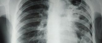
Community-acquired pneumonia (synonyms: home, outpatient) is an acute disease that arose in a community setting, accompanied by symptoms of lower respiratory tract infection (fever, cough, chest pain, shortness of breath) and “fresh” focally infiltrative changes in the lungs in the absence of obvious diagnostic alternatives.
The reasons for the development of an inflammatory reaction in the respiratory parts of the lungs can be either a decrease in the effectiveness of the body’s defense mechanisms, or a massive dose of microorganisms and/or their increased virulence. Aspiration of the contents of the oropharynx is the main route of infection of the respiratory parts of the lungs, and therefore the main pathogenetic mechanism for the development of pneumonia. Under normal conditions, a number of microorganisms, such as Streptococcus pneumoniae, can colonize the oropharynx, but the lower respiratory tract remains sterile.
In cases of damage to the “self-cleaning” mechanisms of the tracheobronchial tree, for example, during a viral respiratory infection, favorable conditions are created for the development of pneumonia. In some cases, an independent pathogenetic factor may be the massive dose of microorganisms or the penetration into the respiratory parts of the lungs of even single highly virulent microorganisms that are resistant to the action of the body’s defense mechanisms, which also leads to the development of pneumonia.
The etiology of community-acquired pneumonia is directly related to the normal microflora that colonizes the upper respiratory tract. Of the numerous microorganisms, only a few that have increased virulence are capable of causing an inflammatory reaction when they enter the lower respiratory tract.
Such typical pathogens of community-acquired pneumonia are:
- Streptococcus pneumoniae;
- Haemophilus influenzae.
Atypical microorganisms have a certain significance in the etiology of community-acquired pneumonia, although it is difficult to accurately determine their etiological significance:
- Chlamydophila (Chlamydia) pneumoniae;
- Mycoplasma pneumoniae;
- Legionella pneumophila.
Typical but rare pathogens of community-acquired pneumonia include:
- Staphylococcus aureus;
- Klebsiella pneumoniae, less commonly other enterobacteriaceae;
- Streptococcus pneumoniae is the most common causative agent of community-acquired pneumonia in people of all age groups.
The drugs of choice for the treatment of pneumococcal pneumonia are betalactam antibiotics - benzylpenicillin, aminopenicillins, including protected ones; II-III generation cephalosporins. New fluoroquinolones (levofloxacin, moxifloxacin) are also highly effective.
Macrolide antibiotics (erythromycin, roxithromycin, clarithromycin, azithromycin, spiramycin, midecamycin) and lincosamides have fairly high antipneumococcal activity and clinical effectiveness. But still, macrolide antibiotics for this pneumonia are a reserve remedy for beta-lactam intolerance.
Haemophilus influenzae
a clinically significant causative agent of pneumonia, especially in smokers and patients with COPD (chronic obstructive pulmonary disease). Aminopenicillins (amoxicillin), “protected” aminopenicillins (amoxicillin/clavulanate), cephalosporins of the II-IV generations, carbapenems, fluoroquinolones (early ones - ciprofloxacin, ofloxacin and new ones - levofloxacin, moxifloxacin, gatifloxacin) have high natural activity against Haemophilus influenzae.
Chlamydophila (Chlamydia) pneumoniae and Mycoplasma pneumoniae
usually characterized by a mild course. Mycoplasma pneumonia - more common in people under 40 years of age. The drugs of choice for treating these pneumonias are macrolides and doxycycline. New fluoroquinolones are also highly effective.
Legionella pneumophila
usually characterized by a severe course. The drug of choice for the treatment of Legionella pneumonia is macrolide antibiotics (erythromycin, clarithromycin, azithromycin). Early and new fluoroquinolones are also highly effective.
Staphylococcus aureus
an infrequent causative agent of community-acquired pneumonia, but its importance increases in older people, in people who take drugs, abuse alcohol, and after influenza. The drugs of choice for staphylococcal pneumonia are oxacillin; amoxicillin/clavulanate, cephalosporins, and fluoroquinolones are also effective.
Klebsiella pneumoniae
and other enterobacteriaceae are very rare pathogens of community-acquired pneumonia and have etiological significance only in certain categories of patients (old age, diabetes mellitus, congestive heart failure, cirrhosis of the liver). III-IV generation cephalosporins, carbapenems, and fluoroquinolones have the highest natural activity against these pathogens.
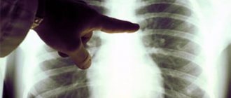
Pneumonia should be suspected if the patient has a fever in combination with complaints of cough, shortness of breath, sputum production and/or chest pain. Patients often complain of unmotivated weakness, fatigue, and severe sweating, especially at night.

If you experience similar symptoms, we recommend
Make an appointment
Signs of pneumonia such as acute fever, chest pain, etc. may be absent - especially in weakened patients and the elderly.
For mild pneumonia, antibacterial therapy can be completed once stable normalization of body temperature is achieved within 3-4 days. With this approach, the duration of treatment is usually 7-10 days. In cases where there is clinical and/or epidemiological evidence of mycoplasma or chlamydial etiology of pneumonia, the duration of therapy should be 14 days. Longer courses of antibiotic therapy are indicated for pneumonia of staphylococcal etiology or caused by gram-negative enterobacteria - from 14 to 21 days.
If legionella pneumonia is indicated, the duration of antibacterial therapy is 21 days. In case of community-acquired pneumonia, it is extremely important to quickly assess the severity of the patients' condition in order to identify patients requiring emergency intensive care. The allocation of patients with severe pneumonia into a separate group seems extremely important, given the high mortality rate, the presence, as a rule, of severe background pathology in patients, the peculiarities of the etiology of the disease and the special requirements for antibacterial therapy.
Late diagnosis and delay in starting antibacterial therapy (more than 8 hours) lead to a worse prognosis of the disease.
Unfortunately, pneumonia can have various complications, such as:
- pleural effusion;
- pleural empyema (accumulation of pus in the pleural cavity);
- destruction/abscessation of lung tissue (formation of limited cavities in the lung tissue);
- acute respiratory failure;
- infectious-toxic shock;
- sepsis;
- pericarditis, myocarditis (heart disease);
- nephritis (kidney disease) and others.
In case of pneumonia, a differential diagnosis must be made with such diseases as:
- pulmonary tuberculosis;
- neoplasms (primary lung cancer, endobronchial metastases, bronchial adenoma, lymphoma);
- pulmonary embolism and pulmonary infarction;
- immunopathological diseases (idiopathic pulmonary fibrosis, eosinophilic pneumonia, bronchocentric granulomatosis, bronchiolitis obliterans with organizing pneumonia, allergic bronchopulmonary aspergillosis, lupus pneumonitis, systemic vasculitis);
- other diseases/pathological conditions (congestive heart failure, drug-induced (toxic) pneumopathy, foreign body aspiration, sarcoidosis, pulmonary alveolar proteinosis; lipoid pneumonia, rounded atelectasis).
In conclusion, it must be said that only a doctor can make a diagnosis, determine the severity of the disease and the prognosis. If the patient has a fever, dry cough or cough with sputum, shortness of breath, chest pain, unmotivated weakness, fatigue, excessive sweating, especially at night, consult a general practitioner.
SM-Clinic’s own laboratory and instrumental base allows you to quickly diagnose and diagnose pneumonia. You will be prescribed timely treatment for pneumonia, individual for each person, taking into account the severity of the disease, age, and concomitant diseases. A therapist will help you become healthy again.
Pleural sinuses, the contour of the diaphragm and the mediastinum on a plain radiograph are normal and pathological.
Diaphragm and subdiaphragmatic space
.
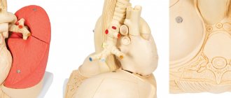
The diaphragm separates the thoracic cavity from the abdominal cavity. The right and left domes have a spherical shape on an x-ray with a convex upward and reach the level of the IV-V ribs. When you inhale, the diaphragm contracts, flattens and moves downwards; when you exhale, it relaxes and rises up. Normally, the domes of the diaphragm are at the same level (on the right above the left by no more than 2 cm), symmetrical.
The dome rises as pressure in the abdominal cavity increases.
On the outside, the chest is surrounded by soft tissues - various muscle groups, subcutaneous fat layer, mammary glands, etc. Below the diaphragm are the abdominal organs: on the right - the liver, on the left - the left lobe of the liver, the vault of the stomach, the splenic angle of the colon and the spleen.
Pleura - consists of two layers - parietal (lining the inner surface of the chest, the upper surface of the diaphragm and the lateral surfaces of the mediastinum) and visceral (tightly covering all lobes of the lung). Between the layers of the pleura there is a closed pleural cavity, in which liquid contents (exudate, etc.) can often accumulate. In the lowest parts of the pleural cavity, in the places where the domes of the diaphragm are attached to the chest, the costophrenic sinuses are located: anterior, external and posterior.
During an X-ray examination, the lung tissue and possible pathological substrate located in the sinuses can be covered by the upwardly convex domes of the diaphragm and become visually inaccessible.
Mediastinum
On an x-ray, in the central part of the chest there is a shadow of the mediastinum, which includes the heart, large vessels (aorta, pulmonary artery, inferior and superior vena cava and pulmonary veins), esophagus, as well as the thoracic spine. The mediastinal organs divide the chest into two halves for the right and left lungs.
In pathology, the mediastinum may be widened. On the left is normal: without going to the left. midclavicular line, to the right no more than 2 cm from the parasternal line.
X-ray syndromes of lung diseases
Extensive darkening - can be observed with:
- compaction (airlessness) of lung tissue of any origin (inflammatory infiltration, cirrhosis, atelectasis). Inflammatory infiltration is often characterized by heterogeneity of the shadow and relatively rapid dynamics.
With atelectasis, the shadow is uniform, the volume of the lung tissue decreases, the mediastinum shifts towards the affected side, the pulmonary aperture becomes narrower, the dome of the diaphragm on the affected side is raised upward.
With cirrhosis of the lung tissue, the darkening is often heterogeneous, the root of the lung can be deformed and pulled to one side, and the pulmonary aperture is narrowed.
- the presence of pathological contents in the pleural cavity - fluid, intra-abdominal organs in diaphragmatic hernias. In this case, a shift of the mediastinal shadow in the opposite direction may be observed.
- the presence of a large neoplasm - the shadow of the mediastinum in such cases can also be shifted to the opposite side.
- absence of a lung - observed with congenital anomalies, after surgical removal. Accompanied by a shift of the mediastinal shadow towards the lesion and a narrowing of the pulmonary aperture.
Limited darkening - may be caused by intrapulmonary and extrapulmonary processes, which can be established during a multi-view examination of the chest.
Intrapulmonary (pneumonia, pulmonary tuberculosis, abscess, tumor, cyst, peripheral tumor, etc.) are usually determined directly in the lung tissue (in all projections) and are displaced during breathing along with the lung tissue.
Extrapulmonary
- fluid in the pleural cavity - when the patient is in a vertical position, the shadow occupies a lower position, merging with the dome of the diaphragm, and the upper contour of the exudate shadow has an oblique border (Ellis-Damoiso line), in the case of the presence of transudate, the upper border of the fluid is horizontal. In the patient’s position “on his side,” the shadow (liquid) takes a lower position, spreading throughout the entire pleural cavity;
- pleurisy
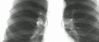
Round shadow. In the lung tissue, shadows of round shape, varying intensity, structure, size are found in: peripheral cancer, tuberculosis, cysts, single metastases, etc.
Tuberculosis infiltrate is a low-intensity shadow of small sizes (1.5-2 cm) with unclear external contours and the presence of small focal dropouts around. When the infiltrate disintegrates, a cavity appears in its structure.
Tuberculoma
– compacted (organized) infiltrate with small calcified inclusions and finely focal screening around.
Cyst
– a homogeneous oval-shaped shadow filled with liquid contents with clear contours. Due to the elasticity of the formation, the shape of the cyst can change with breathing.
Metastases
. A single metastasis is a round, small, homogeneous shadow with fuzzy, uneven contours, which must be differentiated from peripheral cancer, tuberculous infiltrate, etc. Multiple round shadows, as a rule, indicate the presence of metastases.
Extensive clearing is an increase in the transparency of the lung tissue on both sides within one or more lung fields.
It is observed with regional or total (diffuse) emphysema. Diffuse emphysema can be unilateral or bilateral. Unilateral total clearing is most often caused by valvular blockage of the main bronchus.
Extensive clearing syndrome is also observed with total unilateral pneumothorax with a complete absence of a pulmonary pattern on the image and the presence of a collapsed lung at the root.
Change in pulmonary pattern . They arise as a result of circulatory disorders in the pulmonary circulation or lymphatic drainage, fibrosis of the interstitial tissue and can manifest themselves as an increase or depletion of the pulmonary pattern.
Strengthening the pulmonary pattern - an increase in the size and number of elements per unit volume, is observed: as a result of arterial congestion with heart defects; interstitial edema; interstitial fibrosis in chronic bronchitis; pneumoconiosis, sarcoidosis, collagenosis, alveolitis, etc.
Depletion of the pulmonary pattern is characterized by a decrease in the number of elements per unit volume and can be observed as a result of pulmonary hypovolemia with heart defects, pulmonary emphysema, etc. The prevalence of depletion of the pulmonary pattern can be different: limited, total unilateral or bilateral.
Changes in the roots of the lungs can be unilateral or bilateral. They are expressed by changes in: size, shape, structure, density and nature of the contours of the roots. Expansion and deformation of the roots of the lungs can occur due to enlarged lymph nodes, dilation of blood vessels and neoplasms. Blurred root structure occurs with edema and fibrosis; increased density – with calcification of lymph nodes due to tuberculosis or silicotuberculosis. A polycyclic contour of the roots is observed with an increase in the group of lymph nodes (on one side - tuberculous lymphadenitis, on both sides - sarcoidosis, lymphogranulomatosis, lymphosarcoma, metastases, etc.), a tuberous contour - with the central exobronchial form of lung cancer.