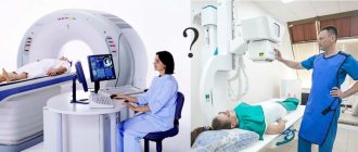
What is fluorography?
It often happens that a person who needs to examine his lungs thinks about how fluorography differs from an x-ray of the lungs. It is worth knowing that fluorography, just like radiography, is based on obtaining an image based on the passage of ionizing radiation through the human body. However, the radiation dose, the technique, and the information content of these methods vary significantly.
The difference between lung radiography and fluorography is that the latter method is considered much more gentle and safe, since the radiation dose at a time is 0.015 mSv. For problems to arise due to excessive radiation exposure, you need to undergo such diagnostics about a thousand times a year.
The history of fluorographic research
Before you understand what fluorography and X-ray of the lungs are, how the methods differ from each other and which one is better, it is worth familiarizing yourself with the history of the fluorographic diagnostic method. It appeared in 1930 and was actively promoted by the Soviet scientist S. A. Reinberg.
The first fluorography devices still required a fairly high radiation dose, up to 2.5 mSv, and such an examination required significant effort on the part of doctors. If you want to know what the difference is between fluorography and x-rays of the lungs, then keep in mind that modern fluorography machines have become much safer and allow you to obtain very high-quality images.
Who needs to undergo the study
The legislation of the Russian Federation has a resolution that determines the frequency of this survey.
Why do you need fluorography of your husband during pregnancy?
The following are required to undergo a fluorographic examination: persons who first came to the clinic, people living together with a newborn, pregnant women, men entering military service or contract, people suffering from HIV infection.
Once a year, every adult should undergo fluorography as a screening for pulmonary diseases. Without this result, doctors do not sign the conclusion of the medical commission. This requirement arose due to the massive spread of tuberculosis.
A single radiation dose for this procedure is within 0.015 mSv. The preventive dose allowed by the Ministry of Health is 1 mSv. This implies the safety of fluorography; excessive radiation exposure can occur only when taking 1000 images per year.
Types of fluorography
You can understand the difference between X-rays of the lungs and fluorography after familiarizing yourself with the main types of the latter. They are widely used to diagnose inflammation and tuberculosis.
Digital fluorography
Lung X-ray or fluorography: what is the difference and advantages of modern research methods? Digital fluorography is a more advanced method and provides minimal stress on the body. As a result of such a study, an image of the shadow of the organs will appear on the computer equipment monitor, which is created thanks to a special chip located in the receiver. The produced beam will linearly pass through the entire examined area, after which special software will reconstruct the image. When choosing between whether fluorography or x-ray of the lungs is better, we can say that the digital research method is much safer.
Traditional fluorography
It is an outdated technique that differs little from x-rays. The photograph is taken on small film. If you have an x-ray or fluorography of the lungs (which is better in this case, it is unlikely to be determined) of the traditional type, the radiation exposure will be the same.
Which method is the safest?
Many people are interested in what is safer, FG or X-ray, and whether diagnostics can be done on children. The first method is more dangerous; it uses a large radiation dose, which causes a risk of complications. R-graphy is characterized by a lower dose, due to which the procedure can be performed on a child. It can be carried out several times to identify the characteristics of the disease.
It turns out that fluorography differs from x-rays of the lungs in terms of safety. However, this is not a cause for concern. Since both cases are characterized by minimal radiation exposure. If you follow certain rules, the occurrence of pathologies is reduced to a minimum.
What is a lung x-ray?
It is not possible to say unequivocally which is better, x-ray or fluorography of the lungs. Although the latter method is more gentle, the resulting images are of lower quality. On an x-ray, shadows measuring two millimeters can be detected, while on a fluorographic image – at least five centimeters.
The difference between X-ray of the lungs and fluorography is also that the X-ray rays pass through the tissue and the film is illuminated accordingly. This type of research creates a high, but very short-term radiation load on the human body.
What to choose - X-ray or fluorography of the lungs?
COVID-19✓ Safe
Many people ask the question: how does x-ray differ from fluorography and which examination should be chosen? X-ray reveals the condition of internal organs, not only the lungs, but almost any part of the human body. Fluorography is done on the chest area, with the help of which you can see the condition of the lungs, heart and part of the spine.
In addition, this examination is most often done for preventive purposes - to prevent the spread of tuberculosis. However, fluorography can reveal cancer and other diseases in the early stages of development, the presence of which the patient does not yet suspect, although he feels healthy.
If, when examining the image obtained during fluorography, the doctor has suspicions, he will refer the patient for re-examination. In this case, to obtain a more detailed picture, you will have to undergo an x-ray or tomography.
Fluorography is considered a more “strict” examination, so it is not prescribed to pregnant women and children under 15 years of age. For young children, if a serious illness is suspected, X-rays are prescribed.
Using X-rays, you can examine a child almost from the first days of life. During the procedure, the child cannot move, so one of the parents must hold him during the examination. To reduce the harmful effects of X-rays on the baby’s body, areas of the body that are not being examined are covered with protective screens.
In general, healthy people do not need to have an x-ray. According to existing sanitary standards, they must undergo fluorographic examination annually in order to prevent tuberculosis and cancer. The date and result of this study are entered in the patient’s outpatient card, as well as in his health record.
More often - once every six months - medical workers, as well as people working in hazardous industries, in the food industry, in children's educational institutions, etc. should undergo fluorography. In addition, people in prison are required to be examined, since they are most likely to become infected with tuberculosis.
The main difference between X-rays and fluorography is that during an X-ray examination, the image is more detailed and larger, and the organ being examined is clearly visible on it. And with fluorography, the image is photographed on photographic film by the device itself. The result is a reduced image. Fluorography is considered a cheaper type of examination, which is why it is used for mass preventive examinations. The image obtained during fluorography is easier to work with, it is easier to store, but its information content is lower than that of an x-ray image.
The results of fluorography and x-rays should not be thrown away; they should be put in a folder and stored. If necessary, they will need to be shown to a doctor, this will help identify changes that have occurred in the lungs.
Telephone
Is X-ray examination of the OGK safe?
Trying to understand the difference between chest x-rays
and fluorography, it is worth knowing that in domestic clinics the radiation dose is several times higher than that permitted in more developed countries. The reason is that Russian clinics often use very old devices. In Europe, the annual dose of X-ray radiation is no more than 0.6 mSv, but in Russia it is more than twice as high - 1.5 mSv.
However, in the event of a threat to the patient’s life, for example from acute pneumonia, doctors will not have to choose whether to do a fluorography or an x-ray of the lungs; they will simply use the available equipment for a speedy diagnosis. Typically, in such a situation, an X-ray examination is performed directly and from the side, and often a targeted X-ray is also needed.
Fluorography. X-rays of light
Duration of the study
: 5 minutes.
Preparing for the study
: No
Contraindications
: No
Preparation of the conclusion
: 30-60 min.
Restrictions
: No
At the Health Clinic, fluorography is performed using high-precision, latest Phillips X-ray equipment, which allows you to create the highest quality images.
Why is it better to have fluorography done with us?
Because our images will allow you to make an accurate diagnosis!
Because you don’t have to stand in line at the clinic, first to see a therapist, then buy a film, and then stand in a huge line for the procedure itself. But that’s not all - after all, you need to come to the therapist again in a few days to get the picture, again wasting a lot of time and nerves in line.
EVERYTHING IS SIMPLE WITH US - CALL, COME, AND GET A GOOD FLUOROGRAPHY CHEAP.
By using X-rays using the equipment of the Health Clinic, you will immediately receive several advantages:
- digital photos (for your convenience, we can save them on a flash drive)
- during the procedure you will not receive any dangerous dose of radiation (in our X-ray machine it is about 0.12 mSv - this is less than if you lay for an hour in the hot sun by the sea on the beach)
- descriptions in Russian and, if necessary, in English. This is an excellent solution both for treatment in clinics abroad and for English-speaking guests of our country.
Our clinic has very affordable prices, convenience and comfort. You don’t have to wait in lines - you will be accepted exactly according to your appointment. There is no need to come back for the photo - you will receive it literally 15 minutes after the procedure. The photo is without defects, on high-quality film. If you lose your photo, we will make a copy because your photo will be stored on our computer.
Patients of the Diagnostic Center are provided with free parking. Reservation of a place for a car is made no later than an hour before arrival at the clinic. Call

Another plus in favor of fluorography in our clinic is that the equipment of the Health Clinic allows you to significantly reduce the dose of radiation during x-rays! That is, you will not be exposed to the same dose of radiation as with conventional X-ray machines. Modern technologies make it possible to undergo fluorography without harm to health.
WE CARE ABOUT YOUR SAFETY AND HEALTH
What is fluorography
Almost everyone knows.
Each of us should undergo fluorography at least once a year. Because only a photo of the lungs will allow you to determine the presence or absence of the most dangerous diseases: tuberculosis and lung cancer. Our equipment and experienced specialists can “catch” the disease at the earliest stages.
And we all know that the earlier the disease is detected, the greater the chances of a complete recovery.
Features of fluorography
Starting from the age of 16, the examination is mandatory annually. First of all, specialists from such professions as medical workers, catering workers, teachers and nannies should undergo it annually.
Fluorography shows:
| _ |
|
Fluorography for corporate clients and organizations. VHI
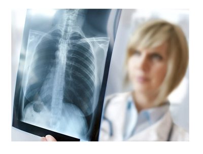
Conduct medical examinations of employees at the Health Clinic because this:
- profitable
- we have very attractive prices for partners; - convenient
- all doctors in one place.
More information about the terms of cooperation.
Consultation can be obtained by phone: +7
You may be interested in:
|
|
|
Cost of fluorography
| Name of service | Price in rubles |
| X-ray of the chest organs (1 projection) | 1300 |
If you do not find a service in the price list, please call us and we will provide you with the necessary information.
Our doctors will help you:
Radiologist CT, MRI
Vasilenko Tatyana Gennadievna
Radiologist CT, MRI
Parfenova Natalya Anatolevna
When is a chest x-ray necessary?
The reason for fluorography (x-ray of the lungs) can be various symptoms, for example, pain in the lung area, hemoptysis, dry cough, causeless weight loss, general weakness, etc.
An X-ray of the lungs is necessary in the following cases:
- diagnosis of inflammatory diseases of the lungs, bronchi and trachea (for example, acute pneumonia);
- detection of inflammatory diseases of the pleura (pleurisy, pleural empyema);
- the presence of fluid in the pleural cavity;
- diagnosis of pneumothorax;
- suspected pulmonary tuberculosis;
- suspicion of tumor processes in the lungs, bronchi and trachea;
- diagnosis of pulmonary embolism (PE);
- chest injuries;
- assessment of heart condition;
- identification of foreign bodies;
- diagnosis of pneumoconiosis;
- examination for diseases of the hematopoietic organs;
- diagnosis of parasitic diseases of the chest;
- diagnosis of diseases of the thoracic spine.
There are no absolute contraindications to X-ray of the lungs. A relative contraindication is pregnancy.
Chest X-ray is indispensable for diagnosing diseases of the main respiratory organs! You can undergo examination at MedicCity every day from 9.00 to 21.00, using the services of a paid emergency room. You can check the cost of x-rays of the lungs and other procedures with our operator by calling +7 (495) 604-12-12 or on the website in the “Prices” section.
