Why is fluorography necessary?
The main goal of diagnosis is to identify the pathological process at an early stage and begin treatment before specific symptoms appear. Here are the diseases that fluorography diagnoses:
- Pneumonia;
- Pleurisy;
- Tuberculosis;
- Malignant tumors of the lungs, mediastinum;
- Emphysema.
If a health problem is detected early, the chances of a favorable clinical outcome increase. This is what can be seen on fluorography:
- Lungs;
- Heart;
- Less often - bones.
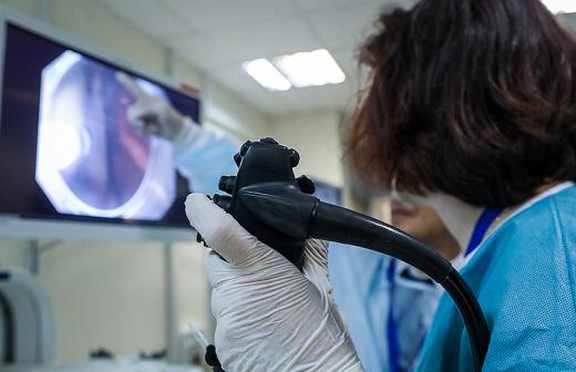
This screening examination shows the inflammatory process, foreign bodies, tumor formations, accumulation of infiltrate, cavities of a non-physiological nature (various cysts). Fluorography even shows if the patient smokes. The result is not the main criterion for making a diagnosis; it allows a more accurate assessment of a specific clinical case.
Lungs of a smoker and a healthy person
Smoking can change the appearance of your lungs. Diagnostic tests such as CT scans and chest x-rays may reveal some changes.
| Healthy lungs | Smoker's lungs |
| Pink colour | Color - Gray or black |
| Normal size | Hyperinflated |
| No inflammation | Foci of inflammation |
| Dome diaphragm | Weakening of the diaphragm muscles |
Smoking not only causes physical changes, but also changes lung function. The changes cause several symptoms that interfere with normal breathing.
What does fluorography give and when to do it?
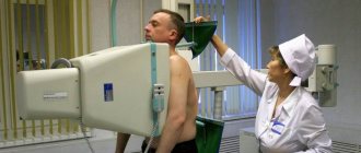
For prevention, persons over 17 years of age are recommended to undergo examination once every 2 years. There are also categories of people who are exposed to x-rays 1 and 2 times a year.
Here's what you can see on fluorography when the picture is already in hand:
- Focal shadows in the lung area;
- Compaction, expansion of roots, as a sign of pulmonary pathologies;
- Fibrous tissue as a result of surgery, trauma, infection;
- Aperture changes;
- Branching of the vascular pattern as a symptom of bronchitis, pneumonia, early stage of oncology;
- Change in the usual shadow of the mediastinum.
Based on the results, it is recommended to contact a phthisiatrician, who, in addition to decoding, will help determine the clinical picture, get examined more quickly, and prescribe treatment.
Does fluorography show that a person smokes?
Young smokers are usually afraid of being detected by X-rays. But since their history of smoking is short, and the degree of lung damage has not reached its pathological level, fluorography will not be able to detect smoking.
Indirect evidence of tobacco use or e-cigarette smoking can be found in changes in the size of the heart muscle. Also, bronchi that are constantly exposed to the destructive effects of smoke may indicate smoking.
Electronic cigarettes or pipes cause the same changes in the structure of the lung walls as regular tobacco smoked in a cigarette or pipe. The difference can be considered the absence of acute or chronic bronchitis caused solely by smoking. This form can be detected if a person smokes for a long time without a break.
Fluorography will show the structure of the lungs, and not the fact of smoking. It is not for the radiologist to decide whether a person is a smoker or a non-smoker.
A fluorogram can show the acute or chronic stages of these diseases. One or more of these symptoms may indicate whether a person smokes or not.
In the video from The Health Channel, you can learn about diagnosing tobacco smoking using the fluorogram procedure.
The difference between healthy lungs and a smoker's
In a healthy person who is not addicted to tobacco, the lungs appear light gray and without any obvious abnormalities. They do not have clearly defined vessels and there is no background darkening in the lungs. The roots of the lungs are straight, without signs of inflammation or edema.
The lungs of a tobacco or hookah smoker who has been exposed to long-term toxic effects of smoke and its decomposition products will have a denser and more pronounced pattern. Small lighter areas will be especially noticeable against the background of a dense structure. With this pathology, the radiologist recognizes bronchiectasis in the lung tissue and the pulmonary nodules themselves. With prolonged damaging effects on the lungs, the retina of the respiratory organs may be exposed.
The fact of smoking tobacco, an electronic cigarette or a hookah is indicated by the fact:
- Black areas on an x-ray. In the affected areas of the lung, there is a significant deterioration in the elasticity of the walls, which in turn can cause gas exchange. This also applies to the heart muscle. An increased volume of the heart in the image indicates the presence of pathologies and changes in tissue structure.
- More pronounced manifestation of the vascular structure. Cells suppressed by the action of nicotine and combustion products die and are not removed from the lungs, forming suckers on the blood vessels. Peripheral parts of the respiratory system can be affected and altered by long-term smoking.
- Dysfunctional tissue resulting from blockage of pores in the lungs with tar. On an x-ray, it will be marked by alternating light and dark areas, which will make the picture of the lung fragmentary. Such changes can disrupt oxygen transport and, as a result, oxygen starvation of the brain.
- Loss of clarity. Indicates the formation and spread of fibrous tissue, an increase in the lumen of blood vessels. In conditions of constant oxygen deficiency and hunger, the human respiratory organs begin to adapt and change their structure.
Mechanisms of adaptation and compensation for oxygen deficiency negatively affect the density of the tissue of the pulmonary sac. The process of change begins in the lower parts of the lung and progresses upward, gradually filling and replacing the lung tissue with a fibrous, nonfunctional protective lining.
Photo gallery
The photo shows the difference between a healthy lung and a smoker's.
If fluorography is bad, what could it be?
The transcript of the study is ready the next day, within 5 minutes after the study. If the result is poor, the person is referred to a TB specialist. It is too early to make a final diagnosis; a comprehensive diagnosis is carried out. Doctors may recommend repeat FLU. For the most part, the transformation of lung tissue is caused by the development of connective tissue in the lungs, which grows and layers.
Depending on the location and shape of the pathology focus, the doctor diagnoses fibrosis, sclerosis, adhesions, radiance, heaviness, shadows, and scar transformations. Based on the results, it is possible to diagnose oncology and emphysema, cysts, abscesses, calcifications, and other neoplasms and cavities.
Diagnostics can be done in every city, both in a private medical center and in a city clinic. The result obtained cannot be trusted 100%; an integrated approach to the health problem is needed.
How to cleanse your lungs and undergo fluorography if you smoke
It is impossible to achieve complete cleansing of the lungs in a few days, or even weeks. However, to improve overall performance, it is recommended to follow a number of recommendations.
What can you do at home?
To cleanse the lungs of toxins and their breakdown products, it is necessary to regularly spend time in a coniferous forest. It is especially useful to do this after rains, when the air is humid. It is recommended to take walks in the coniferous forest at least several times a month. You can combine them with physical activity - running, doing exercises and the like.

At the same time, the oxygen concentration in the lungs increases, the tissue is cleansed, the ciliated epithelium quickly regenerates and restores its evacuation ability. Regular saturation of the body with phytoncide compounds improves the overall tone of the body.
Staying in a high temperature zone helps cleanse the body of toxins and waste. Under their influence, all pores on the skin open, and harmful substances come out along with sweat. To stimulate sweating, you can regularly visit the bathhouse or sauna.
Cleaning the respiratory system
To restore the elasticity of the lung tissue, it is necessary to regularly conduct breathing and cardiac training. The easiest way is to perform special exercises at home: inhale air to the limit, and then gradually exhale it, tensing your abs.
You can enhance the effect by playing sports. For example, aerobics and yoga eliminate tissue fibrosis and restore the elasticity of the lungs while simultaneously developing their volume.
The intensity of training should be increased gradually. As the condition of the lungs improves, the load can also be increased.
Nutrition
It is especially important for people with bad habits to adhere to a properly balanced diet. The diet must include proteins (lean meat, fish) and vitamins. The latter are responsible for maintaining the body in tone and removing the breakdown products of toxic substances.

It is important to stop possible inflammatory processes. Vegetables containing phytoncides - garlic, onions and others - will help cope with this. Dairy products help eliminate nicotine.
Medicines
With long-term tobacco use, dead cells of epithelial tissues and blood vessels appear and accumulate in the lungs. The body cannot remove them from its organs on its own, so it needs medical help. The easiest way is to use inhalers, syrups or tablets. They cleanse the lungs and relieve the side symptoms of smoking – cough and shortness of breath. Doctors prescribe medications based on the results of the examination. Lazolvan, Mucaltin, Ambroxol, Gedelix can be used as drug therapy.
Ozone therapy has a more effective effect. This is an invasive medical procedure to clear the airways. Under the influence of ozone, which is injected directly into the lungs, the blood and respiratory organs are saturated with oxygen. A few cleansing cycles are enough to improve the overall tone of the bronchi and capillary function.
ethnoscience
Teas and decoctions effectively cleanse the body of nicotine. Properly selected preparations saturate depleted tissue with vitamins. As a result, it is possible to improve the overall tone of the lungs and increase the elasticity of the tissue.
One of the most effective recipes suggests the following. You need to mix in equal parts poppy, primrose, plantain, isod, elderberry, licorice, pine buds, horsetail, thyme, elecampane, soapwort, tricolor fragrant violet, fennel. Pour 1.5 tablespoons of the prepared mixture into 1 glass of boiled water, cover with a lid and leave until completely cooled. For 1-2 months, you need to drink a glass of infusion every day before going to bed.
Where and how to check your lungs
You can check your lungs using one of the x-ray methods: x-ray, fluorography, computed tomography. Fluorography is the most accessible and safe method for assessing the condition of the bronchopulmonary system. This procedure is mandatory for every adult. It is carried out in order to detect serious pulmonary diseases: tuberculosis, cancer, bronchitis (chronic and obstructive), cysts, abscesses, emphysema, fibrous formations, as well as indirect signs of problems in the heart (reduction of the lumen between the lungs).
An X-ray of the lungs of a moderate smoker is not much different from an X-ray of the lungs of an ordinary person, provided there are no pathologies.
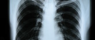
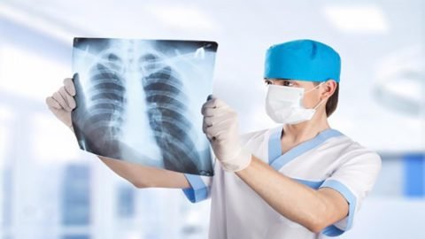
FLG (fluorography) is an x-ray examination. Its results are presented in the form of a small picture of the internal organs. The method is based on the fact that different tissues absorb Ro-rays differently.

There are two types of this procedure:
- classic (film);
- digital.
In the digital method, instead of film material, a CCD matrix and technology for layer-by-layer examination of the chest with radiation are used. It takes longer, but provides more detailed information and minimizes the harm caused to the human body.
The fluorography image is:
- small-frame (from 2.4×2.4 cm to 3.5×3.5 cm);
- large-frame (from 7×7 cm to 10×10 cm).
When it is necessary to examine a specific pathology in detail, a large-frame image is taken.
There are mobile fluorographs, which makes the method in question even more convenient. This procedure can be carried out anywhere. A specially equipped car with such a device can move around the city.
FLG is also used to search for other serious diseases.
No symptoms
“In the early stages, lung cancer is cured up to 80% after surgery. Moreover, now thoracoscopic operations are performed, that is, the chest cavity is pierced and, for example, a lobe of the lung is removed from the puncture,” says Konstantin Titov.
But the first and second stages do not give any symptoms - the tumor grows little by little, but the patient knows nothing about it.
They go up in smoke
Photo: Depositphotos
- Symptoms will appear later - in the third and fourth stages, shortness of breath, chest pain, hemoptysis, weight loss may appear. But while the tumor is small, there will be no symptoms until it spreads to the lymph nodes, grows into some adjacent structures of the chest cavity, or certain metastases appear. Moreover, in our country, lung cancer mainly occurs in smokers, and experienced smokers have a cough and discomfort in the chest, so they simply do not feel the dangerous symptoms and turn to doctors late.
Diagnosis by photo: artificial intelligence will detect ulcers and cancer
The system will identify pathologies from images of the mucous membranes of organs
What does fluorography of a smoker’s lungs show: are changes visible?
Fluorography is an informative research method that allows you to obtain the necessary data on the condition of the patient’s heart and lungs. Such an examination is mandatory and is carried out annually. The main goal is to identify any tumors in the lungs and tuberculosis.
Smoking patients are concerned about the question, what does fluorography of a smoker’s lungs show? Are the consequences of addiction reflected in the results of the study, and can tobacco smoke affect the data obtained? In order to get answers to existing questions, you need to understand the principles of the method.