Take the first step
and make an appointment with your doctor!
Among the most common causes limiting women's reproductive capacity is endometriosis. This pathology is characterized by abnormal thickening and proliferation of the uterine mucosa beyond the organ. Its high prevalence leads to the fact that a significant number of women of reproductive age experience problems with conception, including during artificial insemination. Is IVF possible for endometriosis and what are the chances of getting pregnant for patients suffering from this disease?
Endometriosis: causes and symptoms
To figure out whether IVF is done for endometriosis, you need to understand the essence and causes of this disease. They have not yet been precisely established and are the subject of debate in the medical community. It is believed that an important role in the occurrence of pathology is played by the ability of endometrial cells to “migrate” to other organs and their exposure to hormones that normally regulate the growth of the uterine mucosa within the menstrual cycle. The following factors also contribute to the development of this disease:
- immunity disorders, due to which endometrial cells are not destroyed by the body’s defense system when they penetrate beyond the uterine cavity;
- genetic predisposition - it has been proven that the likelihood of endometriosis is higher in women whose relatives also suffer from this disease;
- genetic abnormalities that lead to endometrial cells becoming more viable and able to survive in conditions to which they were not initially adapted.
In a normal menstrual cycle, the endometrium of the uterus undergoes changes under the influence of sex hormones. In the premenstrual phase (a few days before the start of menstruation), this layer thickens, and the number of blood vessels and glands in it increases. This creates optimal conditions for implantation of the embryo after its fertilization - this is the main function of the endometrium. If conception does not occur, the excess mucous membrane sloughs off and comes out along with menstrual bleeding, after which a new cycle of preparation for fertilization begins. This mechanism allows women to become pregnant all year round, while in many other animals this ability is limited to a fairly short period.
However, in some cases this process can be disrupted by various factors. Thus, the reverse flow of menstrual blood, which occurs as a result of injuries, abnormal structure of the genital tract, sex during menstruation and other reasons, leads to the fact that the desquamated endometrial cells do not come out, but through the fallopian tubes they penetrate into the abdominal cavity and “settle” in other organs that have an intensive blood supply - the ovaries, intestines, abdominal wall, navel, etc. They penetrate into the tissues that shelter them and begin to grow deep into them, forming foci of endometriosis.
The situation is complicated by the fact that endometrial cells, having “settled” outside the uterus, remain susceptible to the influence of hormones. Because of this, the abnormally located endometrial tissue continues to periodically exfoliate, which causes the following negative consequences:
- internal bleeding localized outside the uterine cavity and not having a natural exit from the body;
- inflammatory processes that lead to damage to tissues that “shelter” endometrial cells and, as a result, to dysfunction of the affected organs;
- a high risk of a secondary infection, for which untapped blood and desquamated cells of the abnormal endometrium become a breeding ground.
These processes are manifested by symptoms, which include pain in the lower abdomen and lower back (periodic or constant), more intense and prolonged periods, painful sexual intercourse, and reproductive dysfunction. Symptoms depend on the location and degree of proliferation of endometrial cells - it also happens that endometriosis does not manifest itself in any way, which makes it difficult to detect.
There is also an internal type of endometriosis, in which the endometrium grows not into other organs, but deep into the underlying layer of the uterus, which also leads to disruption of the functioning of this organ. Often both types of the disease (internal and extragenital) are combined with each other, leading to complex damage to the woman’s reproductive system.
Take the first step
make an appointment with a doctor!
Treatment of infertility associated with endometriosis
To predict the likelihood of pregnancy in the natural cycle in women with confirmed endometriosis, the EFI fertility index is used [11]. Low levels of this index, a woman's advanced reproductive age, reduced ovarian reserve, a combination of external genital endometriosis and adenomyosis, and a high risk of relapse of endometriosis are indications for the use of assisted reproductive technology programs.
The low effectiveness of the artificial insemination procedure does not allow it to be recommended for the treatment of infertility in women with endometriosis. That is why the IVF program is considered as the main method of overcoming infertility or subfertility in women with endometrioid disease.
The following factors can increase the likelihood of pregnancy after an IVF program and cryo-protocol in women with endometriosis:
- refusal of wait-and-see tactics and entry into an IVF or cryo-protocol immediately after two-stage (surgical and drug) treatment of endometriosis (6-month GnRH therapy before IVF has no advantages over a 3-month cycle).
- there is convincing evidence in favor of transferring one rather than two or more embryos in women with endometriosis;
- refusal of repeated surgical interventions before the IVF protocol; this can be facilitated by an individualized drug treatment plan designed to prevent recurrence of endometriosis between IVF programs.
Endometriosis and IVF
Pathological proliferation of endometrial tissue has a significant negative impact on a woman’s reproductive ability. The following factors can be identified that accompany this disease and reduce the likelihood of a successful pregnancy:
- Violation of the structure of the endometrium. Endometriosis causes abnormal thickening of the lining of the uterus. Normally, for conception, its thickness is 11-12 mm - if this threshold is exceeded, the tissue becomes too loose, and the fertilized egg either does not attach to it at all, or implantation does not occur completely, which leads to miscarriages.
- Damage to the reproductive organs. The endometrium, growing beyond the uterine region, spreads to nearby tissues. Often this pathology provokes the appearance of endometrioid ovarian cysts, causing dysfunction of these organs, which play a critical role in the formation and maturation of eggs.
- The appearance of synechiae (adhesions). An excessive amount of endometrium leads to the fact that the tubular genital organs (cervical canal, fallopian tubes) are blocked by it. This leads to disruption of the movement of sperm and eggs into the uterine cavity.
Thus, endometriosis is both a cause of infertility, which is treated with IVF, and a contraindication to this procedure, since it affects the function of the reproductive organs. The possibility of both natural and in vitro fertilization depends on the severity of the disease and its location. However, the chances of IVF for endometriosis do not strictly correlate with pathology and can reach 40-45%. In medicine, there are cases of successful artificial conception in women with abnormal endometrial development. Moreover, fertilization is often successful on the first try, and the woman learns about the disease itself after the procedure during routine examinations during pregnancy.
Take the first step
make an appointment with a doctor!
Risk factors during pregnancy with endometriosis.
If a woman does become pregnant while developing endometriosis, there are the following risk factors:
- • Ectopic pregnancy - requires immediate removal of the fertilized egg;
- • Hormonal imbalance, which is associated with low progesterone levels. Sometimes, this leads to miscarriage in the 1st and 2nd trimesters. In order to avoid this, doctors prescribe progesterone analogues;
- • Uterine rupture caused by severe thinning of the muscle layer in late pregnancy. In such cases, a caesarean section is performed;
- • Decreased elasticity of the cervix (caesarean section is also necessary);
* A prerequisite for women suffering from endometriosis is the supervision of a qualified doctor.
Pregnancy management
Is it possible to do IVF with endometriosis?
The implementation of in vitro fertilization is determined by how developed the abnormal tissue is and in which organs of the abdominal cavity it develops. In this regard, timely identification of foci of endometriosis plays an important role, for which the following methods are used:
- gynecological examination, during which tension is revealed in the area of the uterine appendages, the uterus itself and the pouch of Douglas - the area of the peritoneum lying between the posterior wall of the uterus and the anterior wall of the rectum;
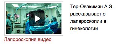
- laparoscopy - an internal examination of the abdominal organs, performed through a puncture in the abdominal wall using a thin tubular device (laparoscope);
- diagnostic examination of the rectal mucosa (sigmoidoscopy) and bladder (cystoscopy);
- Ultrasound of the pelvic organs - the effectiveness of this method in diagnosing endometriosis is limited only by the detection of endometrioid ovarian cysts, since pathological endometrial nodes on other organs are not detected by ultrasound.
The doctor may also prescribe MRI of the pelvic organs and other procedures as additional ones to clarify and differentiate the disease. But the main method in determining endometriosis remains laparoscopy.
Endometriosis identified before IVF is subject to treatment, which uses the following methods:
- Conservative therapy. It consists of taking hormonal medications aimed at reducing abnormally overgrown endometrial tissue that is susceptible to the influence of hormones. Before the advent of laparoscopy, this was the most common and effective method of treating the disease; today it is used as an auxiliary or for the treatment of mild pathology. Also, as part of drug therapy, anti-inflammatory drugs are used to relieve inflammatory processes in the affected organs and the accompanying pain syndrome. Medicines are introduced into the body orally, through injections, or through an intrauterine device. There is no universal drug that is guaranteed to cure the pathology; most often, several medications are used, taking into account the individual characteristics of the patient, the degree of the disease, plans for pregnancy, etc.
- Surgery. Physical removal of endometriosis lesions is the most effective means of their treatment. The operation can be performed through an incision or laparoscopic puncture, in which the abnormally overgrown tissue is excised using laser vaporization, electrocoagulation or resection. Removal of endometrioid cysts is performed completely with the maximum possible preservation of the follicular apparatus. With extensive endometriosis, which also covers the walls of the bladder, uterus, fallopian tubes, and intestines, the affected areas of these organs are often removed.
None of these methods are 100% effective or superior to other treatments for endometriosis. Therefore, the doctor forms therapeutic tactics individually, taking into account the characteristics of the patient’s physiology, the state of her body, the severity and spread of the disease and other factors.
Take the first step
make an appointment with a doctor!
The potential of ovarian function, reflecting the number and quality of follicles in the ovaries at a given moment, is designated by the term “ovarian reserve” [1]. The concept of “ovarian reserve” directly relates to female fertility, therefore all technologies that can disrupt, restore or maintain the patient’s ability to conceive are considered from the standpoint of their impact on the ovarian reserve.
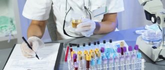
Ovarian reserve is assessed using several markers, the most informative of which is anti-Mullerian hormone (AMH) [2, 3]. In postnatal life, AMH is produced by ovarian granulosa cells, and it is produced most intensively by preantral and small antral follicles and is practically not synthesized at the final stages of folliculogenesis [4]. AMH is more closely related to the number of antral follicles counted by ultrasound than follicle-stimulating hormone or inhibin-B [5, 6]. One of the advantages of plasma AMH levels is its low cycle-to-cycle variability, which distinguishes it from other markers [7], despite slight changes throughout each menstrual cycle [8]. Thanks to these features, determination of the level of AMH in blood plasma is used to assess ovarian damage during surgical interventions, as well as to predict the outcomes of assisted reproductive technology (ART) programs [5, 9-11]. Throughout a woman’s biological reproductive life, the level of AMH in the blood plasma decreases very slowly and reaches undetectable values 5 years before menopause [12], which allows its use as a surrogate marker of ovarian reserve and a predictor of premature ovarian failure (POF) [13].
Not only age, but also other circumstances can influence the functional potential of the ovaries. Pelvic inflammatory diseases, endometriosis, subclinical toxic effects, drug and surgical interventions, and finally, genetic predisposition - all this determines the growing prevalence of POF among modern women.
Surgical interventions on the reproductive organs of the small pelvis can impair ovarian function, both due to loss of the ovarian cortex and as a result of damaging effects on blood flow [14]. The paradox is that the purpose of surgery is often to restore fertility in diseases associated with infertility. The most striking example of such an undesirable outcome is endometriosis, especially ovarian endometrioma.
The relevance of the problem of decreased fertility in endometriosis has given impetus to conducting numerous studies to assess ovarian reserve. The results of these studies are mixed. According to some of them, even minimal/moderate stage I-II endometriosis according to the ASRM classification is associated with a significant decrease in AMH levels in infertile women compared to women with a tubo-peritoneal cause of infertility [15]. In patients with endometriomas, the level of AMH was significantly lower than in women of similar age and anthropometric parameters who were operated on for teratoma [16]. Similar rates were obtained when comparing the rate in women with severe endometriosis (ASRM stage III-IV) and healthy fertile women of the same age in a case-control study [17]. According to other authors [18], a decrease in AMH levels was noted in patients with endometriomas compared to healthy women. However, a large cross-sectional study that included 313 women with diagnosed endometriosis and 413 women without endometriosis (controls) found significant differences in AMH levels only in the group of women who underwent surgery for endometrioid ovarian cysts [19].
Apparently, endometriomas pose a particular danger to the ovarian reserve. The ovarian cortex, which surrounds the cyst and makes up its wall, differs from healthy tissue in its low density of follicles and the loss of cortical-specific stroma [20, 21]. By the quality of this conditional cyst wall, one can indirectly judge the activity of the endometrioid process. It was shown that 3 months after surgery, the level of AMH was significantly higher in those women whose cyst wall contained healthy ovarian tissue, compared with patients in whom such tissue was not found [22].
Thus, today it can be considered recognized that endometriosis can negatively affect the ovarian reserve, to a greater extent this characterizes endometriomas, but we are not yet aware of prognostic markers of unfavorable outcomes of endometriosis in relation to the ovarian reserve.
In contrast to the conflicting results of assessing the effect of endometriosis on ovarian reserve, there are no two opinions regarding the effect of endometrioma removal. It has been established that cystectomy of endometrioma leads to a greater reduction in plasma AMH levels than removal of other benign tumors [23, 24]. Among numerous studies [25, 26], only one, in which the baseline AMH level was lower than in similar trials, did not demonstrate a decrease in hormone concentrations after surgery. The final meta-analyses of studies on the results of surgical treatment of endometriomas showed that cystectomy indeed reduces ovarian reserve in patients with endometriosis [27, 28], which is confirmed by data from a recent cross-sectional controlled study [19].
Measuring serum AMH levels has made it possible to quantify ovarian surgery and generate several issues for discussion. The first of these is related to the frequency and risk factors of postoperative decrease in ovarian reserve. It is likely that laparoscopic cystectomy of bilateral endometriomas causes a greater reduction in AMH levels than unilateral cystectomy [23, 24]. One month after surgery, the relative decrease in AMH from the baseline level was significantly more significant in patients who underwent bilateral cystectomy and also correlated with the ASRM stage of endometriosis [29]. The authors of a year-long study associated the immediate postoperative decrease in AMH levels with loss of cortical volume after excision, and the long-term decrease with other factors, including a reduction in ovarian blood flow [30]. Finally, a study of 193 patients undergoing laparoscopic cystectomy for endometrioma demonstrated a decrease in AMH levels after surgery, regardless of patient age, cyst size, or presence of unilateral or bilateral lesions [31].
Another problematic issue is determining the role of the used surgical technique in causing damage to the ovaries. Cauterization and vaporization of the cyst wall may have advantages in preserving the ovarian reserve [14], as well as the inclusion of hormonal drugs in the complex of treatment measures. One study observed better outcomes in terms of AMH levels when using a three-stage management of patients using irrigation during the first laparoscopy, subsequent administration of gonadoliberin hormone (GnRH) analogues and vaporization of the cyst during the second laparoscopy compared with simultaneous removal of the cyst using stripping [32]. Bipolar electrocoagulation may damage the ovarian cortex, but not all randomized controlled trials (RCTs) support this hypothesis. Thus, in a comparative RCT, the use of bipolar coagulation for the purpose of hemostasis was accompanied by approximately the same decrease in AMH levels 3, 6 and 12 months after laparoscopic cystectomy of bilateral endometriomas, as with suturing [33]. Another RCT comparing the effects of bipolar coagulation and a hemostatic matrix in unilateral cystectomy [34] found that one month after surgery, AMH levels were significantly reduced in the group of women who underwent bipolar coagulation, but after 3 months the differences between groups disappeared. On the contrary, when comparing the outcomes of laparoscopic cystectomy with bipolar coagulation and laparotomic cystectomy with ovarian suturing in an RCT, the AMH level was significantly lower in the laparoscopy + bipolar coagulation group after 6, 12 and 18 months of follow-up with the same initial data [35].
When considering the issues of preserving the ovarian reserve, one must understand that the AMH level does not reflect the number of follicles in the ovary, i.e., not their true reserve, but the quantity and quality of the cohort that has entered folliculogenesis. If the cohort is selected from healthy primordial follicles, then AMH levels that have decreased after surgery may be restored. In a number of RCTs, an increase in serum AMH levels was observed one week, 1 and 3 months after surgery [23, 34, 36]. But, in contrast to these observations, other authors have noted a gradual decrease or maintenance of a stable decrease in AMH levels after surgery [18, 33, 37, 38]. One study noted that the level of AMH in the blood serum after surgery gradually decreases in patients with bilateral endometriomas in cases of initial cyst size equal to or greater than 5 cm [37]. It is possible to predict a decrease in AMH levels in the immediate term after surgery for endometriomas. Over the course of a year, the level of AMH in the blood serum may or may not recover [30], but today it is not possible to unambiguously predict the dynamics of this process.
Measuring AMH levels in the management of patients with endometriosis has the same purpose as in other areas of gynecology - to assess ovarian reserve to predict fertility, ART outcomes and possible negative consequences of medical interventions in the form of premature ovarian failure [39-41]. But forecasting cannot be an end in itself; it is much more important to find ways to preserve the ovarian reserve, which is exposed to the aggressive effects of disease or treatment.
The search for ways to protect the ovarian reserve has begun in the fields of oncology and therapeutics. It is known that treatment of malignant or severe autoimmune diseases in a large number of patients leads to POI, clinically expressed in irreversible amenorrhea and infertility. The risk of POI depends on the cumulative effect of the chemotherapy drug and the woman’s age at the start of treatment and is associated with the death of the ovarian follicular apparatus and depletion of the ovarian reserve [42, 43]. Each month of chemotherapy reduces a woman's reproductive lifespan by an average of 1.5 years [44], with variations depending on the patient's age and type of chemotherapy. Chemotherapy for breast cancer is associated with prolonged amenorrhea in at least 40% of patients. In women 35-40 years old, the incidence of premature menopause reaches 50%, and early menopause after 40 years of age occurs in 85% of patients [45]. Premature/early menopause leads to the occurrence of vasomotor, psychovegetative symptoms, and sexual dysfunction, significantly worsening the quality of life of patients and causing severe psychological distress [46, 47]. Preservation of ovarian function in this category of patients is one of the most pressing problems due to both the preservation of fertility and the reduction of the risk of developing symptoms of estrogen deficiency [48].
One of the strategies for preserving the ovarian reserve in patients suffering from cancer or severe autoimmune diseases is the use of GnRH analogues. This group of drugs has been used for more than 10 years to protect the ovaries in women receiving chemotherapy [49-53]. A meta-analysis that included 7 controlled studies (320 patients) found that the use of GnRH analogues during chemotherapy was significantly associated with the preservation of ovarian function (relative risk - RR 1.7; 95% confidence interval - CI 1.4-2.2) [55]. A recent meta-analysis, which included 11 RCTs (1062 patients), demonstrated a significantly higher rate of restoration of spontaneous menstruation in women using GnRH analogues compared with patients receiving chemotherapy alone (odds ratio - OR 2.57; 95% CI 1.65 -4.01) [56]. In controlled trials involving groups of women with autoimmune diseases (eg, systemic lupus erythematosus) using chemotherapy versus chemotherapy + GnRH analogues, agonists showed significantly better results in preserving ovarian function (significantly higher AMH levels). And although this protection was not complete - AMH levels decreased compared to patients who did not receive chemotherapy [57], the study authors consider the use of GnRH analogues in young women receiving chemotherapy to be appropriate and justified.
The mechanisms by which GnRH analogues are able to maintain ovarian reserve are: cessation of FSH secretion, decreased utero-ovarian perfusion, activation of GnRH receptors, up-regulation of intraovarian antiapoptotic molecules such as sphingosine-l-phosphate, and protection of undifferentiated stem cells [58]. It is assumed that agonists can act on a growing pool of follicles at gonadotropin-independent stages, as evidenced by fluctuations in AMH levels not associated with the levels of gonadotropins, inhibin B and estradiol after administration of GnRH analogues [59]. It is these fluctuations in AMH levels that have given rise to concerns regarding the reduction of ovarian reserve when using GnRH analogues for endometriosis.
No matter how paradoxical this concern may sound in the light of the available data on the prevention of damage to the ovarian reserve using GnRH analogues, the effect of this group of drugs on the ovarian reserve requires discussion. Unfortunately, the current level of knowledge does not allow us to understand the mechanisms by which AMH secretion is associated with GnRH and its agonists; one can only assume that they are realized through factors of autocrine and paracrine regulation at the level of the ovary itself. It is known that GnRH analogues, by binding to specific receptors of types I and II, have a significant tissue effect on the reproductive organs, including the ovaries [60]. The use of these drugs reduces the level of midkine protein, a growth factor involved in the process of ovulation and associated with angiogenesis, chemotaxis, mitotic activity, and inflammation, the overexpression of which in follicular and peritoneal fluids was found in patients with external genital endometriosis [61, 62]. The effectiveness of GnRH analogues is also associated with a decrease in tissue sensitivity to thrombin, which is involved in inflammatory, proliferative and hemostatic reactions [63], a decrease in the expression of vascular endothelial growth factor [64], etc. Significant changes in the expression of various growth factors should undoubtedly affect the secretion AMH, which itself is a member of the transforming growth factor β family. The relationship between growth factors, as well as the functions of AMH, are poorly understood, but taking into account their existence, the dissonance in ideas about the effect of GnRH analogues on AMH secretion and ovarian reserve can be easily resolved: a temporary decrease in AMH secretion when using GnRH analogues does not reflect the death of preantral and small antral follicles (the path to POF), and a decrease in their hormonal activity (the path to maintaining the ovarian reserve).
In the context of the “healing” effect of GnRH analogs on the ovaries and improving endometrial receptivity, the concept of using 3 to 6 injections of these drugs before in vitro fertilization in patients with endometriosis becomes clear [65]. But even outside of connection with ART programs, the use of GnRH analogues in women suffering from infertility associated with endometriosis is completely justified. This is supported by a study demonstrating better surgical outcomes using preoperative GnRH analogues [32], as well as a study that assessed pregnancy rates after endometrioma removal in women who did and did not use GnRH analogues in the second stage of treatment [66]. Postoperative therapy in the form of 6 injections of a GnRH analogue demonstrated significantly better results in such indicators as preservation of the preoperative FSH level (95.3% of women in the study group compared to 82.2% of women in the control group) and the frequency of relapses of endometriosis (12.5 and 24 .7%, respectively) at follow-up. The main result of the study was a significantly higher (57.1) pregnancy rate in women who received GnRH analogue therapy after surgery compared to patients whose treatment was limited to surgery only (36.8%).
GnRH analogues are represented by a large group of drugs that are equivalent in their effectiveness [67]. In particular, buserelin at a dose of 3.75 mg (Buserelin Depot) is widely used in ART programs [68] and the treatment of endometriosis [69]. The data accumulated to date indicate a positive effect of all representatives of the group of GnRH analogues on the ovarian reserve, including in patients with endometriosis who are at risk of POI after ovarian surgery. Reasonable use of GnRH analogues in patients with endometriosis planning childbearing will reduce the risk of infertility and failure of ART programs.
There is no conflict
of interest .
How is IVF done for endometriosis?
If the disease is in an early stage of development and has not seriously affected the woman’s reproductive system, a long and extra-long IVF protocol for endometriosis is used, which includes the following stages:
- Suppression of the patient’s own hormonal levels. Since the growth of the endometrium is caused by the influence of hormones (estrogens) produced by the female body, at the initial stage she is prescribed a course of injections of drugs that reduce or completely block their synthesis. This allows you to simultaneously stop the development of uterine endometriosis during IVF and make the subsequent growth of follicles more controlled. The duration of this phase ranges from 1 month or more.
- Ovarian stimulation. After equalizing the woman’s hormonal levels, she is prescribed a course of follicle-stimulating hormones. They accelerate the development of oocytes, thanks to which the doctor can receive not 1-2, but a larger number of mature eggs in 1 menstrual cycle. The dose of drugs is calculated in such a way as not to cause ovarian hyperstimulation syndrome in the patient.
- Follicle puncture. Once the egg sacs reach the required stage of maturation, their contents along with the eggs are removed for fertilization. To do this, a catheter with a needle is inserted transvaginally into the patient, which pierces the back wall of the uterus and each of the follicles. The obtained eggs are sent to the laboratory for examination and selection of the most viable ones.
- Fertilization and transplantation. Selected 1-2 eggs are mixed in a special incubator with pre-prepared sperm from a donor or the patient’s sexual partner/husband. The fertilized embryos are cultured for 5-6 days to the blastocyst stage and then transferred back into the patient's uterus. After 1.5-2 weeks, she undergoes a routine examination to confirm the successful implantation of the embryos into her endometrium.
Long and super-long IVF protocols for endometriosis are not always used. For example, they are not used if the patient has a high ovarian reserve, which, in combination with intensive hormonal therapy, can lead to ovarian hyperstimulation syndrome. In such cases, a short protocol or IVF is used in a natural cycle, when stimulation is not performed at all. The doctor makes the choice of a specific strategy based on medical research data, the patient’s medical history, her experience of previous pregnancies (including with the use of IVF) and other factors.
Pregnancy after laparoscopy, menstruation after laparoscopy
Laparoscopy is the most effective solution to eliminate such an urgent problem as infertility. Of course, a woman’s first question after surgery is: “When will I get pregnant?”
Typically, pregnancy after laparoscopy occurs in the coming months. But, you should not expect that after you leave the hospital, you will immediately become pregnant. No. This is an individual question. The patient continues to be monitored by her attending physician. Sometimes medication support is required in order to maintain normal hormonal levels during pregnancy. However, much depends on the previous diagnosis. It is also possible that before laparoscopy the woman had certain problems related to her health. Therefore, before undergoing treatment, it is necessary to eliminate them all. But it is worth noting that even healthy women may not become pregnant for a long time. The likelihood of pregnancy after laparoscopy is influenced by the doctor’s competence and the individual characteristics of your body.
It is best to start conceiving 1 month after the operation, that is, from the next menstrual cycle. Maintain sexual rest for 3 weeks to avoid the risk of infection and take care of yourself.
After the operation, two small scars remain. One of them is located in the navel area, and the other on the upper border of the pubis. Over time, they will disappear, since the diameter of the cannulas used to make the puncture is very small.
Within a month, the woman’s body is completely restored, ready to conceive and bear a child. Do not forget that a year is the optimal period for conceiving a baby. If you were unable to get pregnant during this period, we advise you to contact your doctor at the clinic to undergo additional examination. Remember, pregnancy after laparoscopy should be under the strict supervision of your doctor.
In very rare cases, pregnancy does not occur after laparoscopy. But don't despair! Laparoscopy does not cause any harm to the body, so there is no need to worry about it. The body is individual, and if all foci of the disease cannot be eliminated the first time, the operation is repeated.
In fact, the pregnancy rate after laparoscopy is 85%. Therefore, the likelihood that you will find yourself among this percentage of women is very high!
As for menstruation, in the absence of complications, there is no delay. Laparoscopy is usually performed on days 7-10 of the cycle, before ovulation. You may experience heavy discharge after surgery that is similar to your menstrual period. This bleeding can last up to 3 weeks. But even if your period is a little late, there is nothing terrible about it. Since after this period the cycle is restored.
Treat your health with due attention; do not neglect visiting a gynecologist. Therefore, it is better to postpone pregnancy after laparoscopy for at least two to three months. And only then begin to continue the family line. All in your hands!
Have questions about laparoscopy and pregnancy? Call a consultant, 979-00-00 or ask a doctor a question.