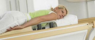
MRI of the lumbosacral region is a diagnostic method that allows you to obtain a comprehensive picture of the area under study with detailed visualization of anatomical structures.
Back diseases have become a real problem in the modern world, where many people lead a sedentary lifestyle. According to some studies, about 80% of the adult population suffers from periodic or constant back pain.
Factors influencing the development of diseases of the lumbar spine are:
- passive lifestyle;
- incorrect posture;
- insufficient physical activity;
- improper exercise in the gym;
- making a careless, sudden movement. For example, lifting weights while twisting the body;
- stress;
- injuries;
- excess weight;
- infectious diseases.
The lower back takes the hit first. It is in this area that hernias, protrusions and osteochondrosis often occur. As a rule, if acute or long-term persistent pain occurs, the patient turns to a neurologist, who may prescribe an MRI of the lumbosacral spine to clarify a possible diagnosis.
What is MRI?
Magnetic resonance scanning is a progressive visual diagnostic method that allows you to obtain many layer-by-layer images of the area under study at one time.
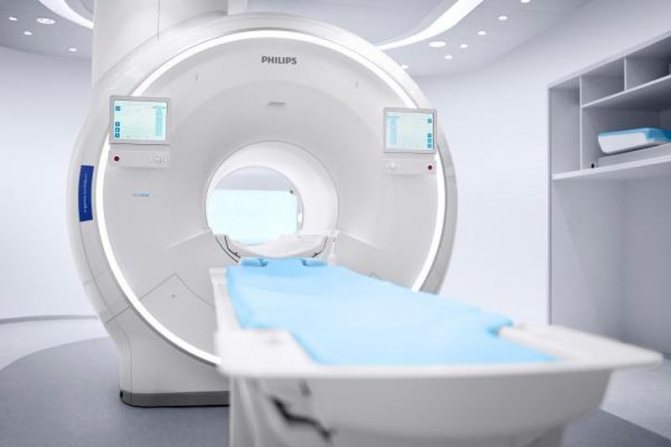
Modern high-field tomograph
Magnetic waves are completely harmless to humans, unlike X-rays. As they pass through the body, they interact with the nuclei of hydrogen atoms in the human body, causing them to move. The tomograph measures the energy emitted by the nuclei and converts it into an image.
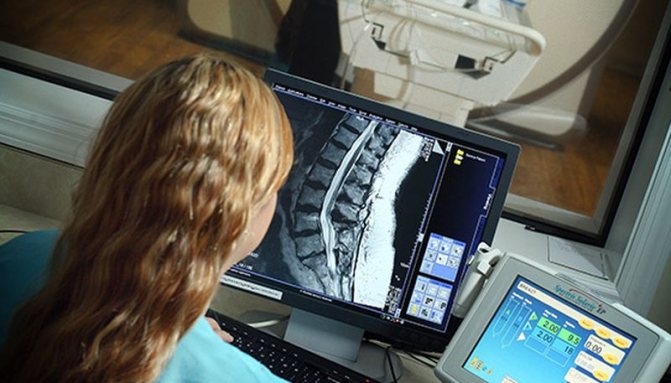
The resulting scans form a detailed picture on the computer monitor screen. At the same time, a layer-by-layer image allows you to examine in detail all areas of the organ or structure under study.
For MRI of the lumbosacral region, the slice thickness is usually set to about 3 mm. This is enough to view the structure of the vertebrae, identify the presence of intervertebral hernias and protrusions, and determine the possibility of finding tumors in the bones or substance of the spinal cord.
MRI diagnostics is the only way today to obtain a full-fledged detailed image of the soft tissues of the lumbar and sacral region. The magnetic radiation used for research is capable of scanning and displaying an image of the organs of the lumbar region, such as the spinal cord, intervertebral discs, nerve processes, and soft tissues surrounding the spine, with the utmost precision.
When using radiography and its more advanced analogue - CT - it is impossible to obtain images of soft tissues. Visualization of bone structures may be sufficient to identify dystrophic changes, injuries and deformities of the vertebrae, but radiography-based techniques are not suitable for diagnosing soft tissue pathologies.
A CT scan allows you to see the gap between the vertebrae, from which the doctor can make an assumption about the presence of a hernia and its size. The intervertebral disc itself cannot be visualized by radiography.
The advantages of magnetic resonance diagnostics also include:
- harmlessness of magnetic radiation to humans;
- no pain;
- speed of obtaining diagnostic data;
- the ability to detect diseases at the earliest stages.
MRI of the lumbosacral region allows you to visualize even the slightest pathological lesions in the spine. This is especially important when diagnosing cancer tumors and metastases to the area under study from other organs.
MRI for low back pain
Pain in the lumbar spine limits motor activity, reduces performance, and worsens quality of life. With radiculitis, patients complain of pain, aching, burning in the back with irradiation to the groin, knee, lower leg, and heel. The pain can be chronic or acute. Discomfort intensifies with load, flexion and extension of the spine, turns of the torso, and in a sitting position.
Traumatic lesions of the lower back, hernias, displacement of the vertebrae, and sudden strain in the lower back are accompanied by acute pain syndrome (lumbago), which patients characterize as a lumbago. More often it occurs against the background of increased load; the attack is accompanied by muscle spasms and limited mobility.
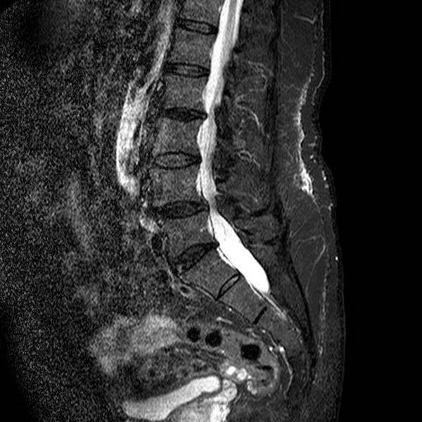
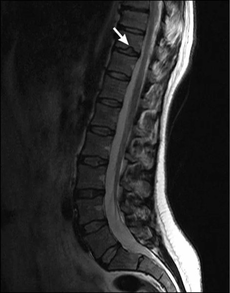
Lumbar spinal stenosis on MRI image
For effective treatment of pain in the lower back, it is necessary to establish the nature of this phenomenon and clarify the localization of the pathological focus. More often in such a situation, the attending physician recommends an MRI of the lumbosacral region, which shows the condition of the morphological elements of the spine and surrounding structures.
The images obtained as a result of magnetic resonance imaging show traumatic injuries to the spinal cord when the integrity of the vertebral bodies and cartilage is damaged. Tears and sprains of muscle tissue and ligaments are determined. In case of compression injuries, MRI visualizes the state of cerebral structures and reveals areas of compression of the nerve roots.
The scan will show the location and size of the tumor, its interaction with surrounding tissues. In the presence of inflammatory and destructive-dystrophic processes that caused pain, MRI helps to determine the extent of tissue damage and the exact location of the lesion.
Tomograms reflect the characteristics of the blood supply to the lumbar region, visualize vascular pathologies and the consequences of impaired functionality of the veins and arteries of the back.
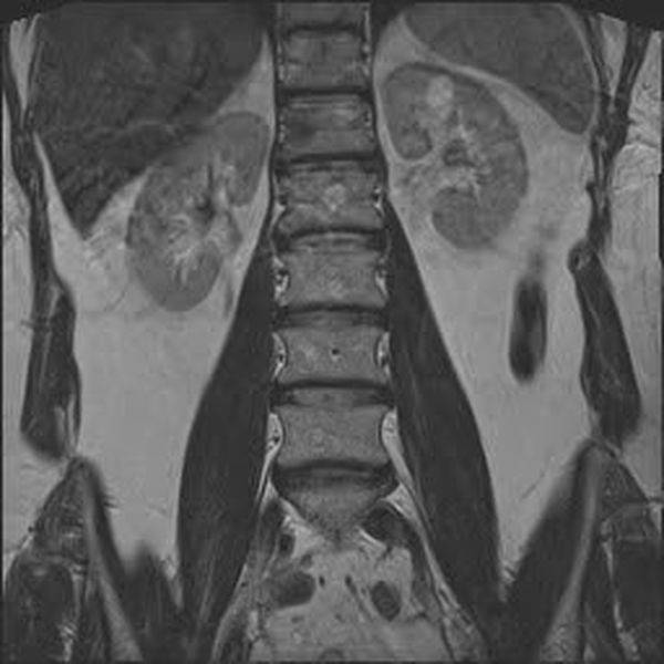
Hemangiomas on an MRI image of the lumbar spine in coronal projection
Indications for MRI of the lumbosacral region
The presence of pathologies of the spine in the lumbar and sacral regions and damage to the intervertebral discs can be manifested by the following symptoms:
- the occurrence of sharp pain in the lumbar region;
- chronic or periodically occurring lower back pain of varying intensity and nature: nagging, sharp, dull, aching, shooting, etc.;
- shooting or aching pain radiating to the thigh, buttock, lower leg, groin, ankle or even collarbone;
- pinching, loss of mobility, inability to straighten after bending or sudden movement;
- urinary and fecal incontinence;
- erection problems;
- weakness in the lower extremities or sensory disturbances.
If the symptoms described above occur, patients in most cases turn to a neurologist, who can prescribe an MRI diagnosis, based on the results of which a further treatment plan will be drawn up. If serious spinal cord lesions or neoplasms are detected, the patient may be referred to a neurosurgeon or oncologist for further consultation.
Open clinic: Medical and rehabilitation center Presnensky
- Street 1905
- Krasnopresnenskaya
- Barricade
- Treadmill
- Exhibition
Price
MRI of the lumbosacral spine 5250 ₽
or call 8 (499) 519-32-71
Schedule
Mon - Fri: 08:00 - 22:00 Sat - Sun: 09:00 - 21:00
Doctors who do MRI
1 4.75
Ilyina Alevtina Alekseevna MRI doctor, radiologist
- Doctor of the highest qualification category (2012)
MRI diagnostician, radiologist. Specializes in magnetic resonance imaging to identify the condition of the patient's vascular systems, organs and tissues. Provides image descriptions and general patient consultation. Annually attends congresses of MRI diagnosticians, forums and conferences.
Anna Alexandrovna, thank you very much for your son. Our baby was born on June 10, 2017. Everything went well. I remember now all these exciting moments, the IVF procedure. First unsuccessful attempt. So many tears and nerves. Thank you very much for always being there and supporting my husband and me. About the clinic "Line...
Make an appointment
JSC "Medicine" Address: Moscow, 2nd Tverskoy-Yamskoy lane, 10
- Mayakovskaya
- Novoslobodskaya
- Belarusian
- Mendeleevskaya
- Pushkinskaya
11 reviews
4.8/5 rating
25 years, experience
8
1 5
Solovyova Anastasia Aleksandrovna MRI doctor
Diagnoses various diseases using magnetic resonance imaging (MRI). Conducts MRI of the abdominal cavity, brain, pelvis, mammary glands, liver, spine, kidneys, heart, joints, neck, etc.
The doctor's constructive approach pleasantly surprised me. Attentive to details. Thank you!
Make an appointment
Moscow Clinic Address: Moscow, Polkovaya street, 12k1
- Marina Grove
- Savelovskaya
- Dmitrovskaya
- Mendeleevskaya
2 reviews
5.0 / 5 rating
4 years, experience
8
1 5
Kulikova Olga Evgenievna MRI doctor, radiologist
- Doctor of the highest qualification category (since 2004)
Radiologist, MRI diagnostician. Specializes in diagnostics of human internal organs using X-ray examination.
Smart, competent, kind. I highly recommend this MRI doctor to everyone. It’s difficult to make an appointment with her, but you can if you want. The doctor will not only make the correct diagnosis, but also find words of consolation.
Make an appointment
Open clinic: Medical and rehabilitation center Presnensky Address: Moscow, 2nd Zvenigorodskaya street, 12с8
- Street 1905
- Krasnopresnenskaya
- Barricade
- Treadmill
- Exhibition
1 review
5.0 / 5 rating
34 years old, experience
8
1 5
Sytnik Konstantin Aleksandrovich MRI doctor, radiologist
- Doctor of the highest qualification category
Head of the CT and MRI department. Conducts MSCT diagnostics of diseases of the central nervous system, lungs, abdominal cavity, musculoskeletal system, MSCT angiography of the arteries of the head and neck, branches of the aorta, arteries of the lower extremities, MRI diagnostics of diseases of the musculoskeletal system and central nervous system. Author of more than 40 published works. In the specialty of radiation diagnostics, he takes part in conferences in Moscow, the Nevsky Radiological Forum, and the European Congress of Radiology.
An excellent doctor, a professional in his field. A doctor with a capital letter. Everything is clear and to the point. Thanks a lot!
Make an appointment
Scandinavian Health Center Address: Moscow, 2nd Kabelnaya Street, 2с37
- Aviamotornaya
- Ilyich Square
- Roman
1 review
5.0 / 5 rating
40 years, experience
8
1 5
Surina Evgeniya Yuryevna MRI doctor, radiologist
Conducts X-ray examinations in pulmonology, cardiology, gastroenterology, urology, gynecology, otolaryngology, neurology, traumatology, rheumatology, endocrinology, pediatrics, surgery, and mammology.
One of the best mammologists: an excellent expert, responsive, thoughtful and emotionally positive doctor. I refer all my relatives only to Elena.
Make an appointment
Moscow Clinic Address: Moscow, Polkovaya street, 12k1
- Marina Grove
- Savelovskaya
- Dmitrovskaya
- Mendeleevskaya
2 reviews
5.0 / 5 rating
14 years, experience
8
Reviews
Andrianov Mikhail Mikhailovich I am very pleased. It is clear that the doctor is an expert in his field. He is very competent and knowledgeable. I immediately understood all my problems, sorted them out and made appointments.
Sazhida
Surina Evgeniya Yuryevna One of the best mammologists: an excellent expert, responsive, thoughtful and emotionally positive doctor. I refer all my relatives only to Elena.
Sytnik Konstantin Aleksandrovich An excellent doctor, a professional in his field. A doctor with a capital letter. Everything is clear and to the point. Thanks a lot!
Hidden
Ilyina Alevtina Alekseevna Alevtina Alekseevna conducted my examination. She was attentive to me, it was a pleasure to talk to me. The doctor is competent and intelligent.
Polyakova Maria Iosifovna Very friendly doctor Maria Iosifovna Polyakova. She provides a very detailed, professional description of the MRI. Although the price is a bit high, I only go to this clinic, since I have never seen such a detailed description in any clinic. I am very grateful to the doctor.
Irina Vladimirovna
Indications
MRI of the lumbosacral spine is prescribed in the following cases: impaired sensitivity of the lower extremities, weakness in the legs; pain in the lumbar area; injury in the lumbar and sacral area; symptoms of a malignant tumor together with pain at the lumbar level; history of cancer and metastases of malignant cells in the lumbar area; congenital defects and anomalies in the development of this part of the spine; infections and abscesses of the spinal cord and bones; disturbances in the urination process.
Preparation
There is no special preparation for MRI of the lower back. An MRI of the lumbosacral region can be done in a child at any age. Tell your doctor if you have any allergies to medications or contrast agents, or if you have any chronic diseases. Contraindications are the presence of implanted pacemakers and hearing aids, metal vascular clips, or previously performed osteosynthesis using metal parts.
Contraindications to MRI of the lumbosacral region
Despite the fact that MRI diagnostics is a modern, painless and harmless procedure for patients, there are still contraindications for its implementation.
The absolute contraindications under which MRI examination cannot be performed are as follows:
- a pacemaker installed in the patient. Magnetic waves can disrupt its operation, which can lead to serious health consequences;
- the presence in the body of structures made of metal that reacts to magnetic radiation. These could be: dentures, dental bridges, vascular clips, etc.;
Modern dentures and implants are often made from inert materials such as titanium. If you doubt that you can undergo MRI diagnostics, show the doctor the passport of the implant or prosthesis, which indicates the material of manufacture.
- first trimester of pregnancy;
Although magnetic waves are harmless to humans, there is not enough data on their effects on the developing fetus. Therefore, it is permissible to conduct MRI in late pregnancy if indicated. Diagnosis in the 1st trimester is carried out only in cases of life-threatening situations for the mother.
- patient weight 130 kg or more, maximum body girth more than 150 cm;
- the patient's inability to maintain a long-term immobile position.
This category of people includes small children and mentally unstable people. Patients suffering from severe pain or severe fear of confined spaces should take painkillers or sedatives before the test. Any movements during the procedure will make the resulting images unclear due to the appearance of multiple artifacts.
What does an MRI of the lumbosacral region show?
Many patients are interested in what an MRI of the lumbosacral region shows. The high detail of images obtained using MRI diagnostics makes it possible to accurately identify the following diseases localized in the lumbar spine:
- protrusion and herniation of intervertebral discs;
- degenerative diseases: osteochondrosis, spondylosis;
- consequences of previous injuries, such as compression fractures, subluxations and vertebral displacements;
- multiple sclerosis;
- neoplasms of primary and metastatic origin;
- osteomyelitis;
- myelitis (inflammation of the spinal cord).
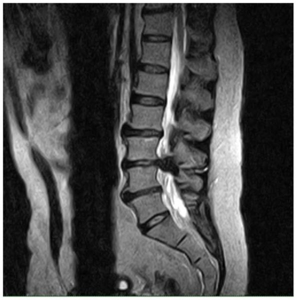
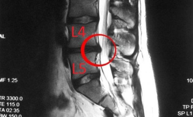
Intervertebral hernia
On MRI you can clearly see:
- thickness and density of intervertebral discs;
- dystrophic changes in the vertebral body;
- narrowing of the spinal canal;
- the condition of the soft tissues in the area under study.
Based on these data, it is possible to draw a clear picture of the course of diseases of the lumbosacral spine.
The most common indication for an MRI examination of the lumbosacral region is suspicion of osteochondrosis, protrusion and herniated intervertebral discs.
Osteochondrosis is a degenerative disease in which deformation of the vertebral bodies occurs, erasure and flattening of the intervertebral discs.
Typical manifestations of lumbar osteochondrosis are periodic aching and nagging pain, numbness and loss of mobility. People who lead a sedentary and sedentary lifestyle are at risk for the incidence of osteochondrosis.
Timely detection of the disease makes it possible to stop degenerative processes and the occurrence of complications. Diagnosis using MR scanning will give an idea of the stage of the disease and the extent of the affected area.
Protrusion is a bulging of the intervertebral disc without rupture of the surrounding fibrous ring.
The disease occurs when degenerative-dystrophic disorders in the lumbar region are advanced. The appearance of protrusions can also be preceded by sudden movement or heavy lifting.
A common symptom of the disease is lower back pain of varying intensity, numbness in the legs and groin area.
MRI of the lumbar region helps to identify even small protrusions in the early stages of occurrence. Further treatment of the disease is usually conservative. The patient is prescribed painkillers, physiotherapy and massage. An important factor in preventing protrusions is physical activity adequate for age and health status.
A herniated disc occurs when the annulus fibrosus ruptures and the disc moves into the spinal canal, compressing the spinal cord.
The disease is characterized by severe pain, which may subside over time and return again. Depending on the location of the hernia and possible compression of the spinal cord roots in a certain area, the patient may experience shooting pains in various areas of the lower extremities, urinary incontinence and severe pain in the lower back, buttocks and thighs.
Timely diagnosis of hernias helps relieve the patient from discomfort and pain. MRI gives a detailed picture of the course of the disease. In the pictures you can see the location of the hernia, its size and the degree of compression of the spinal cord, and determine the inflammation of the surrounding tissues.
Treatment of hernias can be conservative and include therapeutic blockade, avoidance of excessive tension and stress, physical therapy and yoga, and massage. In severe cases of the disease and when the pain syndrome does not subside over time, surgical intervention is performed.
MRI diagnostics of the entire spine: indications and contraindications

There are significantly more indications for MRI than limitations
It is reasonable to perform an MRI of the spine in case of complaints suspicious of a pathological process in the area of interest:
- Cervical region: dizziness;
- short-term loss of consciousness;
- noise in the head;
- instability of blood pressure;
- pain when turning the head;
- weakening of memory, hearing;
- noise in ears;
- facial neuralgia;
- migraine attacks;
- restriction in neck movement;
- numbness of the upper extremities;
- muscle tension in the back of the head.
- pain in the scapular region with irradiation behind the sternum (simulate cardiac syndrome);
- Lumbosacral region: limitation of trunk rotation;
- increased pain after physical activity;
- numbness and weakness of the lower extremities, convulsions;
- difficulties at the beginning of the act of urination;
- feeling of constant fullness in the bladder and rectum;
- incontinence of urine and feces;
- enuresis, etc.
- pain in this area;
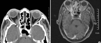
MRI of the cervical spine is often performed if a tumor process in the thyroid gland is suspected.
Urological complaints are initially assessed using ultrasound of the bladder, prostate, and pelvic organs; in the absence of pathological changes on sonograms, an MRI of the lumbar spine is indicated
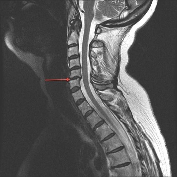
MRI of the cervical spine: spondylolisthesis. Inflammation causes cartilage destruction and bone erosion with anterior displacement of the 6th cervical vertebra (indicated by the red arrow)
Indications for MRI of the spine:
- Osteochondrosis. The most common pathology of the spine, characterized by degenerative lesions of articular cartilage and degenerative processes in the surrounding bone tissue. Osteochondrosis is rarely localized in any one department; changes affect the entire spinal column. Unpleasant symptoms appear with the development of complications: spondylosis, radiculitis, etc.
- Spondyloarthritis. Inflammation, which is typically characterized by damage to the connective tissue of the spinal column, is most often localized in the cervical spine. At the early stage of the pathological process, there are no specific clinical manifestations; symptoms manifest with multiple joint damage.
- Spondyloarthrosis. The disease affects the intervertebral joints and spreads to the ilium and costal bones. Premature degeneration of cartilaginous structures and weakening of the muscular and ligamentous apparatus occurs. A long-term process can lead to disability of the patient.
- Disc protrusion. Due to the loss of strength by the fibrous ring, the core of the intervertebral disc is pressed into the spine. Provoking factors include excessive physical stress on the back, muscle weakness, various curvatures, and infectious and inflammatory processes.
- Intervertebral hernia. As a result of the altering factor, the normal anatomical position or, more often, the physical state of the nucleus pulposus of the intervertebral disc is disrupted, the annulus fibrosus ruptures, which is accompanied by sequestration (displacement and loss) of part of the described structure with damage and an inflammatory process in the spinal nerve roots. More often, intervertebral hernias are found in the cervical or lumbar spine.
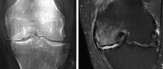
Endplate herniation (Schmorl's) on MRI
- Schmorl's hernia. As a result of structural and anatomical changes in the vertebral bodies, a hernial protrusion is formed, and due to compression, severe pain occurs. Genetic characteristics, postural disorders, and the consequences of bruises and trauma are considered as etiopathogenetic factors.
- Sciatica. The cause of damage to the inflammation of the sciatic nerve is compression of the spinal cord roots; MRI of the lumbar spine is performed to determine the factor that led to the compression. Clinical manifestations vary: from discomfort in the lumbar region and burning in the feet to paralysis of the legs and loss of control over urination and defecation.
- Stenosis. Narrowing of the spinal canal and pinching of nerve endings is often a complication of osteoarthritis. Symptoms depend on the location. More often diagnosed in older patients.
- Spondylosis. Involutional processes as a person ages lead to the growth of bone tissue around the vertebral bodies. The lumen of the canal decreases, traumatization of nerve endings and tendons occurs, and in advanced cases, the vertebrae grow together.
- Ankylosing spondylitis. With ankylosing spondylitis, systemic inflammation develops in the intervertebral joints, up to complete immobility against the background of fusion. Damage to the costovertebral and sacroiliac joints aggravates the process. Pathology is more often detected in men of working age.
- Dorsopathy. A number of degenerative-dystrophic diseases of the spine and adjacent connective tissues at the initial stage are manifested by periodic pain, which typically radiates to the abdominal area, upper and lower extremities. Timely diagnosed pathology can be successfully treated; without drug correction, the condition becomes chronic.
- Kyphosis. Curvature of the spinal column in the sagittal plane can be congenital (including a variant of the anatomical norm) or acquired. The typical location is the thoracic spine; less commonly, kyphosis is detected in the lumbar and neck areas. Curvature develops as a complication of certain diseases or fractures. Poor posture is sometimes so pronounced that it is externally manifested by the formation of a hump.
- Lordosis. The spinal deformity is directed forward and has an arched appearance. Physiological lordosis is not a disease and is formed as the child masters a sitting position. Pathological protrusion is caused by a violation of the musculoskeletal and ligamentous apparatus, and large body weight. Similar changes accompany rickets, hip dysplasia and birth injuries.
- Scoliosis. Three-plane curvature of the spinal column in any part, most often an acquired condition, is accompanied by rotation of the vertebral bodies. Shapes vary: C, S, Z-shaped.
Endplate herniation (Schmorl's) on MRI
- Myositis. Trauma, infectious-inflammatory process, autoimmune pathology initiate damage to muscle fibers, which often ends in complete atrophy.
- Tumors. Neoplasms of the spine and spinal cord account for 10-15% of all neoplastic processes. MRI with contrast with the greatest reliability allows us to assume the nature of the pathology: benign or malignant. Without undergoing a qualitative study, it is difficult to assess symptoms that resemble those of osteochondrosis or pelvic inflammatory diseases.
- Osteoporosis. An metabolic disorder characterized by a loss of bone tissue structure, which leads to fractures even with minimal trauma. The most significant reason is hypocalcemia due to the natural processes of aging of the body. Osteoporosis, expressed to one degree or another, is registered in every second woman over 50 years of age.
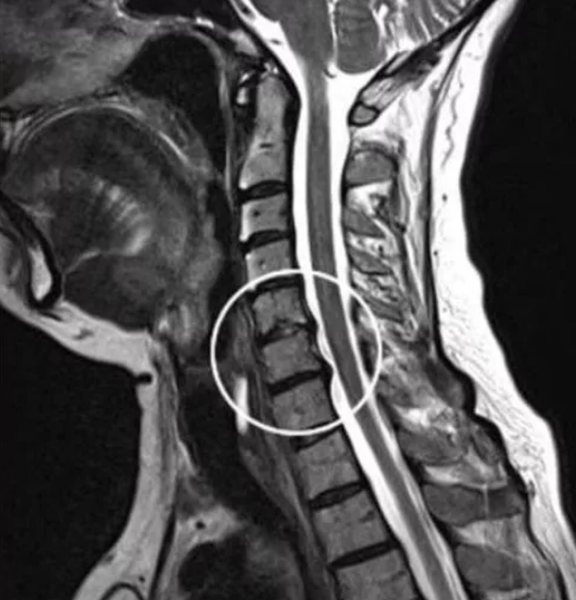
MRI of the cervical spine: osteochondrosis with herniation in the area of 5-7 cervical vertebrae (the pathological area is highlighted in a white circle)
- Fractures. An MRI of the spine is more reasonable for assessing the consequences of the injury; initially, preference is given to computed tomography. In this case, MRI will show the condition of the spinal cord and its membranes, the degree of involvement of muscles, blood vessels, spinal nerves, and the severity of cerebral compression.
Diagnosis of spinal diseases using MRI data is the most informative and safe method of research today.
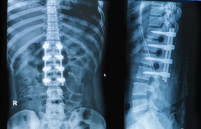
X-ray of the spine: metal structures installed during orthopedic surgery as fixations for a fracture are a contraindication to MRI.
Contraindications for MRI of the spine:
- significant curvature in any of its parts, preventing prolonged lying on the back;
- metal parts in the body: installed orthopedic structures, pacemaker, vascular clips, cochlear implants, insulin pumps, etc.;
- pregnancy.
For claustrophobia and severe pain, magnetic resonance imaging of the spine is performed after the administration of sedatives and painkillers.
Contrasting also has its contraindications:
- early pregnancy;
- end-stage chronic renal failure;
- generalized allergic reaction to contrast agent.
Is it necessary to use a contrast agent when performing MRI of the lumbosacral region?
Magnetic resonance imaging of the lumbar spine usually does not require additional use of contrast; they are well visualized on images without the use of contrast agents.
If the indication for an MR examination is the suspicion of the possible presence of a tumor or metastatic screenings, the use of a contrast agent is justified. The staining substance helps to assess the size of the formation and its possible growth into the spinal cord tissue
How is an MRI of the lumbosacral region performed?
If it is planned to conduct a magnetic resonance scan of the lumbar region using contrast, it is recommended to follow a rational diet on the day of the procedure.
In other cases, no preliminary preparation for the study is required.
Before the scan, the patient will be asked to remove all metal items. This could be: accessories, jewelry, clothing containing metal fittings, glasses, removable hearing aids.
You must bring with you to your appointment your previously taken photographs, if available. The radiologist questions the patient about the presence of contraindications to the study and concerning symptoms.
The patient lies down on a couch, which is placed in the tomograph tube.
You cannot move during the procedure, so the area being examined can be additionally secured with straps, although this is usually not required. The device produces quite loud sounds, but at the DiMagnit medical center they take care of the comfort of patients and provide special headphones to minimize the unpleasant sensations from the sounds of the operating device.
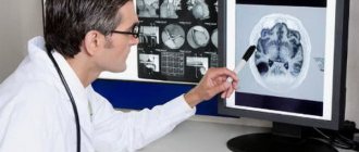
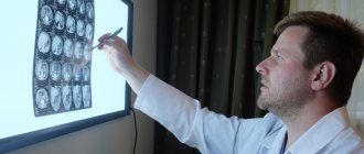
The radiologist carefully studies the images before making a conclusion
Once the scan is completed, results can be obtained in as little as 30 minutes. The radiologist draws up a detailed report in which he describes all the anatomical structures of the area under study, including those in which no abnormalities were detected. Possible pathologies are also described in detail, indicating their size and location in relation to other organs. The images are recorded on disk and sent to the patient along with a paper report.
How long does an MRI of the lumbosacral spine take?
MRI diagnostics of the lower back and sacrum will last no more than 20 minutes. If you need to administer contrast, the duration of the procedure will increase by another 15 minutes.
MRI of the lumbosacral region makes it possible to identify frequently diagnosed spinal pathologies such as intervertebral hernias, protrusions and osteochondrosis, as well as other, less common diseases. The study helps determine the exact location of the lesion and its size. MRI of the lumbosacral region is safe and painless, and the results of the study can be obtained immediately upon completion of the procedure.
| MRI of the spine, what does it show? |
| What does an MRI show? |
| What does an MRI of the cervical spine show? |
| MRI of the sacroiliac joints, which shows |
| MRI of the sacroiliac joints |
| MRI of the coccyx |
Benefits of MRI of the sacrolumbar spine
Today, magnetic resonance imaging is considered the most highly informative diagnostic method used in medicine. The main strengths of this study include:
- absolute safety;
- the possibility of repeating the procedure;
- minimal number of contraindications;
- high quality and detailed results.
MRI of the lumbosacral region is an advanced diagnostic method that makes it possible to obtain a layer-by-layer image of the area under study. As a result, specialists receive three-dimensional images of the spine, with the help of which they can easily recognize almost any pathology.
The study can also be performed with contrast. In this case, the patient is given an intravenous substance immediately before the procedure begins. Unlike contrasts used in other diagnostic methods, the product does not contain iodine. Therefore, the risk of an allergic reaction in people to this drug is minimal.