Indications for CT scan of the thoracic spine
- hernial protrusions of discs between the vertebrae at the level of the thoracic segment of the spine;
- suspicion of fractures of individual vertebrae, including compression fractures;
- congenital structural anomalies of the vertebrae;
- diagnosis of rheumatological diseases, for example, ankylosing spondylitis;
- stenosis of the spinal canal at the level of the thoracic segment;
- painful sensations in the back of unknown origin;
- paresis of the upper limbs, paralysis, decreased sensitivity and other symptoms characteristic of spinal cord diseases;
- clarification of data from other research methods;
- signs of radicular syndrome in the form of numbness in the limbs or “lumbago”;
- vertebral neoplasms of various types, including metastatic ones;
- damage to the spinal cord and nerve roots;
- inflammatory pathology of the spine and spinal cord.
When is the study scheduled?
Doctors recommend doing a CT scan of the thoracic spine in the following situations:
- numbness in the fingers and other sensitivity disorders;
- if you have had a back injury;
- spinal surgery is planned
- painful discomfort regularly occurs along the ribs and between the shoulder blades;
- changes in posture;
- frequent pain in the forearms and shoulder blades.
In addition, computed tomography is prescribed if there are contraindications to MRI.
Contraindications to CT of the thoracic spine
The main contraindication for performing a CT scan is the period of gestation, regardless of how far along the pregnancy is. Scientists have proven that even small doses of X-ray radiation can cause the development of congenital anomalies in the fetus.
CT scan of the thoracic spine is not performed on patients whose weight exceeds 200 kg.
There are certain limitations that apply to performing CT scans using contrast. Contrast studies cannot be performed in the following cases:
- the patient has intolerance to iodine-containing components;
- severe renal failure, in which the removal of contrast from the body is impaired;
- breastfeeding, as the contrast agent may pass into breast milk;
- the patient has serious illnesses that require emergency care, in which the loss of time for a CT scan can harm the patient’s health;
Risks
Risks of CT scans include:
- Exposure to radiation
- Allergic reaction to contrast agent
- CT scans expose the patient to a fairly large dose of radiation, but a single scan is virtually safe.
- Some patients are allergic to contrast dye.
The most common type of CT contrast, injected into a vein, contains iodine. People with an iodine allergy may experience symptoms such as:
- nausea or vomiting
- sneezing
- itching or hives
- If the patient is allergic to iodine, the doctor may prescribe antihistamines (such as Benadryl) or steroids before the scan.
- The kidneys help remove iodine from the human body. Patients with kidney disease or diabetes need to take additional fluids after a CT scan with contrast, which will help remove iodine from the body more quickly.
- Quite rarely, contrast can cause anaphylaxis.
Alternative Methods
To detect osteoporosis, osteochondrosis, pathological fractures due to tuberculosis infection and sepsis, radiography can be used as an alternative to CT.
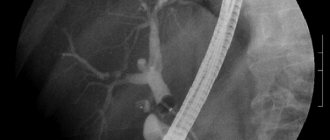
To visualize the soft tissues of the spinal area, such as ligaments, muscles or blood vessels, MRI is a more informative method. This technique is a good alternative to CT for detecting vertebral tumors that have grown into nearby tissues. If for any reason the patient is unable to undergo an MRI or CT scan, then myelography may be an alternative method for imaging the spinal cord and spinal canal. The principle of this technique is that the doctor injects a contrast agent into the spinal canal and takes a series of pictures. The disadvantage of myelography is that the study can lead to various complications. Therefore, it is performed only in exceptional cases.
Advantages and disadvantages of the method
MRI of the neck and other parts allows you to visualize any part of the body layer by layer, creating three-dimensional contrast images. The method provides a clear image of blood vessels, soft tissues, the spinal cord and brain, and a detailed state of the internal organs, which is not what you would expect from ultrasound and radiography.
Other benefits:
- The method is non-invasive, so during the examination the patient does not feel pain or discomfort;
- the ability to take pictures in different planes;
- unlike computed tomography (CT) and radiography, it does not irradiate the body;
- gives the best images of tumors and other diseases of the central nervous system;
- captures an image of moving blood, which angiography is not capable of (thanks to this, vessels can be examined without a contrast agent);
- MRI accurately measures the amount of tumor perifocal edema (fluid accumulation in soft tissue).
So, MRI has advantages over other diagnostic methods, but there are also disadvantages:
- shows acute hemorrhages less clearly than CT;
- cannot be performed on patients with implants (pacemaker, etc.) and metal foreign bodies;
- the closed chamber of an MRI machine causes attacks of claustrophobia (suffocation, panic) in some people;
- the high cost of the procedure.
Also, if a long-term study is necessary, respiratory movements of the chest or arrhythmia may somewhat distort the results.
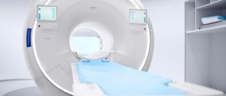
How is a CT scan of the thoracic spine performed?
Before undergoing the examination, the patient must remove all metal objects from himself. This is necessary so that no artifacts are created in the images when performing a CT scan.
The study is carried out with the patient lying on his back. During the examination, the patient must remain motionless, so for people with hyperkinesis, it is sometimes necessary to use additional fixation. When performing a CT scan, contrast is administered intravenously.
After all the preparatory procedures, the doctor sets up the tomograph and selects the required scanning mode.
During a CT scan, the patient is alone in the room. If the need arises, he can always contact medical personnel using two-way communication.
The procedure is accompanied by rotation of the tomograph ring around the table on which the patient is located. During this, the tomograph makes a slight noise.
Specifics of CT scanning
CT scan of the cervical spine is a rather complex procedure, the result of which depends on proper preparation of the patient before the upcoming study. If it is planned to study the area using contrast, the patient is prohibited from consuming a number of foods per day. The last meal should be 8 hours before the start, even drinking water is not recommended.
Main features of CT scanning:
- Before starting, the specialist must provide an extract from the medical record, a referral for the procedure, and the results (along with images) taken earlier using a CT scanner.
- As in the case of an MRI, all existing metal jewelry is removed from the body, everything is removed from the pockets, to avoid distortion of the image.
- The patient is fixed in one position on a special couch and does not move until the operator allows him to do so.
- The reproductive organs are protected from negative radiation using a special lead apron. If the study is carried out on a child, then the same robe is put on the parent who will hold him during the process.
- The scan is carried out within 10 minutes, in rare cases it takes 25 minutes. The diagnostician and assistants are at this time in the next room, observing the process through transparent glass.
We advise you to study - Schmorl's nodes of the vertebrae - what is it
An alternative to this study is multislice computed tomography (MSCT), which is characterized by a lower radiation dosage and a higher speed. Some time after the completion of the session, the patient is given a medical form describing the information received and an electronic medium containing the images received.
A specialist deciphers the received images, and it takes some time to write a conclusion. They are usually given to the patient 1-2 days after the examination. But it is worth understanding that the specified data is not the basis for making a diagnosis, since it is made by a doctor based on the clinical picture, symptoms and medical history of the patient.
Computed tomography is an absolutely painless procedure
Contrast enhancement
To diagnose spinal tumors or vascular pathology, a CT scan with contrast is performed. Omnipak and Yopamiro are used as a contrast agent. These substances are administered intravenously immediately before the procedure. After administration, the doctor waits for the contrast agent to accumulate in the tissues. When this happens, pictures are taken in the recommended projections.
At the moment when the drug is administered to the patient, he may feel warmth or notice redness of the skin at the injection site. This reaction is considered normal. If any other sensations appear, the patient must immediately tell the doctor who is performing the procedure.
Physics of computed tomography
Computed tomography, unlike MRI, is a radiological method. The method is based on the ability of body tissues to retain X-rays differently depending on the density of the organ and is measured in Hounsfield units (HU). Air has a density of -1000 HU, water -0 HU, bone +1000 HU.
Depending on the type of device, computed tomography can be linear, spiral and multislice (multi-slice).
We advise you to study - Instability of the cervical vertebrae
With linear tomography, the thickness of the slice is determined by the step of the tomograph, which is on average at least 3 cm. Scanning of the slice is carried out when each new layer step by step falls into the scan area. This method has many disadvantages
- it is impossible to examine small details, because the layer thickness is too large to detect them
- significant radiation exposure to the patient due to the long duration of the procedure
- the patient has to be completely still, since movement will blur the already few photographs
With the advent of spiral tomographs, the era of step-by-step linear tomographs came to an end. During spiral computed tomography of the spine, the table deck with the patient enters the Gantry aperture gradually but continuously, as opposed to step-by-step. In this case, one row rotates around the patient, receiving radiation not retained by the body, from which a CT image of the spine is formed, showing the density of healthy and pathological tissues.
With multislice tomography, the essence of scanning is the same, the only difference is that there is not one row of receiving detectors, but at least two; now 32, 64 slice devices are most often found, you can find 128 and even 256 slice devices. This means that for one revolution of the emitting tube, not one slice is made, but at least 2 (maximum 256 slices per revolution). Such tomographs allow you to make sections of 2-3 mm, the scanning time is reduced to 2-5 minutes, it is possible to construct three-dimensional models of the vertebrae, their discs, the spinal canal, and their relationship to surrounding tissues. In other organs, the construction of three-dimensional structures makes it possible to avoid painful colonoscopies and bronchoscopy and replace them with virtual ones.
However, increasing the number of cuts is not always necessary and is always harmful. The fact is that the radiation exposure during scanning is directly proportional to the increase in the number of slices, while the information content of 32 slices is quite enough to fully formulate a conclusion. Research on 128, 256 cutting devices is shown only when searching for the smallest formations measuring 1 mm. Enlargement of sections should be strictly indicated to the patient; the diagnostic value should always exceed the risk of ionizing radiation received during scanning!