- examining the patient for the presence of degenerative, demyelinating diseases;
- clarification of the severity of pathologies and stages of their development;
- to select a treatment method that is appropriate for the patient;
- to determine the need for surgical intervention and to monitor the postoperative process.
In addition, tomography is a fairly effective tool for the early detection of degenerative processes, for example: osteochondrosis, spondylosis, osteophytes, osteoarthrosis.
MRI of the thoracic spine
MRI of the lumbosacral region
MRI of the cervical spine
MRI of the craniovertebral junction
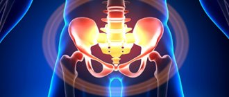
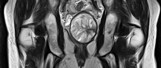
MRI of the sacroiliac joints
MRI of the sacrococcygeal area
MRI of the entire spine
MRI of 3 parts of the spine
Advantages of the diagnostic method
The main advantage of MRI of the cervical spine
is information content and safety. The equipment produces accurate images of the examined area, while soft tissues are examined simultaneously with the spinal column. The procedure is harmless and can be performed simultaneously for several departments. The tomograph takes pictures in different planes and creates a three-dimensional image. The advantage of MRI of the cervical spine is that the device shows the entire structure of the region being studied: vertebrae, neck vessels and muscles. This does not involve x-ray radiation; the method can be used several times to clarify the diagnosis and monitor the progress of the disease.
Indications
- Severe back pain;
- Trauma, bruise of the spine;
- Exclude malignant or benign neoplasm;
- Inflammatory process.
MRI should be taken regularly for people with diagnoses such as osteochondrosis, protrusions, hernias, and vertebral displacements. If the patient has ever had a birth or congenital injury in any part of the spine, this diagnosis will also be very useful to monitor the consequences of birth pathology.
Free diagnostic consultation
If in doubt, sign up for a free consultation. Or consult by phone
+7
Who is indicated for MRI?
The study is prescribed for dizziness, numbness of the hands, and headaches of unknown origin. Indications:
- neck injuries;
- stiffness and discomfort in the cervical spine;
- blurred vision, loss of consciousness;
- diseases of the vertebrae, nerve roots, discs, muscles;
- osteochondrosis;
- suspicion of degenerative-dystrophic changes;
- short neck syndrome.
The procedure is prescribed before neurosurgical intervention to make orthopedic diagnoses.
What is an MRI of the brain and why do this procedure?
To begin with, we will briefly describe this technique; its essence is to study any part of the human body by using an electromagnetic field that comes from all atomic nuclei, which is possible at high voltage. The possibility of capturing images appears due to the fact that the tissues of the human body contain a large amount of hydrogen.
Information always first gets onto the monitor to specialists, and this technology makes it possible to ultimately obtain even a multi-layered image, which can later be recorded by saving it on some medium; they often resort to simple printing on paper. Among the advantages of the method over similar procedures, for example, over CT or over simple X-rays, it is worth highlighting its complete non-invasiveness, as well as the absence of pain in the patient during its implementation and safety, which is achieved only thanks to the technology itself, briefly discussed earlier.
In most situations, MRI is used to diagnose various pathological processes in the body; here are the main diseases, if the development of which is suspected, the procedure is most often prescribed:
- strokes;
- various vascular anomalies, for example, aneurysm;
- pathological processes associated with the organs of vision, as well as with the inner ear;
- tumors, and we are talking about both malignant and benign neoplasms;
- diseases associated with the pituitary gland;
- injuries of various types;
- chronic diseases that affect the nervous system, for example, multiple sclerosis;
- headaches that appear constantly and are quite severe, and in most situations doctors cannot assess their etiology.
It is also worth mentioning that sometimes magnetic resonance imaging of the head is required to diagnose pathological processes that occur during dementia, that is, acquired dementia.
Contraindications
The tomograph creates a magnetic field, so patients who have an insulin pump, pacemaker, hearing aid, or cardioverter-defibrillator in their body do not undergo the procedure. Scanning is not performed for people who have metal objects in their bodies (Ilizarov apparatus, staples and wires, stents on blood vessels). The procedure with contrast is contraindicated for pregnant women and nursing mothers. Contraindications include renal failure. For technical reasons, the study is not possible if the patient weighs more than 120 kg.
Algorithm for the procedure
Best materials of the month
- Coronaviruses: SARS-CoV-2 (COVID-19)
- Antibiotics for the prevention and treatment of COVID-19: how effective are they?
- The most common "office" diseases
- Does vodka kill coronavirus?
- How to stay alive on our roads?
The principle of testing with and without contrast is not very different. In any case, the person will be placed on a medical couch, which, when the equipment is activated, will move under the scanner, where there is a magnet.
Since modern devices usually have a wide design, most of the body will not be under the arch, which will help those suffering from claustrophobia to survive the panic. For convenience, the subject’s head is fixed so that during subsequent manipulations it will be easier for him to maintain immobility. Some lab technicians even suggest using a head roller.
Since during operation of the tomograph it constantly emits sound signals. Patients, especially children, are recommended to wear special vacuum headphones. And if an unforeseen situation occurs during testing, you can always contact the x-ray technician via internal two-way communication.
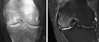
What does the cervical spine look like on a scan?
In the absence of diseases, the cervical vertebrae are smooth and intact, the discs are the same size with a flat and smooth surface. In this case, the joints are without roughness, the structures are located symmetrically. A healthy spine has no obstacles to the exit of the spinal nerves; there are no neoplasms, inflammations or infectious foci on it. If there is a fracture, the fracture line, deformation and displacement are visualized on the image. Using MRI, the causes of pathological fractures are determined: chronic diseases of the spinal column. It is possible to determine whether the spinal cord is affected by injury. If the image shows a decrease in the height of the disc, this indicates a herniation. When the marginal areas with the surface of the nerves grow, osteochondrosis is diagnosed.
What does diagnostics show?
The results of the study provide specialists with the opportunity to visualize tissues in all the smallest details, thanks to which they can easily examine their structure, size, location, dimensions, and any anomalies.
MRI of the soft tissues of the neck shows:

- inflammatory diseases;
- osteochondrosis;
- stenosis;
- neoplasm and presence of metastases;
- pathologies of the spine structure;
- consequences of injuries;
- diseases of the thyroid gland, lymph nodes.
MRI makes it possible to determine the presence and location of protrusions and hernias. Despite the fact that hernias in the neck area are extremely rare, their presence can cause spinal cord displacement.
Tomography is very often prescribed to detect the presence of foreign bodies that simply cannot be recognized on ultrasound or radiography.
From the above, we can draw a logical conclusion that MRI is a comprehensive diagnosis that shows the presence of many diseases and pathological conditions, so doctors do not have difficulty making the correct diagnosis.
How to prepare for scanning?
Special preparation for MRI of the cervical spine
not required, there is no need for dietary restrictions (if the MRI will be performed without a contrast agent), no need to stop taking medications. If contrast will be used, the doctor should be informed of any allergic reactions. It also needs to be said about pregnancy and the presence of claustrophobia. Before the procedure, metal objects, including piercings, removable dentures and hearing aids, must be removed. If you are using a contrast agent, you should not eat anything five hours before the procedure.
How long does an MRI of individual organs and body parts take?
When examining organs using MRI, a person is placed on a movable table, which slides into a magnetic capsule. A strong electromagnetic field is directed to individual parts of the body so that information about the condition of the tissue can be obtained. The time it takes to obtain a clear picture depends on both the organ and the disease.
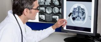
MRI of the brain
The examination lasts 30-40 minutes. The three-dimensional picture obtained during this time allows us to establish the cause of dizziness, pain, loss of consciousness, ringing in the ears, blurred vision, mental disorders and other signs of brain diseases. In the resulting image you can see dangerous dilations of blood vessels (aneurysms) and tumors. Detection of metastases, inflammatory processes, and congenital anomalies usually requires a detailed study using contrast agents that are injected into a vein.
To study Almag with osteochondrosis of the cervical spine, does deception help?
Detection of metastases, inflammatory processes, and congenital anomalies usually requires a detailed study using contrast agents that are injected into a vein
MRI of the liver
The procedure is carried out within 20-25 minutes. A preliminary diagnosis of the presence of diffuse changes, tumors, traumatic or toxic injuries in a patient is established with great accuracy. Before examining the liver, as well as the rest of the abdominal organs, it is necessary to take an antispasmodic agent (No-Shpa) and an anti-gas agent (Espumizan). This is done 40 minutes before the test.
MR tomography of joints, spine
Diagnosis requires inspection of damage from several sides and in different positions. If the patient experiences pain, then preliminary anesthesia is performed, the procedure is carried out under anesthesia. It may take 30-40 minutes to prepare and conduct an MRI. Then you need to wait for the patient to come out of anesthesia and make sure that he is feeling normal. This may take several hours.
MRI examination of the breast
MR imaging allows one to distinguish a malignant lump from a benign neoplasm. An examination within 20-25 minutes allows you to determine the complexity and degree of danger of a neoplasm in the breast. Thanks to the contrast agent, you can see the condition of the tissues around the seal. In most cases, no additional measures are required. You may need to consult a gynecologist and endocrinologist, which will increase the duration of the examination.
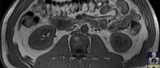
Kidney examination
Before MR imaging of the kidneys, it is usually necessary to donate blood for analysis to ensure that there is no severe inflammatory process that would prevent the use of contrast agents.
The total duration of an examination using magnetic resonance imaging is from half an hour to several hours.
In the modern diagnostic process of diseases of the human body, magnetic resonance imaging or MRI for short is often used - the most informative type of study of human internal systems. This diagnostic method has a number of positive characteristics that are not inherent in other medical examinations.
Medical clinic NACFF
- Ugreshskaya910 m
- Dubrovka1.4 km
Moscow, st. Ugreshskaya, 2, building 7
The NACFF medical clinic is a modern multidisciplinary medical complex that offers its patients a full range of services in the field of diagnosis and treatment of diseases, prevention, and rehabilitation for adults and children. The clinic provides services in the field of oncology, surgery, cardiology, neurology, orthopedics, gynecology, urology and many others. The Oncology Department deserves special attention, where highly professional doctors work, using in their practice the latest world developments in the field of treatment of oncological diseases, which allows them to effectively fight tumor diseases and achieve stable remission. The capabilities of the Oncology Department of the NACFF clinic allow us to perform: • Laparotomy operations • Laparoscopic operations • Chemotherapy • Immunotherapy • High-dose chemotherapy • Provide palliative care
Among the oncologists of the clinic there are gynecological oncologists, urological oncologists, oncodermatologists, doctors working with malignant formations of the gastrointestinal tract, as well as with malignant formations of soft tissues. First of all, when contacting our clinic, doctors carry out a full range of diagnostic measures in order to make an accurate diagnosis. Using modern equipment and many years of experience, NACFF clinic specialists can make a diagnosis on the day of treatment. Such efficiency has a beneficial effect on the length of hospitalization, and as a result, on the success of treatment! The clinic is equipped with a comfortable hospital with round-the-clock monitoring of the patient’s condition, which allows prompt provision of medical care to all applicants. For over 10 years, the NACFF clinic has enjoyed an impeccable reputation among its patients, who value a high level of service, an individual approach, as well as the friendliness, sensitivity of the staff and comfortable conditions.
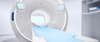
Open type tomograph, 1.5T