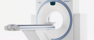
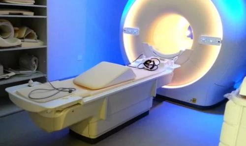
What is an MRI of the craniovertebral junction?
This is a non-invasive diagnostic method that assesses the condition of the spinal cord at the junction of the skull and the upper vertebrae. MRI of the craniovertebral junction: what does it show? The equipment creates visualization of the base of the skull and the cervical spine to assess the condition of the spinal cord, blood vessels, structure and structure of the vertebrae, and intervertebral discs. A thorough examination on a tomograph allows us to identify old and recent injuries, as well as pathological abnormalities in development. Anomalies of the upper vertebrae in most cases are diagnosed only a long time after the injury, resulting in complications and diseases. To avoid this, after an injury or when characteristic signs appear, it is recommended to undergo magnetic resonance imaging.
MRI examination to detect platybasia
The craniovertebral junction is the boundary between the base of the skull and the upper cervical spine. The normal relationship of these bony structures provides adequate space containing the lower parts of the brain stem and the cranial portion of the cervical spinal cord.
There are a number of acquired and congenital developmental anomalies that contribute to the narrowing of this space and lead to intermittent or constant compression of brain structures.
One of these anomalies, often encountered in MRI diagnostics, is platybasia.
It is a flattening of the base of the skull, mostly of the posterior cranial fossa. Platybasia can be either congenital (with Down's disease, in combination with Arnold-Chiari anomaly, etc.) or acquired (with fibrous dysplasia, osteomalacia, or as a consequence of long-term intracranial hypertension in childhood).
Clinically, this dysplasia is asymptomatic in most cases.
Platybasia rarely occurs in isolation. In most cases, this anomaly is combined with basilar impression.
Basilar impression is an impression of the base of the skull into the cavity of the posterior cranial fossa. Manifests more often by the age of 15-25. Manifested by headaches, brainstem and cerebellar symptoms: ataxia, nystagmus, paresis of the caudal cranial group. Pathological reflexes are possible. When the spinal cord is compressed, pyramidal disorders of varying severity occur.
Of clinical interest is also such a pathology of the craniovertebral junction as assimilation of the atlas, i.e. partial or complete fusion with the occipital bone. This leads to limited movement in the upper cervical spine and instability in the lower cervical spine. In addition, compression of the brain structures in this area is possible with a corresponding clinical picture.
Anomaly of the cranio-vertebral junction: assimilation of C1, occipital bone, odontoid process of C2 vertebra; basilar impression. Rotational displacement of the C1 vertebra, hypertrophy of the lateral masses of the C1 vertebral body. Foramen magnum stenosis. Focal myelopathy at the level of the C2 vertebra.
Fusion of the right sections of the lateral masses of the atlas and the base of the occipital bone (synostosis of the right atlanto-occipital joint).
In addition, when studying the craniovertebral junction, it is possible to identify various types of space-occupying formations affecting the membranes or substance of the brain and spinal cord, as well as abnormalities in the development of the brain structures of the posterior cranial fossa.
Space-occupying lesion on the anterior surface of the posterior arch of the atlas (meningioma)
Intramedullary mass formation of the cervical spine. Small form of the Dandy-Walker variant.
Arnold-Chiari malformation.
MRI examination of the atlanto-dental joint
The atlanto-dental joint is a strong connection of the first two cervical vertebrae: the atlas and the axis. Together with a strong ligamentous apparatus, they form a strong osteoligamentous joint, preventing excessive mobility of the atlas or odontoid process.
Nevertheless, damage to this joint due to various diseases and pathological processes contributes to a violation of the strength of the joint and the development of pathological mobility. The fusion of the ossification points of the axis and the odontoid process occurs at 4-6 years of life, and complete growth of the tooth occurs at 8-10 years. However, there are often cases of incomplete fusion of the odontoid process - a developmental anomaly. This situation contributes to the pathological displacement of the atlas along with the odontoid process with minor mechanical impact.
Variant of incomplete fusion of the odontoid process.
Possible traumatic damage to the atlantodental joint with the formation of dislocation, subluxation or fracture of the odontoid process. Fractures develop, as a rule, from a fall from a height onto the head, a “whiplash” injury, or blows to a bent head.
Clinically characterized by limited mobility (patients hold their head with their hands), pain in the neck and occipital region.
With displaced fractures, neurological disorders are observed: tetraparesis, numbness of the limbs, dysphagia, respiratory failure.
Transligamentous dislocation of the odontoid process.
Tooth fracture of C2 vertebra
Arthritis of the atlantodental joint
The atlantodental joint is often damaged in patients with rheumatoid arthritis. Inflammatory changes in the joint contribute to the development of erosive changes in the odontoid process, decalcification and weakening of the ligamentous apparatus. This can provoke the development of subluxation or dislocation of the joint, including compression of the brain structures.
An MRI study clearly visualizes the position of the odontoid process, the condition of the soft tissue structures, the degree of pannus, the consequences of subluxation or dislocation (the condition of the spinal cord and medulla oblongata, surrounding soft tissues).
Rheumatoid arthritis
Indications for MRI of the craniovertebral junction
The procedure is prescribed in the presence of injuries and the following symptoms:
- fractures, dislocations of cervical vertebrae;
- stiffness and pain in the upper neck;
- soft tissue and ligament injuries;
- developmental anomalies;
- damage to the occipital condyles, bruises;
- intervertebral disc injuries;
- inflammation and neoplasms;
- congenital diseases of the spinal canal and spinal cord.
Using the procedure, old injuries of the first vertebrae are identified, causing abnormalities in the functioning of the brain and spinal cord.
What can be seen with an MRI of the craniovertebral region?
Using MRI of the craniovertebral junction area, you can see the general condition of individual vertebrae and ligaments in this area. When using resonance testing, specialists have the opportunity to examine pathologies with maximum accuracy and detail of the picture. Thanks to MRI, you can determine the presence of such diseases:
- Dislocations and subluxations.
- Offsets.
- Damage of a cervical nature, that is, concussion, whiplash fractures, etc.
- Fusion of the bodies of the cervical structures.
- Concrescence of vertebrae of ankylosing spondylitis nature.
- Scheuermann Mau disease.
The first stages of the disease can be distinguished using a magnetic resonance examination of the required area of the body in a three-dimensional plane with a section thickness of less than 3 mm on a modern device whose power is 1.5 Tesla. If the treating expert believes that the patient has a tumor, then he will write out a referral for an MRI session with a contrast agent, which will allow a more effective study of all structures susceptible to the harmful process. The high success rate of the presented method for diagnosing the craniovertebral junction in combination with a correctly drawn up therapeutic treatment plan eliminates the development of the pathological process in the future.
That is why it is very important to consult a neurologist in a timely manner and perform a resonance examination. To quickly and easily sign up for an MRI session, use the convenient search service. On the official website you can apply online for an appointment at any local clinic, get information about the cost of the test, and also find out about the availability of vacancies in the coming days. Using the portal and the selection system is easy and intuitive, and if you need to find out some information, you can call the company’s support service, where they will provide you with comprehensive answers to any questions and provide professional advice.
Decompression of the craniovertebral junction for Chiari malformation
The range of methods of surgical treatment of patients with Arnold-Chiari malformation is represented by more than 20 types of surgical techniques. Unfortunately, most neurosurgeons, based on contradictory literature data and their own experience, “adjust surgical tactics to the patient.” In our opinion, the strategy for surgical treatment of patients should be based on a deep understanding of the pathophysiology of the disease. Taking into account modern views on the cause of the disease, based on the results of a completed dissertation devoted to this problem, surgical intervention, in addition to eliminating direct compression of neural structures, should be aimed at restoring normal liquor circulation at the level of the craniovertebral junction.
The main treatment method for Chiari malformation remains surgical correction of imbalances at the level of the craniovertebral junction. The range in the degree of surgical “aggressiveness” varies from methods accompanied by resection of the tonsils to minimally invasive endoscopic techniques of bone decompression. In recent years, there has been a clear trend towards more gentle methods of surgical treatment, although some surgeons are currently promoting radical methods.
The “gold standard” is decompression of the craniovertebral junction with dural plastic surgery. The essence of this method is to eliminate pressure on the cerebellar tonsils and create “comfortable” conditions for the normal process of liquor circulation in this area.
For this purpose, through a small linear incision in the cervico-occipital region, most of which is located in the hair growth area, access is made to the structures of the craniovertebral junction. In order to eliminate compression, a small section of the occipital bone and a fragment of the first cervical vertebra, which does not play a key role in the biomechanics of the cervical spine, are removed. Already at this stage we begin to use an operating microscope. An important step in achieving maximum decompression is excision of the thickened atlanto-occipital membrane, which exerts direct compression on the descended cerebellar tonsils. After this, the dura mater and its plastic are dissected. The modified semicircular configuration of the sheath incision allows us to use the entire length of the wound without causing technical difficulties at the stage of its plastic surgery. Shell plastic surgery means sewing in a “patch” from the patient’s own tissues, taken in advance at the stage of soft tissue incision. The advantages of using the patient’s own tissues are their inertness, elasticity and rapid “implantation” into surrounding tissues. The use of “local” tissues allows us to avoid additional incisions and ensure maximum tightness with minimal cosmetic defects. The most common complications when using foreign synthetic materials are infectious complications, the possibility of adhesions and poor healing due to the body's immune response. After completion of the main stage, layer-by-layer, maximally airtight suturing of soft tissues and cosmetic, intradermal sutures are carried out on the skin. If the postoperative period is favorable, the patient can be discharged from the hospital with detailed recommendations as early as 4-5 days after surgery.
What causes craniovertebral anomalies?
Congenital pathologies of the craniovertebral junction are formed during the prenatal period under the influence of infection, intoxication caused by metabolic disorders, increased radioactive background, smoking, and alcoholism. In addition, there is a genetic predisposition to the occurrence of anomalies in this department.
Acquired disorders of the cerebrovascular accident occur as a result of injuries, including birth injuries, as well as diseases such as:
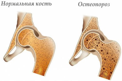
- Osteoporosis;
- Rickets;
- Osteitis deformans;
- Hyperparateriosis;
- Osteomyelitis;
- Tuberculosis;
- Bone tumor;
- Syphilis;
- Actinomycosis.
MRI of the craniovertebral region with an additional amplifier
If there is a possibility of a tumor-like object forming inside the body, experts in the field of neurology prescribe an MRI session with intravenous administration of a contrast agent. The composition of the required drug includes iodine elements, which, when entering the blood supply system, are able to reach a given perimeter. The main advantage of contrast is its ability to brightly color various types of soft tissue, which significantly improves the resulting image. Using this technique, it is possible to identify cancer pathologies in the first stages of development and begin timely treatment.
However, there are certain limitations to using this scanning option:
- Pregnant women are not allowed to use the presented testing method, since particles of the substance can penetrate the placental membrane and adversely affect the fetus.
- Nursing mothers undergo an MRI in case of urgent need, but for 48 hours the baby will have to be switched to artificial nutrition or the amount of breast milk must be expressed in advance (the liquid can be stored in the refrigerator in special sterile bags).
- People with individual intolerance to this medication.
- If the patient suffers from renal or liver failure, it is forbidden to make enhancements, since stable functioning of the urinary system is necessary to remove the iodine component (if this instruction is neglected, the person receives intoxication, which can cause serious consequences).
- MRI is not done if the individual has thyroid disorders, diabetes mellitus or heart problems.
You can select a medical center and make an appointment using the search portal. Using the official website or a call center operator, you can apply for an MRI session for craniovertebral anomalies and other abnormalities that affect general well-being. Using the company’s services, our clients receive favorable discounts and hot promotional offers that significantly save diagnostic costs. In addition, in a special section you can read real reviews from visitors to many clinics and quickly decide on the choice of place.