Article navigation: What CT shows Capabilities and advantages of CT Application of computed tomography in various areas of dentistry Indications Contraindications Preparation How they do Cost of CT in INTAN
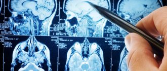
In any field of medicine, diagnosis is critical to prescribing treatment. Correctly diagnosing a disease in dentistry is often difficult, since it is impossible to examine the hidden part of the tooth and oral cavity. For a more detailed study, X-rays and panoramic photographs of the teeth are used. With the development of technology, new diagnostic methods are being introduced into medical practice, and today dental computed tomography is increasingly used in dentistry. What opportunities open up, what CT scan shows and whether there are contraindications for its implementation – this worries many patients.
What does a CT scan show?
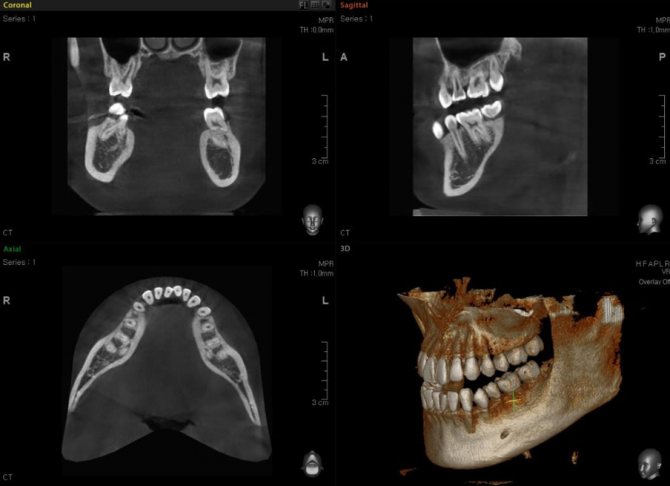
A visit to an orthodontist, implantologist, maxillofacial or plastic surgeon most often ends with a referral for a tomography. A 3D dental image gives a complete picture of the condition of the patient’s jaw, because the doctor gets the opportunity to examine the object in three dimensions, rather than in a flat image. The difference from an orthopantomogram is precisely this: the doctor sees a three-dimensional image, without any distortion.
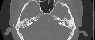
The picture shows a kind of virtual layer-by-layer slice, available for inspection in any projection. You can examine soft and bone tissue layer by layer without performing traumatic additional procedures. The picture is complete enough to accurately assess the condition of the jaw and teeth and establish a diagnosis for the patient.
Advantages
The advantages of the computed tomography method include:
- lack of special training;
- suitable for children;
- painlessness (the device only makes a quiet sound, no discomfort, pain, or unpleasant sensations);
- minimal radiation (the dose is 40 - 60 microsieverts, so up to three procedures can be done per day);
- the possibility of obtaining a voluminous, high-quality model.
The advantages also include a short list of contraindications:
- first trimester of pregnancy;
- lactation period.
Some patients consider the price of the procedure to be a disadvantage of dental computed tomography.
What capabilities and advantages does tomography have?
Compared to X-rays or panoramic photographs, dental computed tomography has a number of advantages, which lead to its increasingly widespread use in dental practice. Using tomography data, it is possible to plan in detail operations to install implants and various orthopedic structures, even if there are no teeth at all.
A specially developed computer program allows for detailed processing of the examination results, after which a spatial template is made for precise implantation of implants into the patient’s jaw. The presence of a template image makes it possible to monitor all stages of implant installation and place them at the required angle in the required areas. This ensures not only aesthetics, but also the durability of the orthopedic structure.
Compared to previously used CT techniques, CT has a number of advantages:
- no special preparation is required for the procedure
- The patient receives radiation exposure to a minimum, without harm to health
- the procedure takes no more than 30 seconds
- produces a high-quality three-dimensional image
Computed tomography of teeth is widely used in various areas of dentistry:
- In therapeutic dentistry, CT is used to assess the condition of the tooth, the number of canals in the roots of the tooth in the treatment of periodontitis and pulpitis
- In periodontology, it is used to determine the condition of periodontal tissues and periodontal pockets.
- In implantology, CT scans are used to determine the location of implants and measure the thickness of bone tissue at the site of implantation.
- In orthodontics, tomography is the best method for determining the possibility of extracting an incompletely erupted (impacted) tooth and correcting the dentition
- In endodontic therapy, CT allows one to evaluate the curvature and length of tooth canals
A high-quality 3D image of the tooth and jaw obtained during computed tomography allows you to establish the diagnosis as accurately as possible, without which it is impossible to prescribe treatment in any field of dentistry.
The CT technique gives a complete picture of the condition of the patient’s oral cavity, without leaving the slightest deviations unattended. This allows you to prescribe the correct effective treatment.
How harmful is CT scanning to health?
The second most popular question among patients after “what is a dental CT scan” is “is it harmful?” People are wary of any method based on x-rays, and that's normal. Of course, the radiation dose is also present in computed tomography, but the decisive factor is its level, and it is minimal!
X-ray radiation standards are strictly limited by the requirements of SanPiN 2.6.1.1192-03. They indicate that in one CT session the patient receives only 40-50 μSv (microsievert), and the permissible dose is 3500 μSv per year. Even a large 3D dental CT scan emits 10 times less radiation than conventional radiography. It turns out that with such a minimal load it is possible to perform almost 100 CT procedures per year without the slightest harm to health.
If your doctor has prescribed you to have a CT scan of your teeth before implantation, extraction or other intervention, you should not refuse. The radiation received during the study is negligible, but the harm from neglecting the best diagnostic method may be too great.
When is a dental CT scan indicated?
It is often quite difficult to visually determine damage resulting from trauma. Computed tomography of the teeth must be carried out after any injuries, because it is necessary to identify in time whether there is a fracture or displacement of the jaw, as well as damage to the roots of the tooth, which can lead to its loss. The list below gives a complete picture of the indications for CT scanning:
- Any mechanical damage, the consequence of which may be a fracture or displacement of the jaw. Invisible during external examination, these injuries are revealed by tomography. Injuries can also damage the roots of the tooth, which subsequently leads to its loss. Tomography will allow you to detect damage and prescribe timely treatment
- Deviations in the development of teeth, anomalies of the dentition. The orthodontist prescribes a tomography before starting treatment for the patient. Determine the ability to withstand the load of the bracket system. CT scan of teeth allows you to accurately determine the time and direction of growth of wisdom teeth
- If it is necessary to create a bracket structure, it is important to correctly model the future systems. A special role is played by spatial modeling in the manufacture of internal systems and aligners. Individual production of such products is provided, and it is tomography that gives a complete picture of the structural features of the teeth and jaw of each patient. This contributes to the speedy achievement of positive results from orthodontic treatment.
- If there is a suspicion of complications after endodontic treatment. The doctor cannot visually check the quality of the therapy performed. Only a dental CT procedure makes it possible to monitor and timely detect relapses or improper treatment
- If tumors are suspected or already present, the use of computed tomography will help to detect a tumor in time or clarify the condition of an existing one. The jaw bones and soft tissues of the oral cavity are susceptible to the appearance of neoplasms after injuries and other damage. To avoid the development of a tumor and the negative consequences of this, it is recommended to remove the tumors
- A planned surgical operation must be carefully prepared, and computed tomography is of great importance in this regard. If implantation or particularly complex tooth extraction is necessary, a CT scan will help the doctor identify factors that complicate the operation and determine how long the operation will take, given its complexity
- Monitoring and checking the effectiveness of treatment in many cases is impossible without CT scanning. Just as it is sometimes difficult to diagnose a disease without tomography, after a course of treatment it is not always possible to correctly evaluate the treatment. It will help determine whether all damaged tooth tissues have been removed or whether partial roots remain after a complex tooth extraction
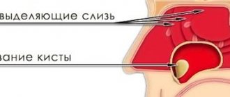
- If sinus lifting or bone grafting is planned, then a high-quality 3D image is necessary. Bone tissue can also be seen on an x-ray, but the condition of soft tissues, mucous membranes, canals and blood vessels can only be seen on a three-dimensional high-quality image
Purpose of the procedure
They resort to it to qualitatively treat teeth and identify anomalies of the dental system. Indications for CT scanning:
- damage to the jaws and dentition due to trauma;
- the need to obtain a picture of abnormally located and developing teeth and jaw bones;
- diagnosis of changes in primary teeth in children;
- damage to the maxillary sinuses by diseases;
- upcoming surgery;
- the appearance of tumors of tooth roots, bone and soft tissues of the face, jaws;
- when there are complications after cleaning or filling root canals;
- you need to determine the direction, curvature, depth of the channels;
- if you plan to install prostheses;
- to select the desired type and size of pins (the degree of their controllability and the service life of the implant depend on this).
A CT scan is also prescribed to determine the presence and location of hidden carious cavities and to draw up a plan for orthodontic treatment with braces and plates. In addition to diagnostics, CT scans are performed to monitor the quality of ongoing dental treatment.
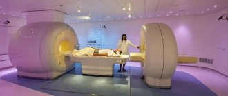
How is a CT scan done?
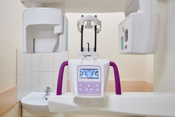
A dental computed tomography machine resembles an orthopantomograph. The process itself is simple: the person stands or sits. The patient's neck and chest are protected with a special apron with a lead layer. The subject rests against the frontal support, which, together with the chin and temple supports, provides reliable fixation of the head; hands on the handrails; firmly holds a special contact segment with his teeth. Based on the required shooting mode and frequency, the patient must remain motionless in this position for 15-25 seconds, during which the device’s planar sensor will make a full revolution around the subject’s head. In these few seconds, up to two hundred photographs are taken in various projections.
It takes several minutes to process the received information and build a multidimensional model; the CT scan is recorded on disk. After 5-10 minutes, accurate data on the current condition of the teeth and jaws is ready, the doctor can, based on it, make a final diagnosis and prescribe treatment or monitor the therapy already completed.
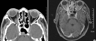
What jaw diseases does MRI detect?
Magnetic resonance imaging allows you to diagnose inflammatory, degenerative, dystrophic processes. The method is used to determine the nature of the blood supply to the lower, upper jaw and TMJ, and to clarify the localization of tumors.
MRI reveals:
- hilar granulomas, cysts;
- periostitis;
- abscesses, phlegmon of the maxillofacial area;
- benign and malignant tumors;
- traumatic injuries to the soft tissues of the face and oral cavity;
- osteomyelitis;
- congenital and acquired anomalies of the structure of the jaws and joints;
- teething disorders;
- vascular diseases;
- arthritis, synovitis of the temporomandibular joint;
- arthrosis;
- pathological conditions of the facial nerves (neuritis, neuralgia);
- myositis, etc.
Contrast-enhanced MRI is used in the diagnosis of neoplasms and to assess the condition of the blood vessels of the maxillofacial system. A gadolinium solution is used as a “coloring” substance. The drug fills the bloodstream and intercellular space, visualizing the slightest changes in the vascular system and showing formations ranging in size from 3 mm.
Cost of CT scan?
Modern dental clinics at INTAN use high-tech equipment that allows you to make three-dimensional CT projection in 20 seconds, and X-ray images in nine! This acceleration of the procedure makes it safe, minimizing the radiation exposure to the patient’s body.
You can clarify the possibility of carrying out such a procedure in the center you are interested in by calling us.
The cost in our centers for this procedure starts from 1,530 rubles.
What can dental tomography show?
Dental computed tomography allows:
- determine the position of the teeth;
- identify damage under fillings and fixed dentures;
- assess the condition of the dental canals;
- identify tumors, cysts, fistulas, foci of inflammation in the gums;
- detect the consequences of injuries (fractures of varying complexity, etc.)
Contraindications for 3D dental imaging
The radiation exposure of CT scans ranges from 0.045 to 0.06 mSv. This is relatively little, given the recommendations of SanPiN, which indicate the upper possible threshold of radiation exposure for research purposes equal to 1 mSv per year. However, if you compare a CT scan with a dental x-ray, where radiation occurs in the range of 0.002 - 0.003 mSv, a 3D photograph of teeth no longer seems so harmless. In this regard, there are a number of contraindications to CT scanning. If we are talking about simple tomography, then, as a rule, it is not done during pregnancy, especially in the first trimester. The doctor will always consider the need for radiation exposure for a pregnant patient based on the balance of benefit to the mother and risk to the fetus. If tomography with contrast is necessary (for better visualization of soft tissues and blood vessels, it is rarely used for CT of the jaw), then the number of contraindications expands. It should not be given to pregnant women, nursing mothers, or to persons suffering from thyroid disease, severe diabetes mellitus, renal failure, or an allergy to iodine. If you have any allergies or have ever experienced drug intolerance, be sure to tell your doctor.
What are the contraindications?
Computed tomography has few contraindications. However, the procedure is not performed during pregnancy. Even despite the negligible radiation exposure, it is better not to risk the baby’s health. In this case, the examination is allowed to take place during breastfeeding, but this also has its own nuances. For example, when a contrast agent is administered, the patient will have to take care to switch the child to another type of feeding for at least 48 hours.

The procedure is not recommended for pregnant women
Another contraindication to CT is an allergy to the contrast agent - in this case, the examination can be performed without any additional drugs. During the procedure, you must remain still, which may be somewhat difficult for young children.
How to prepare for a CT scan
If you are about to undergo a CT scan of the jaw, no special preparation is required. There is no need to limit yourself in food or drink. Be prepared to stand or sit still for some time. Directly at the appointment, you will be asked to remove all metal jewelry - this is necessary so that the metal does not interfere with the operation of the device and does not distort the diagnostic results.
“I remember when I underwent a CT scan, they immediately asked me to remove all metal jewelry. It was necessary to stand still; in my opinion, it was impossible to even swallow saliva. But it’s only half a minute, so it’s tolerable. I didn’t see anything wrong with the procedure itself. Yes, the device made a buzzing noise, but it didn’t scare me. Personally, I took a picture before implantation, but I did not go to the clinic, but to the diagnostic center...”
Marina_74, Samara, from correspondence on the forum www.32top.ru.
By the way, you cannot undergo such an examination with metal braces in your mouth - the picture will turn out to be of poor quality, and the fastenings of the braces may become loose. In this case, it is better to choose an alternative diagnostic technique.
Which is better - panoramic or 3D photography?
The difference between a CT scan and a standard orthopantomogram is the production of a three-dimensional image. This diagnostic option is more effective, but in some cases it is not necessary. An orthopantomogram can be analogue or digital. The latter is of higher quality, and pictures can be saved to various media. This procedure is also fast, as the entire scanning process takes a few seconds.
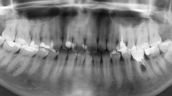
Standard orthopantomogram
Panoramic photography can be taken by everyone, including small children, but not often. Moreover, it is of paramount importance for the child, because it helps to visualize the dentition to prevent problems with bite and early diagnosis of diseases.
Carrying out tomography
During a dental scan, the patient can stand or lie down, depending on the equipment used. The mouth can be either open or closed. While the machine is operating, the X-ray detector will rotate around the area being examined, and you must remain stationary during this time. It takes no more than 50 seconds when scanning the entire jaw and about 10 seconds when obtaining images of several teeth.
In a cone beam computed tomography (CBCT) scan of the mandible (and maxilla), the machine rotates around the head. One revolution produces up to 200 2D images, which are then combined into a 3D image.