- Home /
- Branches /
- CT scan /
- CT scan of the facial part of the skull
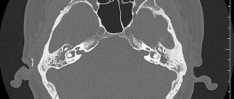
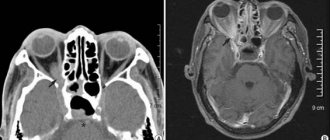
At the Clinical Hospital on Yauza, a computed tomography scan of the bones of the facial part of the skull . CT performed in our clinic, taking into account the use of iterative reconstructions, can reduce radiation exposure by up to 80% and improve the quality of studies. This technique significantly increases the efficiency of diagnosing diseases and injuries of the facial part of the skull, including hidden ones.
Computed tomography of the skull
Computed tomography (CT) is a diagnostic method based on weak X-rays that allows multi-layered images to be taken from different angles with an interval of 1 to 1.5 mm, which are then converted into a three-dimensional model. This study, compared to other methods (conventional X-ray, MRI), provides a more accurate and informative picture of the condition and structure of bone tissue and cartilage. In addition to hard structures, soft tissues, as well as vessels of the face and brain, can be examined.
Get a CT scan of the facial part of the skull in St. Petersburg
A CT scan of the facial part of the skull can be done at Medical in St. Petersburg. The procedure helps in identifying pathologies and prevents the development of serious diseases. The diagnostic cost includes:
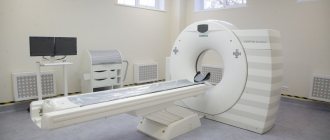
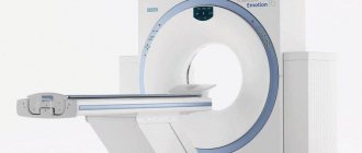
- Examination using an expert-class computed tomograph Aquilion Lightning TSX-035A (Canon Japan).
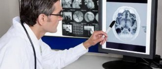
- The patient receives a detailed, comprehensive conclusion made on the basis of the images by a highly qualified radiologist. The description of the study is checked by the chief physician.
- 24/7 access to your personal account to view all your research and conclusions
- Internal research quality control
- 100% guarantee of photo quality
- Detailed information about prices can be found in the “Prices” section.
- You can find out about the benefits and ongoing promotions in the “Promotions” section.
CT scanning of the facial and cerebral parts of the skull in St. Petersburg can be done by appointment. Call us to undergo a study using the best tomograph from Japan!
In what cases is a CT scan of the skull prescribed?
Typically, a computed tomography scan of the skull is prescribed in the following cases:
- if it is impossible to accurately diagnose the disease, the presence or absence of anomalies and deviations from x-rays, or it is impossible to conduct an x-ray examination;
- if the patient is contraindicated for MRI due to the presence of an insulin pump, pacemaker or fixed metal prostheses in his body;
- severe traumatic brain injuries with unclear consequences (suspicion of a fracture or displacement of the skull bones, hematoma, edema);
- sudden deterioration of vision or hearing, headaches, nausea, fainting, dizziness of unknown origin;
- differentiation of malignant and benign tumors;
- bruises, injuries to the facial part of the head;
- suspected stroke or cerebral hemorrhage;
- hydrocephalus.
Listed above are only the most basic indications for performing a CT scan of the skull bones, including the facial area. Based on the individual situation and the totality of symptoms, the decision to prescribe a procedure is made by the attending physician (traumatologist, ophthalmologist, otolaryngologist or other specialist)
What you need to know when preparing for research
For a CT scan of the skull, metal objects, including jewelry, glasses, dentures, and hairpins, should be left at home or removed before the examination, as they can negatively affect the CT images. You may also be asked to remove hearing aids and removable dental work.
You need to tell your doctor about the medications you are taking and whether you have any allergic reactions to medications or, as previously known, contrast materials and dyes.
If there is a history of this, your doctor may prescribe medications (usually steroids) to reduce the risk of allergies or prevent them altogether.
The radiologist will need to know if you have asthma, diabetes, myeloma, or heart disease, kidney disease, or thyroid problems. Any of these listed conditions may increase the risk of adverse effects on a person's overall health.
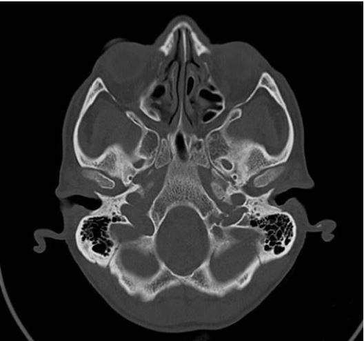
What does a CT scan of the skull show?
According to the results of tomography, it is possible to detect foreign objects, cracks and fractures, displacement of bones, tumor growth into the soft tissues of the face, the spread of metastases, circulatory disorders, any abnormalities and pathologies in the development of the brain and skull bones. High resolution images make it possible to diagnose cancer and their severity. Also, a CT scan of the skull shows how effective drug therapy or surgery was.
CT diagnostic services at CELT
The administration of CELT JSC regularly updates the price list posted on the clinic’s website. However, in order to avoid possible misunderstandings, we ask you to clarify the cost of services by phone: +7
| Service name | Price in rubles |
| CT scan of the cerebral arteries and screening examination of the brain | 14 000 |
| CT pituitary gland | 7 000 |
| CT scan of the brain | 6 000 |
All services
Make an appointment through the application or by calling +7 +7 We work every day:
- Monday—Friday: 8.00—20.00
- Saturday: 8.00–18.00
- Sunday is a day off
The nearest metro and MCC stations to the clinic:
- Highway of Enthusiasts or Perovo
- Partisan
- Enthusiast Highway
Driving directions
How is a CT scan of the skull performed?
As a rule, no special preparation is required before performing a CT scan of the skull bones. If the procedure is performed with the use of a contrast agent, then you need to refrain from eating and drinking 3-6 hours before the examination. Immediately before the procedure, you must remove all metal objects (hairpins, glasses, elastic bands, earrings), then lie down on a special couch that rolls into the tomograph ring. The radiologist takes images from an adjacent room, but can always be contacted via feedback. During the examination, the tomograph ring rotates around the head with slight noise and crackling. Depending on the complexity, the procedure can take from 1 to 30 minutes. Results with a radiologist’s report can be obtained within a few hours after the examination.
CT scan of the facial skull
Basically, a CT scan of the facial skull can be prescribed for: facial injuries with damage to soft tissues, edema and tumors of the paranasal sinuses and orbits, clarification of the location of unerupted teeth, and also before surgery (including during dental operations). The photographs can be used to study the bones of the facial skull and adjacent soft tissues.
CT skull base
Tomography of the bones of the base of the skull is performed for various head injuries, as well as for diagnosing oncological and other types of tumors. Once accurate and prompt results are obtained, timely and effective treatment of identified diseases can begin, especially in the early stages.
When is a CT scan of the facial part of the skull prescribed?
- for injuries of the skull bones;
- for bruises and head injuries;
- if benign or malignant formations are suspected;
- in order to clarify the results after other diagnostic procedures;
- when planning operations in the nasal cavity and sinuses;
- for detailing small bone fractures;
- before dental implantation and prosthetics - to determine the place where the implant should go.
Based on the results of a CT scan of the facial part of the skull, bone tissue augmentation is sometimes prescribed for successful implantation.
Contraindications for CT scan of the skull
Due to the low radiation exposure, when the attending physician prescribes tomography for pregnant and lactating women, as well as young children, it is necessary to compare the risk and the expected benefit. An overweight person (more than 150 kilograms) may not disable the device. In this case, you should first find out about the maximum possible load on the tomograph.
A CT scan of the skull with contrast may be contraindicated in the following cases:
- allergy to contrast agent;
- renal failure;
- thyroid diseases;
- a set of other diseases that, in the opinion of the attending physician, are unacceptable for examination using a contrast agent.
Contraindications for CT scan of the skull bones
Scanning of the skull bones is prohibited for pregnant women, even though the radiation only affects the head. After all, even the smallest dose can harm the unborn child. If the doctor plans to administer contrast, then computed tomography is prohibited for patients who are allergic to iodine, endocrine disorders and renal failure,
There may also be difficulties in assessing patients who are overweight. Typically, the equipment is designed for patients weighing up to 150 kilograms.
Features of scanning for pregnant women
Although the upper part of a pregnant woman's body is exposed to x-rays, the developing baby may be negatively affected by the rays. When performing a CT scan of the facial skull, the level of radiation exposure exceeds what is relatively safe for the fetus, which negatively affects the development of the fetus. The cells of the embryo are actively dividing; when they are exposed to radioactive radiation, developmental defects may form, and in severe cases, fetal rejection.
Conducting a CT scan of the facial skull during pregnancy is strictly contraindicated; it is permitted only in cases of extreme necessity in the later stages, as prescribed by the attending physician, using special means of protecting the fetus and setting the MSCT to a minimum radiation dose. It is optimal to postpone the procedure, carry it out after delivery, or prescribe an alternative examination method - magnetic resonance imaging.
Research methodology:
The examination is carried out lying on your back, clothing should be free of metal objects. A scan of the head bones is performed. The scanning procedure lasts about 2 minutes
Dose load – 0.6-2 mSv
Use of contrast agent:
When performing a CT scan of the head bones, the administration of a contrast agent is not required.
Volumetric constructions (reconstructions):
after the end of the study, volumetric reconstructions of bone structures are constructed at the study level (images in 3D and 4D, MPR (multiplanar reconstructions)).
Advantages over X-ray and MRI:
- CT allows visualization of the eyeball and optic nerve
- speed of research
- three-dimensional and four-dimensional modeling of the bone structures of the head make it possible to more clearly imagine the nature and extent of the pathological process
- conducting a study of patients with internal and external fixation devices (especially large metal structures), including those with a pacemaker
The results of computed tomography allow doctors to make the most accurate, correct diagnosis and timely prescribe treatment. Computed tomography is a non-invasive, painless diagnostic method. Carrying out CT scans very often reduces the required number of studies to a minimum and eliminates the advisability of using invasive techniques.
CT scan:
— allows you to study the state of the bone structure, soft tissues, blood vessels and individual organs layer by layer; - often used in emergency situations, for post-traumatic disorders, for suspected damage to internal organs, bleeding; — can be used if the patient has implanted medical devices of any kind; — used for control during biopsy of organs and pathological areas of the human body. X-rays from a CT scan have no immediate side effects.
Estimated risks and contraindications
The effective dose of radiation from computed tomography is minimal, however, it is always present. The radiation dose is usually specified in the study protocol. In the interests of accurate diagnosis, especially in acute cases, this risk is mitigated. Women are required to inform the referring physician and radiologist about the possibility of pregnancy. Because of the potential risk to the baby, CT scans are not recommended for pregnant women unless there are specific medical indications. It is extremely rare, but allergic reactions to a contrast agent that contains iodine do occur. In such cases, PATERO CLINIC has all the necessary means to stop such reactions.
Manufacturers of intravenous contrast media do not recommend that mothers breastfeed for 24 to 48 hours after receiving or administering contrast media.
Additional studies if necessary:
- in case of injury to the eye or orbit - CT is supplemented by ultrasound
- MRI is indicated in the subacute period in patients with unclear damage to soft tissues, orbits, and suspected brain damage
CT scan of the skull base with contrast
Computed tomography supports the use of contrast agents. This is an advanced examination technique that involves introducing a non-ionic iodine solution into the patient’s bloodstream. The contrast, distributed throughout the body, penetrates all organs and tissues, selectively accumulating in those places where the blood-brain barrier is broken, or there is inflammation, infection, or tumor. Iodine, which is part of the contrast agent, blocks almost 100% of X-rays, which causes the effect of “illuminating” the pathology, facilitating the diagnosis of the corresponding disease.
CT scan of the skull base
Contraindications for CT scan of the skull base with contrast
CT with contrast is contraindicated in the presence of the following pathologies and conditions:
- Renal failure: Contrast is primarily excreted by the kidneys. If their function is impaired, the removal of the contrast agent is disrupted, which is accompanied by acute damage to the kidney tissue. Clinically, this is manifested by a progressive increase in serum creatinine levels within 1-2 days after the examination. This pathology is known as contrast-induced nephropathy. Risk factors: advanced age, chronic kidney disease, diabetes mellitus, dehydration, high doses of contrast agents. A mandatory blood test for creatinine allows you to identify hidden renal failure and prevent complications;
- Allergy to iodine: in most cases, allergic reactions do not pose a health hazard and occur in the form of itching, skin rashes, nausea, and vomiting. However, in patients with anaphylaxis, the administration of radiocontrast drugs can provoke swelling of the upper respiratory tract, loss of consciousness, and respiratory arrest. For this reason, an allergy to iodine is a contraindication for the administration of contrast;
- Individual intolerance to radiocontrast drugs: clinically manifested as a severe reaction to the administration of contrast and is accompanied by vomiting, loss of consciousness, and coma. It is an absolute contraindication for contrast-enhanced CT. It is extremely rare;
- Diabetes mellitus during treatment with metformin, which is used to lower blood glucose levels, which, in combination with contrast, can provoke ketoacidosis - a pathological increase in the concentration of nitrogenous metabolic products and ketone bases. This terrible complication is observed in patients with diabetes mellitus and is accompanied by loss of consciousness and coma. To prevent such a complication, you should contact an endocrinologist who will change the treatment regimen;
- Thyroid diseases accompanied by hyperthyroidism: iodine is the main component of thyroid hormones. When contrast is administered, their levels in the body increase sharply, which can serve as a provoking factor causing thyrotoxic crisis - a dangerous complication of thyroid diseases, accompanied by nausea, vomiting, cardiovascular failure, impaired renal function, fever, acute psychosis, coma;
- Breastfeeding: Contrast agents pass into breast milk. Excess iodine in them can cause dysfunction of the thyroid gland in infants. For this reason, when performing a CT scan of the base of the skull with contrast, it is necessary to: temporarily switch to artificial feeding or feeding with pre-expressed breast milk. The contrast is completely eliminated from the body within several hours - during this time it is necessary to express excess milk at least twice to prevent lactostasis.
Safety of the CT method of the facial skull
During the examination, the patient is in a specialized room, under the constant supervision of a radiologist, who is in the next room during the procedure. Communication is carried out via a special microphone. Single radiation exposure to MSCT is minimal and absolutely safe for the patient. For clearer visualization of soft tissues, focal inflammatory processes and differential diagnosis of cysts from soft tissue formations, iodine-based contrast agents are used, which practically do not cause side effects. Occasionally, after their use, mild dizziness occurs, which quickly passes. The procedure itself lasts several minutes, minimizing patient exposure to radiation.