Second planned ultrasound during pregnancy
The second trimester of pregnancy is the time for the second ultrasound examination of the fetus. This examination is routine and mandatory for every pregnant woman. The second ultrasound during pregnancy (at what stage the procedure is performed will be discussed below) is more informative than the first. It helps to accurately understand whether the fetus is developing normally, whether there is a threat of miscarriage, whether there are indications for medical abortion, and so on.
Together with ultrasound, a biochemical blood test is performed, as well as other diagnostic studies.
At what time should I do the second ultrasound?
The main question that worries every expectant mother is: “At what stage of the 2nd trimester during pregnancy is it necessary to do a second screening ultrasound?” There are specific dates for conducting this examination - 20-24 weeks of pregnancy. In cases where the first ultrasound showed any abnormalities in the development of the fetus or the results of a blood test are abnormal, the second screening can be carried out earlier than 20 weeks (for example, at 16-17 weeks).
Standard indicators for the second trimester
Fetometric parameters
This group of indicators includes a large number of fetal measurements, which are important in the diagnosis of defects and developmental anomalies.
Summary table of basic fetometric values.
| Fetometric indicators, cm | Gestation period, weeks | |||||
| 19 | 20 | 21 | 22 | 23 | 24 | |
| Biparietal size | 3,58-5,19 | 3,91-5,58 | 4,22-5,91 | 4,47-6,29 | 4,81-6,59 | 5,11-6,81 |
| Fronto-occipital size | 4,79-7,02 | 5,32-6,44 | 5,61-7,92 | 6,11-8,34 | 6,49-8,72 | 6,91-9,04 |
| Average breast diameter | 3,12-4,89 | 3,55-5,32 | 3,81-5,64 | 4,02-6,06 | 4,31-6,34 | 4,62-6,65 |
| Average abdominal diameter | 3,51-5,35 | 3,88-5,61 | 4,11-6,04 | 4,34-6,41 | 4,67-6,71 | 5,03-7,13 |
| Femur length | 2,13-3,50 | 2,32-3,79 | 3,80-4,21 | 3,91-4,40 | 4,12-4,74 | 4,30-5,03 |
| Tibia length | 1,89-3,12 | 2,10-3,41 | 2,43-3,82 | 2,63-3,89 | 2,81-4,09 | 3,02-4,30 |
| Fibula length | 1,78-2,90 | 1,97-3,22 | 2,20-3,52 | 2,51-3,77 | 2,72-4,09 | 2,94-4,24 |
| Humerus length | 2,09-3,40 | 2,42-3,59 | 2,63-3,88 | 2,81-4,13 | 3,07-4,34 | 3,30-4,61 |
| Radius length | 1,72-3,11 | 1,91-3,20 | 2,08-3,39 | 2,30-3,63 | 2,49-3,78 | 2,69-4,02 |
| Ulna length | 1,81-3,10 | 2,03-3,30 | 2,22-3,63 | 2,41-3,78 | 2,61-3,89 | 2,81-4,10 |
| Head circumference | 14,4-18,2 | 15,6-19,4 | 16,8-20,6 | 18,0-21,8 | 19,1-22,9 | 20,2-24,1 |
| Abdominal circumference | 11,6-16,6 | 12,7-17,6 | 13,9-18,9 | 15,0-20,1 | 16,1-21,2 | 17,2-22,1 |
| Exhaust/coolant ratio | 1,05-1,23 | 1,04-1,25 | 1,06-1,23 | 1,05-1,22 | 1,05-1,23 | 1,01-1,20 |
| Distance between the outer edges of the eye sockets | 2,41-3,52 | 2,62-3,76 | 2,78-3,89 | 3,01-4,15 | 3,12-4,21 | 3,29-4,41 |
| Eye socket diameter | 0,71-1,11 | 0,79-1,21 | 0,83-1,25 | 0,91-1,35 | 0,92-1,36 | 1,01-1,41 |
| Interhemispheric size of the cerebellum | 1,61-2,02 | 1,81-2,20 | 1,93-2,31 | 2,02-2,63 | 2,11-2,69 | 2,31-2,89 |
Biparietal and fronto-occipital size, average diameters of the chest and abdomen are used to clarify the duration of pregnancy, determine the weight of the fetus, the correspondence of development to the gestational age, as well as to determine the symmetry and proportionality of development. Circumferences of the chest, head and abdomen are used to diagnose malformations and intrauterine growth retardation.
Measuring the length of the tubular bones of the limbs is carried out on both sides and is used to identify hypoplasia and aplasia of the limbs.
The interhemispheric size of the cerebellum is used in the diagnosis of brain abnormalities.
The distances between the edges of the orbits and the average diameter of the orbits are of an auxiliary nature and can be used in the diagnosis of defects of the facial skull.
Fetal anatomy
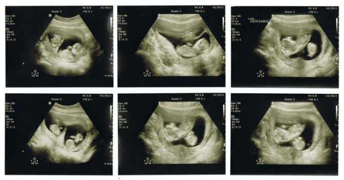
During the second ultrasound scan during pregnancy, the main task is to assess the structure of the internal organs and identify malformations that were not detected during the first ultrasound diagnosis.
Each specialist has his own principle of examination: from top to bottom, according to organ systems, haphazardly. The quality of the study does not depend on this; the main thing is that all organs are analyzed.
Brain and spine
Studying the structure of the brain using 2 ultrasounds usually begins in the axial plane, conducting a series of sequential sections. This allows us to study the structure of the lateral ventricles, cerebellum, optic thalamus, and interhemispheric fissure.
If pathologies are detected during this scan, then the study is carried out in other planes. At this stage, it is possible to identify the following defects:
- ventriculomegaly,
- hydrocephalus,
- hypoplasia of the corpus callosum,
- cerebellar abnormalities.
The standard study is complemented by a color Doppler study, which makes it possible not to miss developmental disorders of the vascular bed.
The spine is assessed along its entire length in two perpendicular planes. The evaluation criteria are structure, spinal curves, integrity of vertebral arches, muscle defects. This allows you to identify hernial protrusions (spina bifida).
When examining facial structures, it is important to evaluate the condition and size of the eye sockets, nasal bones, and the condition of the nasolabial triangle.
Respiratory system
Diagnosis consists of assessing the condition of the bronchi and lung tissue. During examination, important importance is given to the division of the bronchi, the echogenicity of the lung tissue and its homogeneity, as well as the correspondence of the size of the lungs to the period of pregnancy. The latter parameter is assessed based on measuring the chest circumference and calculating the ratio of the chest circumference to the abdominal circumference.
A formula for calculating lung volume has also been developed, but its use is limited due to the large error. Taking into account the fact that the main stage of maturation of lung tissue occurs in the third trimester, it is possible to identify only gross malformations during the periods discussed.
Heart
Prenatal diagnosis of heart defects is particularly difficult to diagnose. The standard protocol includes evaluation of only the 4-chamber section of the heart. If there are changes in this position, then detailed fetal echocardiography is recommended, which is a separate study.
Digestive system
The protocol deals with the stomach, intestines and liver as separate points. Diagnosis of pathology of the stomach and intestines does not cause difficulties due to the presence of anechoic contents, which allows one to evaluate the structures of organs and exclude atresia (closure of natural orifices) at any level.
When analyzing the structure of the liver, it is important to assess the condition of the liver vessels and the bile duct system.
Measurement of the organ is necessary only if hepatomegaly (pathologically large liver) is suspected. The main pathology is cysts, which are easily differentiated.
urinary system
Examination of the kidneys does not cause difficulties, since their location and appearance are typical. The bulk of the pathology is occupied by aplasia, hypoplasia and pyeloectasia.
The bladder is visualized due to the presence of anechoic urine in the lumen, and if it cannot be determined, the examination should be repeated after 10-25 minutes to exclude its aplasia.
Assessment of the structures of the uterus and the state of the provisional system (umbilical cord, amniotic fluid, placenta) does not differ from screening ultrasounds at other times.
Ultrasound in the second trimester of pregnancy is of key importance in deciding on the further management of pregnancy, so the study should be carried out using expert equipment by high-class specialists.
What does the second ultrasound show during pregnancy?
Screening ultrasound of pregnancy 2nd trimester
shows:
- Features of fetal fetometry (sizes of its head, abdomen, limbs, etc.).
- Features of the anatomy of the fetus (structure and structure of its brain, heart, liver, kidneys, spine, lungs, etc.).
- Features of the structure of the placenta and umbilical cord.
- Congenital features of fetal development (if any). At this stage, chromosomal disorders such as Down Syndrome, defects of the facial part of the skull, such as cleft lip or cleft palate, etc. can be detected.
- The gender of the unborn child (this criterion is not mandatory for inclusion in the ultrasound protocol).
What do they look for on an ultrasound in the second trimester?
There are a number of certain fetometric indicators that are necessarily studied during the second ultrasound screening:
- BPP is the biparietal size of the fetal head. At 20-23 weeks of pregnancy, it is normally between 36 and 43 mm.
- LZP – fronto-occipital size. At week 20, this parameter should normally be 56-68 mm. By the 24th week, the LZR can reach 71-85 mm.
- Fetal head circumference. At the 20th week of pregnancy, the normal OG can be 154-186 mm. By the 24th week, OG reaches a value of 201-237 mm.
- Fetal abdominal circumference. At 20-24 weeks this figure averages 144-193 mm.
- Cephalic index, that is, the ratio of BPR to LZR. This index is not always prescribed in the ultrasound protocol, although for a more complete assessment of the development of the fetus (in particular its central nervous system), it is necessary to know it. The cephalic index allows you to accurately identify the type, size and structure of the fetal head.
- The length of the tibia and fibula, the length of the femur, humerus, ulna, radius.
In addition, the second ultrasound examines the location of the placenta. It can be located on the anterior or posterior wall of the uterus. You also need to evaluate its location relative to the internal pharynx.
Purpose of the event
Ultrasound of the second stage of screening is performed to identify the following indicators and assess the condition of the fetus:
- estimate the number of fetuses (multiple pregnancies may not be detected at the first ultrasound);
- presentation and position of the fetus, its weight and size;
- fetal heart rate;
- condition of the facial bones of the skull (partially excludes Down syndrome), lengths of paired bones, assessment of the chest, spine;
- volume of amniotic fluid;
- assessment of the condition and maturity of the placenta;
- anatomy of all internal organs of the unborn child (heart, brain, lungs, kidneys, liver);
- identification of developmental defects;
- umbilical cord assessment, identification of entanglement;
- assessment of the tone of the uterine walls and the condition of the cervix.
In the 2nd trimester of pregnancy, it is already possible to determine the sex of the child, but this is not included in the ultrasound examination protocol and is not a mandatory indicator.
How is the procedure done?
As a rule, a transabdominal ultrasound
method of research. The woman lies on the couch on her back and slightly bends her knees. A special gel is applied to her stomach.
The duration of the ultrasound procedure may vary. It all depends on the position of the fetus in the uterus, the presence of obvious signs of pathology, as well as the professionalism and experience of the doctor conducting the diagnosis. On average, the procedure takes from 15 to 30 minutes.
Preparation for ultrasound and methodology
No special preparation is required for a second trimester ultrasound. The intestines, regardless of the amount of gases it contains, are displaced by the enlarged uterus, and the filled bladder is replaced by amniotic fluid.
Ultrasound examination in the 2nd trimester is carried out using the transabdominal method, that is, the doctor examines the uterus and fetus through the anterior abdominal wall. To perform an ultrasound, a woman only needs to expose her stomach, which the doctor treats with a special gel and moves a sensor over the skin of the abdomen.
Ultrasound screening 2nd trimester: interpretation of results
Interpretation of ultrasound results is carried out immediately after it is performed. The screening test protocol is filled out by a doctor or nurse in the ultrasound diagnostic room. It is then transmitted to the attending physician, who, based on the data provided, either prescribes corrective therapy or informs the patient that her pregnancy is proceeding without complications.
Ultrasound standards for the second trimester
In addition to the parameters listed above and the values of their norms, studied at the second ultrasound screening (at 20-24 weeks of the second trimester), there are other indicators that need to be assessed:
- The weight of the fetus is a standard value of 300 to 600 grams.
- The thickness of the placenta is 25-45 mm.
- The amount of amniotic fluid (with index determination).
- Number of umbilical cord vessels (three or two vessels may be present).
- The contraction frequency of the fetal heart muscle is 140-160 beats/min.
Peculiarities

Previously, a second ultrasound was not prescribed to all women, but only to those who were in the so-called risk group. It includes those who are over 35 years old, who have a history of infectious diseases, miscarriage, and genetic pathologies in the fetus identified in the past. Now the second scheduled screening period is assigned to all pregnant women, who must undergo it without fail.
Many mothers involuntarily wonder what they look at on an ultrasound in the 2nd trimester. The study confirms what was recorded as a result of the first screening, and also shows how much the baby has grown and strengthened since then, and whether factors have appeared that could negatively affect his condition.
The peculiarity of ultrasound in the 2nd trimester is that it does not have a strict framework. The Ministry of Health recommends sending pregnant women for the procedure from 18 to 21 weeks, but the gynecologist can revise the period and schedule screening for 17 or 22 weeks.
After IVF pregnancy

All pregnant women as a result of in vitro fertilization will undergo examination in the 2nd trimester. Only, unlike those mothers whose pregnancy took place naturally and was not overshadowed by threats of miscarriage, for women with IVF this will not be the second screening. After all, as you know, almost the entire artificial insemination procedure is carried out under constant ultrasound control. In such patients, special attention is paid to Doppler studies - assessing blood flow in the vessels of the uterus and placenta.
Pregnancy with twins or triplets
During pregnancy with twins or triplets, the timing and procedure for performing ultrasound are the same as for singleton gestation. The only difference is that the procedure itself will take a little more time, because the specialist must carefully study the development parameters of two or even three babies. The indicators of each of them will be recorded in the examination protocol.
Second screening during pregnancy – is it necessary?
Second ultrasound screening during pregnancy
mandatory for a period of 20-24 weeks. Neglecting doctors' recommendations and avoiding ultrasound can have disastrous consequences. In the second trimester, ultrasound allows you to determine intrauterine growth retardation in the early stages, identify oxygen starvation, impaired blood flow and other deviations from normal development. Untimely detection of such disorders can lead to the development of serious pathologies in the unborn child or even to his intrauterine death.
Ultrasound in the second trimester
Ultrasound examination is the main screening procedure, which is carried out at 17-19 weeks. At this time, the baby’s internal organs are formed, as well as the structure of his body. An ultrasound is performed transabdominally, so before the procedure the mother needs to follow some rules:
- do not eat three hours before the examination;
- Do not under any circumstances eat fatty foods, citrus fruits, seafood, or soda.
The ABIA Clinic invites expectant mothers to undergo this painless and very important examination, which allows timely detection of possible abnormalities in the health of the fetus. What could be more important than knowing that everything is fine with the baby? We also take photos of the fetus and record an ultrasound video on electronic media - this is a very pleasant memory of the exciting period of pregnancy.
In what cases is an unscheduled ultrasound performed?
All 3 planned ultrasounds performed during pregnancy are carried out during its normal course and are designed for dynamic monitoring of the development of the fetus and the woman’s condition. However, there is often a need for additional examination using an ultrasound machine.
Unscheduled ultrasound is performed at any stage of pregnancy and has only medical indications:
- Suspicion of ectopic pregnancy;
- Frozen pregnancy;
- Bleeding from the genital tract;
- Multiple pregnancy;
- Pregnancy as a result of artificial insemination;
- The woman has a history of miscarriages;
- Previous caesarean section;
- The presence of chronic diseases of the genital area (inflammatory, endometriosis);
- Endocrine diseases in women (diabetes, hypothyroidism and others);
- Woman's age over 40 years;
- Detected abnormalities of the fetus or placenta at the 2nd ultrasound.
Interestingly, many parents prescribe additional ultrasound for themselves , turning to paid clinics that have modern color three-dimensional equipment. It is quite natural for them to want to see and capture the future baby, his movements and even facial features and facial expressions. It won't hurt him or his mom.
What to do if a woman missed the second ultrasound examination?
For various reasons, a pregnant woman may miss her second ultrasound and there can be a huge number of reasons for this: travel, illness or others that are not related to problems on the part of the doctor, since almost always, all examinations are scheduled on time, especially when the pregnant woman regularly visits the women's clinic consultation.
Of course, you should not deliberately not come for regular examinations and ignore the doctor’s recommendations, however, even if, for some reason, this happened, you should not be afraid to contact us. Depending on the period, the pregnant woman will still be scheduled for an examination, because there is also a third screening ultrasound during pregnancy.