How dangerous is x-ray for humans and why? To understand why X-ray radiation is dangerous for human health, you need to remember from your school physics course what X-rays are and what properties make them dangerous.
X-rays (X-rays, X-rays, X-Ray) are a special type of electromagnetic radiation, which is a stream of photons moving at the speed of a light beam. X-ray photons carry no electrical charge.
The wavelength of X-rays on the electromagnetic wave scale falls between the gamma spectrum and ultraviolet rays.
X-rays have a number of characteristic properties:
- invisible to the human eye;
- penetrate living tissue and non-living structures that normally do not transmit sunlight;
- cause the glow of certain chemicals (green glow of cadmium and zinc plates, blue glow of tungsten plate);
- decompose silver halide compounds in photographic emulsions (used to take photographs);
- ionize molecular structures (cause the disintegration of neutral atoms into positive and negative particles);
- cause radiation damage (non-healing soft tissue burns).
In medicine, X-rays are used for radiation diagnostics (radiography, fluoroscopy, fluorography, computed tomography, angiography) and radiotherapy (proton therapy, brachytherapy).
Passing through living tissue, X-rays transfer their energy to atoms, causing them to be excited and ionized. The higher the radiation dose, the more pronounced the properties of the rays are. In small doses, x-rays cannot cause stable adverse reactions, but in high doses, the likelihood of negative health consequences appears.
The biological properties of X-rays depend on the sensitivity of living tissues to ionizing radiation. Different cells have different radiosensitivities and depend on many factors:
- stages of cell division (cells are most sensitive at the stage of DNA synthesis);
- level of oxygen saturation of cells (the less oxygen in a cell, the less its sensitivity to rays);
- degree of cell differentiation (poorly differentiated (for example, stem) cells are more sensitive to irradiation than specialized ones);
- the state of the cell at the time of exposure to rays;
- external factors (ambient temperature, water and oxygen content in the surrounding space).
Taking into account the combination of factors influencing the radiosensitivity of cells, it becomes clear why the tissues of the human body that are most sensitive to ionizing radiation are:
- gonads (testes in men, ovaries in women);
- placenta (in pregnant women);
- thyroid;
- Bone marrow;
- lungs;
- retina;
- stomach and large intestine.
In terms of radiation dose, the most powerful of all types of X-ray examinations are fluoroscopy, computed tomography, scintigraphy, and fluorography. Fluoroscopy examines the organ in dynamics, so the procedure lasts several minutes. As a result, the patient receives a large dose of radiation. Computed tomography involves taking many slices, which are essentially x-rays. Accordingly, the more sections, the higher the radiation dose.
Fluorography produces small-sized images. To obtain them with great image clarity, it is necessary that the power of the X-ray machine for the procedure be significantly higher than for conventional radiography.
Scintigraphy is a combined radiation study that involves intravenous administration of radioactive isotopes and a series of images. Radioisotopes selectively penetrate the organ planned for examination (for example, the thyroid gland, kidneys or adrenal glands), resulting in the organ appearing in contrast on the pictures.
To minimize possible negative consequences from procedures performed using X-rays, it is necessary to know what dose of ionizing radiation the patient received one-time and in total, that is, to conduct dosimetric monitoring.
When is an x-ray necessary?
Doctors are convinced that the study is done for children only for indications. No doctor will prescribe an x-ray “just in case” or to “play it safe.” How often the procedure can be performed will be determined by the indications. There are clear recommendations for performing x-rays:
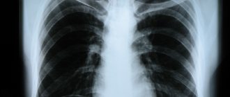
- A chest x-ray is performed to diagnose pneumonia and tuberculosis infection, as well as if there is a problem with the heart. It is used to visualize an organ, for example, the heart, and estimate its size.
- Examination of the gastrointestinal tract organs is carried out in case of severe digestive disorders, as well as in case of penetration of foreign bodies.
- An examination of the genitourinary system is carried out in case of urination disorders indicating the presence of congenital pathologies.
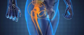
- Orthopedics and traumatology account for the lion's share of all studies in children. This is caused by congenital anomalies in the structure of the organs of the musculoskeletal system, birth injuries, as a result of which the child receives dislocation and other injuries, etc.
- In neurology, they try not to conduct examinations, but indications for mandatory examination are signs indicating the presence of a tumor, as well as skull injuries - bruises, concussions, soft tissue damage, etc.
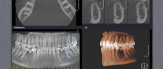
- In dentistry, X-rays are used to examine tooth germs, to decide on tooth extraction, and also if periodontitis has developed. They also do research when treating canals.
Little ones
X-ray - how dangerous it is for kids, when it needs to be done, read all the details related to these issues below. All parents have heard at some point in school about the harmful effects of x-rays, and when a child is prescribed x-ray examinations , parents are very worried about this.
Is it really impossible to do without an x-ray, is it possible to do it often and how to prepare and carry out the procedure in the least dangerous way. What is an X-ray?
X-rays are a special type of radiation that has the property of passing through the body at the speed of light. It is used in medicine as one of the key diagnostic tests because of its special property, which is not characteristic of the light that we see with the eye. It penetrates the body depending on its density, and if this is reflected on a special film, it will be visually distinguishable. So bones transmit the least amount of x-rays and appear the lightest on x-rays, while soft tissues appear the darkest. Any compaction in soft tissue will also cause lightening. Where cavities contain air, the film will generally be transparent.
It is this property of X-rays that is used in medicine to diagnose serious disorders in the skeleton - fractures, displacements and disorders in soft tissues - lungs, liver, intestines. X-rays passing through the body with varying intensities decompose special substances on the X-ray film with varying degrees of activity, resulting in very long-lasting high-definition images that a doctor can see with his eye and make a diagnosis.
Everything would be fine if not for one thing, one of the properties of x-ray radiation is the ability to form ions of different charges in tissues, which is unsafe for the tissues of living beings with prolonged exposure. Large doses of X-ray radiation can cause mutations in genes, abnormalities in the development of offspring and tumor transformations. But so that parents don’t get scared, these are absolutely the wrong doses and the wrong duration of irradiation. To get a similar effect, you need to take 10 pictures every day for at least a month in a row.
It is more difficult for the staff of X-ray rooms, but modern devices are protected by special casings and lead plates - lead does not allow x-rays to pass through. And today, the latest digital devices are used with minimal impact on children.
What are the methods of conducting
Conventional radiography using a digital or film device is the acquisition of one or more images on a computer monitor or X-ray film. The usual radiography, which almost everyone has had done at least once in their lives. There is also a fluoroscopy method - obtaining images of moving objects in x-rays with image processing on a computer monitor; this is essentially an examination of a real patient in passing x-rays. It is used extremely rarely in children, as it provides a large dose of radiation per study.
Linear tomography is layer-by-layer X-ray images, in which the X-ray tube and film cassette move in different directions. As a result, an image is obtained at a certain depth from the skin surface - for example, 4, 8 or 10 cm.
X-ray computed tomography is scanning an object with a very narrow and directed X-ray beam and collecting a complete image on a computer monitor screen. Used for a comprehensive assessment of the structure of internal organs and skeleton. You can scan any organs and tissues from head to toe with a CT scan.
Fluorography and electroradiography are not children's examination methods; the body is still growing and hard X-ray rays are more dangerous for it than for an adult. Therefore, these methods are not used for children under 16 years of age.
When is an x-ray indicated?
X-rays are done at any age, starting from the neonatal period, the most basic criterion should be the need for this study to make an accurate diagnosis. That is, without x-rays it is impossible to make a correct diagnosis under these conditions. But for any x-ray, a referral from a pediatrician, surgeon or doctor of another specialty is required, justifying the need for the method. When prescribing an x-ray, the doctor must explain to parents the need and expediency of this method, and the consequences for diagnosis and treatment if they refuse an x-ray. According to the law, doctors do not have the right to perform any manipulations on the child, including photographs, without your consent. A parental consent or refusal form with their signatures and their transcript is pasted into the child’s card.
So, x-rays are indicated for:
- any suspicion of bone damage, their diseases, such as osteomyelitis, tumors, osteoporosis, rickets and others. In newborns, x-rays are indicated to exclude birth injuries in the form of bone fractures or skull cracks; in older children, in case of falls, head injuries, suspected concussions, etc. In case of an accident, strong falls, beating. - X-rays are also indicated for suspected infectious and inflammatory diseases of the lungs and bronchi, pneumonia, abscesses, as well as lung lesions - atelectasis (collapse of areas). - an x-ray is indicated if there is a suspicion of metal (or in combination with metal) foreign bodies of the digestive and respiratory system; - an x-ray will be taken if tuberculosis, damage to the thymus or thoracic lymph nodes is suspected. - X-ray is indicated for malformations of the digestive system or damage to the intestines - it clearly shows intestinal obstruction and its level based on the presence of gases. Often, if there are problems in the digestive system, studies with contrast agents are performed. - X-rays are performed for heart defects, especially during preparation for surgery and rehabilitation after it. — X-rays with contrast are performed for damage to the kidneys and urinary tract, for vascular problems and many other diseases.
Thanks to the advent of x-rays, the diagnosis of children's diseases and saving their lives has significantly improved. X-ray sometimes sees what is hidden from the eyes of the doctor and other research methods. For diagnosis, sometimes one image in a direct projection is enough - that is, when the child is facing or with his back to the device, and sometimes several images are required from different angles or targeted images of a separate area - the head, neck, chest, legs or arms, abdomen.
X-ray scanning is rarely prescribed, mainly during operations to combine bone fragments, and some problems with the intestines for health reasons. CT scanning is rarely prescribed and only in unclear and serious cases, it must be done for a long time and the child must lie still, so it is usually performed under light anesthesia. This is usually a study of the heart or lungs before surgery, tumor processes and other severe pathologies.
How to prepare a child for an x-ray?
In most cases, x-rays do not require special preparation; the child is asked to remove clothing and metal objects in the area where the x-ray will be taken. Getting into the field of research into appliqués from clothing. Metal objects can distort the picture and cause an incorrect diagnosis.
One of the important conditions for conducting the study is complete immobility, which is often very difficult for children. For this purpose, the child is taken into the office with an adult, and he is carefully fixed using special devices. Older children need to be persuaded and told that it is not painful and not scary. During the examination, parents help the child follow the radiologist’s commands.
To conduct studies in the area of the intestines or kidneys, especially with the introduction of x-rays, special preparation is required before the study - the intestines must be freed of food and gases through a special diet and an enema in the morning before the study. Three days before the x-ray, the child will be prescribed a low-fermentation diet with enzymes and espumisan.
How to protect your baby from x-rays.
When conducting research, special rules must be observed - all areas that do not participate in the research. They are protected from X-rays by special screens, usually lead aprons or screens, or other means of protection. This gives less radiation exposure to a fragile body. If the baby is held by a parent during the examination, he is also given special protection.
What do you need to take with you to the X-ray room?
For x-rays, you need a child’s card (from a clinic or hospital), a referral from a specialist and permission from parents for x-rays. For babies, you need to take a diaper with you, especially if the x-ray will be done while lying down; very little ones will need a pacifier (if they suck). A little older baby can take a small plastic toy, but they should not interfere with the child’s position in the device.
The time it takes to take pictures will depend on the complexity of the study and the behavior of the child and his parents. The less he twitches and is capricious, the faster everything will pass. If the picture is of poor quality, it will require repeating. And this is an additional burden on the child.
The time for developing and describing the image will depend on the power of the X-ray laboratory. After receiving the image, the doctor needs to study and describe it, and this will take some time - it will depend on the complexity of the disease, the amount of work and the doctor’s experience. In emergency situations, the description is made as quickly as possible and can be done initially, even in words, with subsequent recording in the chart; sometimes a council of doctors is brought in to study the images. For various illnesses of the child, his photographs must be stored and presented to the doctor if necessary.
http
(10911)
No similar posts
Negative effects of X-rays
X-ray equipment produces high-quality images when patients are irradiated with low doses. Such studies will not harm the child’s health. When studying the question of whether x-rays are harmful to a child, you need to know that the child’s body is susceptible to x-rays. Therefore, doctors are careful when performing the procedure on children, so as not to have an adverse effect on the growing body.

There is no need to be afraid of the study; a negative effect is only possible when exposed to large doses of radiation for a long time, which does not happen with an x-ray examination.
The harmfulness of X-rays for children is seen by doctors in the following facts:
- a child’s body reacts to x-rays much faster than an adult’s, therefore the risk of developing abnormalities inherent at the genetic level increases;
- the consequences are not visible immediately, but after a while;
- The child’s internal organs are located compactly, so when one organ is irradiated, the rays may hit the neighboring one;
- Radiation exposure provokes non-standard reactions that are impossible to predict.
X-ray machines use rays with low doses, and even those affect the body for a short time. All this speaks in favor of conducting an x-ray examination, since even a series of procedures will not harm the child, since the level of radiation received is too low.
If we talk about the likely impact, then the blood and hematopoietic system are at risk. The consequences of X-ray irradiation can be:
- leukemia is a mutation of bone marrow cells that do not mature into normal leukocytes, but degenerate into cancer cells that are aggressive to the body;
- erythrocytopenia is a reduced content of red blood cells in the blood, which leads to a natural decrease in hemoglobin levels. With low hemoglobin, the oxygen supply to cells suffers, which negatively affects the child’s body;
- Thrombocytopenia is a decrease in the level of platelets in the blood, which are involved in the blood clotting process. If there is an insufficient number of platelets, increased bleeding and clotting problems occur;
- malignant degeneration of cells in the body - X-rays affect the process of cell division and growth, which leads to oncological pathologies.
Doctors say that you should not be afraid to take x-rays; these possible adverse effects do not always develop, only if the body is irradiated many times over a long period of time. X-ray examinations, which are carried out in clinics, are safe and do not affect the child’s health.
How dangerous is radiation diagnostics?
There is no reliable evidence that radiotherapy causes cancer. However, some studies of large populations exposed to radiation have shown a small increase in cancer risk even at low levels of radiation exposure, especially in children. Leading national and international organizations involved in radiation risk assessment agree that even minimal levels of radiation can increase the risk of cancer. In the interests of safety, even small doses of radiation must be considered dangerous.
The risk of cancer resulting from radiation diagnostics must be compared with the average statistical risk of developing cancer. The lifetime risk of dying from cancer is approximately 20-25%. For every 1000 children who have never undergone radiation diagnostics, 200-250 will die from cancer. The proportion of increase in a person's lifetime cancer risk after a single computed tomography (CT) scan remains controversial and is approximately 0.03-0.05%. This risk is for the general population and does not pose a direct risk to a specific child. Another way to assess the relative risk of CT is to compare the theoretical risk of abdominal CT with other risk groups. It is estimated that the risk of one CT scan of the abdomen is comparable to the risk of getting into an accident with:
• travel 12,000 km by car
• traveling 1600 km on a motorcycle
(estimates based on US traffic accident statistics)
These data show that, despite errors in estimating radiation dose, the risk of cancer after a single CT examination is very small. However, available research suggests that some risk exists and can accumulate.
Precautions when taking x-rays
Children are given a reduced dose to get the desired x-ray, and there are ways to get an x-ray for a child with minimal consequences. Here are some expert tips:
Scanning fluorograph (the safest and most modern diagnostic method)
- You should not conduct the study on newborns; if necessary, do it from the age of three months;
- older children are prescribed when other diagnostic methods fail to make a diagnosis;
- if it is necessary to undergo an x-ray of the lungs or other organs, you need to choose modern low-dose equipment, where the radiation exposure is less than others;
- For children under one year of age, the body is covered with a special lead apron, leaving the area being examined unprotected. For older patients, sensitive areas are covered - the thyroid gland, eyes, genitals;
- to avoid repeating the examination if the first one was carried out with errors, doctors pay attention to the need to completely immobilize the small patient;
- A radiologist must interpret the X-ray images in order to make the correct diagnosis and begin treatment. With a high-quality image, the doctor’s lack of knowledge negates the results of the study.
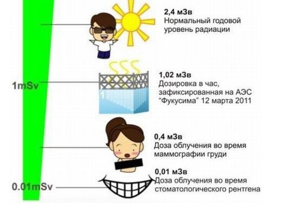
Note to parents! If circumstances force you to undergo an examination, pay attention to an X-ray machine; it must be a digital matrix or scanning one. In the latter option, the dose is 0.03-0.05 mSV. The natural background of Moscow is 0.02 mSV. An old-style analog film device, the dose is 0.1-0.5 mSV. In this case, refuse the examination. This information can be obtained from the x-ray technician.

How harmful is x-ray for a child?
X-ray radiation in large doses is dangerous for humans. Especially for young children, since their bodies are still in the process of development.
Long-term exposure to X-rays can cause:
- changes in the hematopoietic system;
- deterioration of vision and skin condition;
- disruption of metabolic processes in the body;
- pathologies of development of the genital organs;
- development of cancer.
Modern digital X-ray machines can reduce the radiation dose received by the patient. To obtain one x-ray, the radiation dose is 0.1–0.6 mSv .
This is not enough to harm the child's body. But doctors recommend adhering to the permissible number of x-ray procedures per year:
- No more than 2 for newborns and infants.
- No more than 6 children under 12 years old.
But the maximum permissible dose of radiation is considered by the doctor individually for each child - depending on the physiological characteristics, nature of the disease and the need for diagnosis.
When can doctors order an x-ray?
Since the procedure may be unsafe, doctors give referrals for examination of children only in extreme cases. No one can tell you how many times you can do an x-ray; it all depends on the indications. It can be:
- falling from a height, injuries during outdoor games, road accidents;
- birth injuries;
- problems with bone growth and mineralization of bone tissue, which manifests itself as rickets and osteoporosis;
- entry of foreign objects into various cavities and organs (ear, nose, stomach);
- skull injuries;
- neurological disorders;
- asymmetry of bone formations, congenital abnormalities in the formation of the facial skeleton;
- signs of pulmonary tuberculosis;
- as preoperative preparation on any organ.
These factors are good reasons to have your child x-rayed for diagnostic purposes. It is not carried out for the purpose of prevention.
At what age can X-rays be taken?
It is not recommended for children under three months of age, and at older ages they try to replace the diagnosis with other research methods.
For example, to determine tuberculosis, they do the Mantoux test or Diaskintest, but with several persistent positive answers, they have to resort to x-rays and the patient’s age will not matter.
The same applies to diagnosed severe pathologies, the treatment of which needs to be monitored. In case of pneumonia, the child needs to undergo an x-ray two weeks after the end of treatment to check the effectiveness. X-rays are just as important for a child under one year old as for an older one. It will be much more reliable to perform the procedure, because a focus of pathology may remain in the lungs, which threatens relapses. In this position, age does not matter since x-rays are a necessity.
With some exceptions, even infants are examined if there is such a need.
Which parts of the body are most exposed to radiation?
As practical radiology shows, in children the lion's share of images is taken from the musculoskeletal system - this area is most often irradiated. In childhood, injuries and injuries are common - dislocations, bruises, fractures. When a child enters the clinic with complaints of a blow or obvious signs of pathology, an x-ray is prescribed.
Congenital pathologies of the musculoskeletal system are also added to the percentage of statistics, when children are diagnosed with dislocations and subluxations in large joints, which requires x-ray diagnostics and further monitoring.
In second place in popularity are the chest organs, since if the Mantoux reaction is positive, an x-ray is taken. A positive Mantoux can be either a demonstration of pathology or a banal irritation and scratching of the sample area, but in any case, diagnosis is required.
X-ray alternative
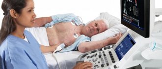
Ultrasound and magnetic resonance imaging are considered alternatives, but there are disadvantages. With ultrasound, for example, the quality of the image does not always allow a reliable diagnosis. It depends on how the doctor “read” the results of the study, so ultrasound can be perceived as an additional diagnosis for some pathologies, but not the main one.
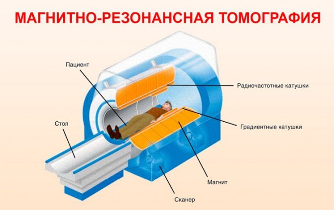
The image with MRI is much better, there are some peculiarities here - small children are given drugs for sedation so that they lie still and do not move. In this case, high-quality results are obtained. For example, at 3 years old it is difficult to do this; it is easier to take an x-ray.
X-ray examination of children is an effective diagnostic non-invasive method. With its help, you can identify pathologies and begin treatment. Parents do not need to worry whether x-rays are harmful to a child under one year old if there are indications for it - the value of the procedure is much higher than possible adverse results.
If my child's doctor thinks it is necessary to perform a CT scan, should I allow it?
As with any medical procedure, the benefit should always outweigh the risk. CT is a very valuable diagnostic test that can improve treatment and diagnose a number of diseases that other diagnostic methods do not detect. CT can provide the best course of treatment, avoid other tests or surgeries, and improve treatment outcomes. It is important to know that if your child has a serious illness that requires a CT scan, it is necessary to overcome the fear of radiation. In many cases, the merits and benefits of the test clearly outweigh the risk of radiation exposure to your child. If CT is the best test in your case, ask the staff if they are using proper techniques to minimize radiation dose.
How do I know that my diagnostic center is using proper techniques to minimize radiation dose?
Some centers that perform CT scans on adults do not use dose minimization techniques for pediatric patients. You will never know unless you ask about it. You have the right to this information. Your diagnostic center must provide you with information about how they reduce radiation doses. It is also necessary to find out whether the center has the appropriate certification for performing CT scans, for example, in the USA - accreditation from the American College of Radiology.
Alternative diagnostic methods
A CT scan may be the best way to obtain the diagnostic information needed to make clinical decisions in your child's treatment. In some cases, the doctor may decide that
It is safer to place the child for observation instead of performing a CT scan. Waiting while being monitored may be difficult for you and your family, but can provide the same end result without exposing your child to an unnecessary dose of radiation. Ultrasound (ultrasound) and magnetic resonance imaging (MRI) are diagnostic methods that do not use radiation. They can sometimes provide similar diagnostic information and can be a useful alternative. MRI is a lengthy procedure and sometimes requires anesthesia, which carries its own risks. A CT scan may be the only way to obtain the information needed to make clinical decisions about the best treatment for your child. You should ask your doctor and radiologist if there are alternative testing options for your child.