In what situations should you undergo a cardiac ultrasound?
- Changes in ECG indicators.
- Heart murmurs.
- A slight increase in body temperature in the absence of inflammatory processes in the throat, nose, ear or kidneys.
- Arrhythmia.
- An x-ray of the heart showed a change in its shape and size, as well as in the vessels extending from it.
- Hypertension.
- Predisposition to hereditary heart diseases.
- Possible changes in cardiac structures.
- Fainting, swelling, shortness of breath, dizziness.
- Pain in the left half of the chest and behind the sternum.
- Risk of heart tumors.
- After myocardial infarction.
- Effusion pericarditis.
- Angina pectoris.
- Diagnosis of cardiomyopathy and its type.
- To detect true or false cardiac aneurysm.
It is extremely important to carry out this procedure after a myocardial infarction to determine how many muscle cells have died.
ECG and ultrasound of the heart should be performed by people in stressful situations, experiencing severe emotional and physical stress.
EchoCG has no age restrictions. It is prescribed to children if a congenital heart defect or a change in its structure is suspected, which often occurs during the period of active growth of the child.
EchoCG is prescribed for pregnant women, because allows you to identify heart defects in the fetus in the womb. The procedure is painless and does not cause harm.
Heart ultrasound is a mandatory procedure during pregnancy if:
- There was a history of spontaneous abortions.
- Diabetes.
- Hereditary predisposition to heart disease.
- In the 1st and 2nd trimester, the woman was forced to take antiepileptic drugs and antibiotics.
- The analysis revealed high titers of antibodies to rubella, or the woman had it during pregnancy.
Fetal echocardiography is prescribed at 18-22 weeks. Children undergo it when the above symptoms appear.
Objectives of cardiac ultrasound during infarction
When carrying out the procedure, the doctor sets a goal - to identify those parts of the heart that do not participate in its contraction. If such areas are detected, this is an indication to confirm the diagnosis and begin treatment.
What parameters does the doctor pay attention to during ultrasound during myocardial infarction:
- location of the source of the disease;
- degree of damage, presence of contractile function, blood supply;
- for the presence of a blood clot;
- development of complications;
- condition of blood vessels, presence of atherosclerosis.
Who is prescribed ultrasound to prevent myocardial infarction?
In order to prevent heart attacks, doctors have identified a group of people for whom cardiac ultrasound is recommended:
- people over 45 years of age;
- workers in industries that are “harmful” to the heart;
- people interested in extreme sports;
- a history of atherosclerosis;
- hypertension;
- obesity;
- bad habits;
- genetic predisposition.
Methods for performing echocardiography of the heart
- M – method (one-dimensional). With its help, the dimensions of the heart chambers and the work of the ventricles during their contraction are accurately determined. The indicators are presented in the form of a graph.
- B – method (two-dimensional). Allows you to see tumors, blood clots and aneurysms. With its help, the thickness of the heart walls and valves is measured, and the degree of contractility of the ventricles is determined.
- Electrocardiography with Doppler. The procedure is necessary to diagnose heart defects and other serious disorders.
Ultrasound can be performed through the esophagus or during periods of active physical activity.
Echocardiography through the esophagus is prescribed in situations such as:
- Infectious endocarditis.
- Routine examination before installation of an artificial valve.
- After a stroke and in cases of cerebral circulation disorders and atrial fibrillation.
- Cardio version.
- Defect of the septum between the atria.
- The impossibility of traditional ultrasound of the heart. This occurs in the presence of costal ossification and other disturbances in the structure of the chest.
It is prohibited to perform cardiac echocardiography through the esophagus if there is:
- Varicose veins of the esophagus.
- Strong gag reflex.
- Radiation therapy of the esophagus.
- Osteochondrosis of the cervical spine in acute form.
- Enlarged hernia on the diaphragm.
- Gastric and intestinal bleeding.
- Intestinal perforation.
- Spasms, tumors, intestinal diverticula.
- Pathological mobility of the cervical spine.
Rules for preparing and conducting echocardiography through the esophagus:
- Stop eating and drinking at least 4 hours before the ultrasound
- Pull out dentures and gastric tube
- Before the procedure, the patient's mouth and throat are irrigated with lidocaine.
- The patient lies on his left side, a mouthpiece is inserted into his mouth and an endoscope is carefully inserted into the esophagus, through which ultrasonic waves penetrate
- The procedure takes from 15 to 20 minutes and is recorded on video.
Preparation for the procedure
No special preparation is required before an ultrasound examination. The patient should behave calmly, be positive, and completely abandon physical activity. You can even drink something calming. Immediately before the test, you should not eat chocolate, drink strong tea or coffee, or take drugs that stimulate heart activity. You should not overeat before the procedure, because this will not only increase the load on your body, but may also distort the examination data. The last meal should be no later than 3-4 hours before the examination. And in general, nutrition has a great influence on the functioning of the heart, so it is worth following the diet prescribed by your doctor.
In what cases is stress echocardiography prescribed?
- To diagnose coronary artery disease.
- To assess the extent to which narrowed coronary arteries in patients with coronary artery disease affect their quality of life and exercise endurance.
- To assess the degree of blood supply to the myocardium.
- To monitor the effectiveness of treatment.
- Uncomplicated heart attack or chronic ischemic heart disease.
- Risk of negative consequences during complex operations.
It is prohibited to perform echocardiography with stress if:
- Acute myocardial infarction (less than a month).
- Aortic aneurysm of dissecting type.
- Respiratory, cardiac, renal and liver failure.
- Blockage of blood vessels by blood clots.
The normal ultrasound of the heart with a load is considered to be:
- Evenly moving walls of the left ventricle (LV) during exercise.
- Decrease in end-systolic volume (ESV).
- Raising the exile faction.
- Absence of disturbance in the functioning of the walls during movement, if a disturbance was recorded at rest.
- Increase in wall thickening in systole.
Negative indicators are an enlarged right ventricle (RV), the appearance of new areas with inactive walls, and a decrease in the ejection fraction to 35%.
Acquired heart defects. Common diagnostic errors
As is known, among the causes of the development of chronic heart failure (CHF), the leading place is occupied by coronary heart disease (CHD), while the role of acquired heart defects is very modest and amounts to 6–14% in various countries.
According to the Framingham Study, during the period 1950–1987, valve defects as a cause of heart failure decreased by 45% in men and by 32% in women. At the same time, the proportion of coronary heart disease increased by 41% in men and by 25% in women. However, despite the fact that acquired heart defects are a disease with a well-studied clinical picture, errors in their detection are quite common. At the same time, the main causes of errors are usually the same. Next we will talk about true valvular heart defects, when the basis of the disease is damage to the valve leaflets themselves (their shortening, wrinkling, fusion along the commissures, or a combination of pathologies) in contrast to situations when the valve itself is not affected, but there is regurgitation, the causes of which can be very varied. This is, first of all, relative valve insufficiency with sharp dilatation of the corresponding chamber of the heart (of any etiology), as well as prolapse of the valve leaflets, most often due to myxematous degeneration of valve tissue or connective tissue dysplasia of the entire valve apparatus. Accessory chordae in various parts of the left ventricle in the inflow or outflow zones can also simulate valvular disease. This list of situations simulating valvular damage can be continued indefinitely (let’s add to this list of causes of noise ruptures of tendon threads, damage to the papillary muscles of various etiologies, etc.).
Unfortunately, many doctors do not have a sufficiently clear understanding of the diverse etiology and true frequency of certain defects. It is traditionally believed that acquired heart disease is a consequence only of rheumatic fever or infective endocarditis (which is beyond any doubt), but they do not take into account the possibility of developing heart disease (usually valve insufficiency) with diffuse connective tissue diseases (systemic scleroderma, systemic lupus erythematosus, occurring with secondary antiphospholipid syndrome, rheumatoid arthritis, ankylosing spondylitis, primary antiphospholipid syndrome, etc.), as well as the development of cyanide calcification of the mitral or aortic valve in elderly people. The listed situations lead, as a rule, to mitral or aortic valve insufficiency (stenoses are extremely rare, moreover, their possibility is generally questioned). On the other hand, currently rheumatic fever has a latent course, with minimal activity of the inflammatory process, which eludes the attention of the doctor and the patient himself, and the doctor interprets changes in the sound picture that are detected many years later in a completely different way. In addition, it is generally accepted that isolated aortic and mitral stenoses are quite rare defects, which is completely untrue.
A significant reason for the failure to detect acquired heart disease is the idea that this disease is extremely rare in elderly and senile people. Other diseases developing by this period of life - coronary heart disease (CHD) and arterial hypertension (with all the inherent symptoms and complications) lead the doctor to think about the possibility that the patient also has valvular damage, and therefore adequate diagnostic measures are not carried out. Or the altered auscultatory picture is interpreted by the doctor from a completely different perspective (especially since the patient may not remember the former attacks of polyarthritis in his youth).
Another reason for errors is the idea of valvular heart disease as a “frozen” situation. Meanwhile, the progression of valvular damage (mitral or aortic) causes a certain staged course. Therefore, there may be patients whose valve defect is quite fully compensated by hyperfunction and hypertrophy of the heart section(s) adjacent to the affected valve. Such persons may not even know about their disease, especially since they have no complaints. At the same time, there are patients who complain of shortness of breath (expressed to varying degrees) due to pulmonary hypertension, and as the disease evolves, signs of right ventricular failure of varying severity.
One of the main mistakes is the idea that the patient must have all possible signs of heart disease. However, even V.Kh. Vasilenko said that with aortic stenosis, all the symptoms of this heart defect are observed only in 25% of cases. In this regard, it is appropriate to recall the need to divide the signs of heart disease into “direct” and “indirect”.
“Direct” signs are caused by dysfunction of the valve and disruption of blood flow through the corresponding hole; they are also called “valvular”; These signs include changes in tones and the appearance of noise. The presence of these symptoms indicates damage to one or another valve and the type of this damage (valve insufficiency, narrowing of the corresponding hole, or a combination of both). “Valve” signs are detected using auscultation and ultrasound examination of the heart (visualization of the movement of the valve leaflets and blood flow through the corresponding openings).
“Indirect” signs are caused by: 1) compensatory hypertrophy and hyperfunction of certain chambers of the heart; 2) circulatory disorders in the pulmonary and systemic circulation; 3) decreased cardiac output; 4) sharp fluctuations in pressure in the arterial part of the vascular system. Their presence indirectly indicates the severity of the valve defect and the circulatory disorders it causes in various vascular areas (pulmonary circulation, arterial (including coronary and cerebral regions) and venous parts of the systemic circulation).
Practical experience shows that the initial defect in examining a patient (who undoubtedly has valvular heart disease) is poor-quality auscultation (failure to detect altered tones, as well as murmurs and additional tones) or incorrect interpretation of correctly identified auscultatory symptoms. Taking this into account, when starting auscultation, the doctor must remember that “any person has the right to have a heart defect,” even if he: 1) feels well and does not present any “heart” complaints; 2) has a high tolerance to physical activity; 3) does not have any “rheumatic” history; 4) comes to the doctor not at all in connection with “heart” complaints.
“Poor quality” of auscultation consists in: 1) listening to the patient only in an upright position; 2) non-use of special techniques, such as listening to the patient in the knee-elbow position, on the left (right) side with holding the breath during the exhalation phase; 3) listening to the patient against the background of severe tachy- or bradycardia (optimal data are obtained at a heart rate of 70–80 beats/min); 4) paying attention only to noise while ignoring the nature of changes in heart sounds; 5) listening to the heart only at standard points (no active search for sound epicenters is carried out). Meanwhile, the “mitral melody” can often be heard at the level of the anterior or even mid-axillary line, and the “aortic melody” - in the area of the right sternoclavicular joint or in the jugular fossa.
To diagnose valvular heart disease, it is necessary to detect exactly “direct” (valvular) signs, while “indirect” signs may be absent (or very slightly expressed). The discovery of murmurs in a patient who does not present any “cardiac” complaints at all prompts the doctor to look for an explanation for his findings in areas very far from the true cause of the murmurs.
On the other hand, incorrect assessment of murmur in a person who does not have a valve defect leads to overdiagnosis of heart disease. As is known, the appearance of noise is caused by a number of reasons:
1) disruption of the functioning of an anatomically unchanged valve;
2) violation of the coordinated functioning of the unchanged valvular and subvalvular apparatus of the heart;
3) a change in blood flow through unchanged valvular structures of the heart in healthy individuals or in individuals suffering from some other heart disease (but not valvular disease).
In this regard, the “accompaniment” of such noise becomes extremely important, namely: 1) the presence (or absence) of enlarged heart chambers; 2) the state of the 1st and 2nd heart sounds, the presence of additional sounds in diastole (III sound, the “opening” sound of the mitral valve); 3) changes in noise under the influence of body position and breathing phases; 4) characteristics of the noise itself (timbre, conduction to other areas). In differential diagnosis, taking into account the nature of heart sounds is of exceptional importance.
Thus, the presence of a preserved first sound in combination with a systolic murmur detected at the apex of the heart practically eliminates valvular mitral insufficiency. A rough systolic murmur to the right of the sternum in the presence of a sonorous second tone excludes valvular stenosis of the aortic mouth.
Another reason for failure to detect a heart defect is a defect in ultrasound examination (equipment with low resolution, inexperienced operator). It should be remembered that recording an ECG at a low speed of the tape mechanism (25 mm/s) does not allow identifying atrial hypertrophy syndrome. Finally, a routine X-ray examination, if carried out with sufficient quality (with mandatory contrast of the esophagus), can significantly help in identifying indirect signs of a defect (not only determining the enlargement of certain chambers of the heart, but also the type of pulmonary hypertension).
It has already been mentioned that in elderly and senile people with valvular heart disease, the appearance of ischemic heart disease, arterial hypertension, obstructive pulmonary emphysema, atherosclerosis of various localizations can “push” the signs of heart disease into the background, as a result of which auscultatory signs will be interpreted completely differently. These difficulties increase even more when the patient develops complications of his “new” diseases - acute myocardial infarction, cerebral stroke, complex rhythm and conduction disorders (especially those that require the installation of a pacemaker).
Thus, it is possible to avoid misdiagnosis of valvular heart disease with a certain degree of alertness, remembering that every patient may have a heart defect (although he does not know about it and does not present “cardiac” complaints). Instrumentalization of the examination, in its negative sense, also does not contribute to improving the diagnosis of heart defects. Relying only on ultrasound examination disarms the doctor and deprives him of the skills of so-called “bedside” diagnostics.
Of course, in some medical institutions, ultrasound examinations are performed very well, but we do not have many such institutions. Probably, the key to solving the problem lies in instilling in students and young doctors a “taste” for direct examination of the patient and, in particular, in teaching them correct auscultation.
What does an ultrasound of the heart show?
The resulting parameters allow you to determine:
- hypertrophy of the LV walls;
- regurgitation of blood through the valves;
- tumors, blood clots, scars, aneurysms;
- blood flow condition;
- valve condition;
- degree of myocardial contractility;
- size of the heart cavities;
- state of the LV pumping function in a dynamic state.
It is considered normal if:
- there is no fluid in the pericardium;
- the size of the final section of the pulmonary artery is within 1.8-2.4 cm; trunk - up to 3 cm;
- ejection fraction is 55 – 60%;
- the volume of the pancreas at the final stage of relaxation of the heart (diastole) ranges from 0.9 to 2.5 cm;
- thickness of the interventricular septum in the final stage of diastole from 0.5 to 1.12 cm
- the final section of the aorta in diameter is 2 – 3.7 cm
- the thickness of the posterior wall of the LV at the final stage of relaxation of the heart (diastole) varies from 0.6 to 1.12 cm
- the change in the movement of the posterior wall of the LV towards contraction (systole) is 0.91 – 1.41 cm
- the size of the LV cavity in the final stage of relaxation (diastole) ranges from 3.51 to 5.7
- cardiac output (MCV) must be no less than 3.5 and no more than 7.5 l/min
- cardiac index ranges from 2 to 4.1 l/m2
- the diameter of the terminal part of the pulmonary artery is from 1.8 to 2.4 cm, its trunk is up to 3 cm
- the blood flow speed through the carotid artery is 22 cm/s with an error of +-5 cm/s
- there are no manifestations of regurgitation and dysfunction of the papillary muscles, growth on all valves.
The degree of discrepancy with normal values is determined by a sonologist, but the final diagnosis is made by a cardiologist taking into account the information received and based on other symptoms. The disease cannot be diagnosed by ultrasound alone.
Indications
There are quite a few pathologies for which echocardiography is necessary, and all of them are related in one way or another to the differential diagnosis of heart or aortic diseases. Urgent indications for transthoracic echocardiography include:
- sharp, stabbing, cutting pain in the heart area, which the patient feels for the first time;
- pressing, squeezing pain that arose for the first time or increased in intensity and duration;
- sudden shortness of breath.
In addition, periodic examination of the heart is mandatory in case of chronic diseases and in the presence of risk factors:
- coronary heart disease, including angina pectoris, previously developed myocardial infarction;
- myocarditis;
- bacterial endocarditis (inflammation of the inner lining of the heart);
- heart defects - acquired and congenital;
- prosthetic valves;
- some minor anomalies of heart development (bicuspid aortic valve, mitral valve prolapse);
- cardiomyopathy;
- arterial hypertension;
- heart tumors;
- aortic diseases;
- heart failure;
- heart murmurs;
- heart rhythm disturbances;
- chest injuries;
- professional sports.
How is cardiac ultrasound performed?
During echocardioscopy, the doctor uses various modes of the ultrasound machine:
- one-dimensional (M-mode),
- two-dimensional (B-mode),
- Doppler mode (assessment of the speed of blood flow in chambers and vessels),
- color Doppler - CDK (to determine the direction of blood flows and identify pathological ones),
- power doppler (records the very fact of the presence of blood flow in the vessels),
- tissue Doppler (a deeper assessment of myocardial contractility, based on a study of the nature of the movement of the walls from the sensor and to it),
- 3D echocardiography (it brings maximum benefit before operations on the valves - they are almost completely visualized before the intervention, which is important for the surgeon to determine tactics).
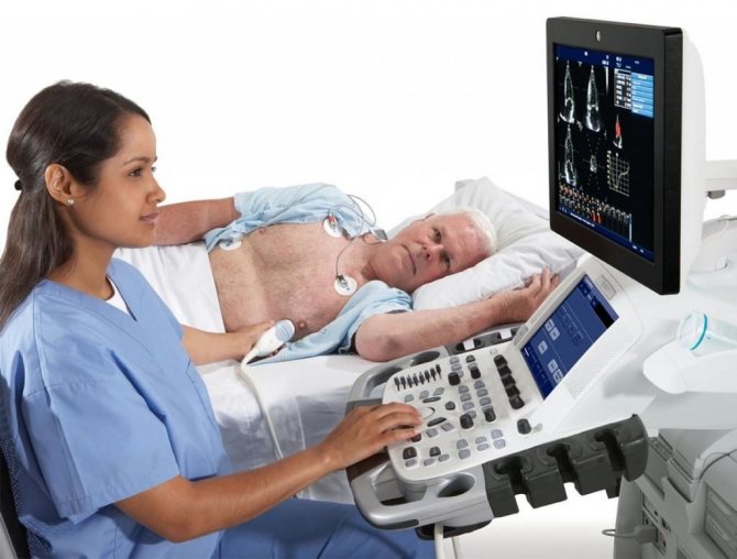
Transthoracic ultrasound
Echocardiography through the chest is performed in an ultrasound or functional diagnostics room. Sometimes in a hospital, due to the severity of the patient and the impossibility of transportation, a portable ultrasound machine is used to examine him. The nurse or doctor asks the patient to undress from top to waist, including women who need to remove their underwear. Next, the patient needs to lie on the couch on his left side and place his left hand under his head. In this case, the head end of the couch is slightly raised - this way, the maximum distance between the intercostal spaces is achieved for better visualization of the heart.
The position of the patient in relation to the doctor can be different, it all depends on the preferences of the latter and the arrangement of the office. The patient can be turned with his face or back towards the doctor, with his head towards or away from the device.
The specialist lubricates the sensor with ultrasound conductive gel for better contact with the skin, applies it to the left side of the chest, visualizes the heart, displaying its standard positions for measurements.
Transesophageal echocardiography
Transesophageal ECHO is performed strictly on an empty stomach in an ultrasound or functional diagnostics room. They take the patient’s consent to conduct the study, having first explained its entire essence and course of events. The throat is irrigated with lidocaine spray, the patient is asked to remove dentures and lie on the couch on his left side, bending his knees and placing his hands under his cheek or on his stomach. A mouthpiece is inserted into the mouth to prevent the patient from biting the probe. The doctor then inserts the endoscope. At the beginning, he asks the patient to make swallowing movements to move the device easier. Having reached a certain position, the doctor begins the examination itself. It lasts 10-20 minutes.
Echocardiography with contrast
When using a contrast agent, it is injected into one of the veins or arteries on the thigh - depending on the type of drug and the purpose of the study. In this case, the ultrasound sensor remains on the patient in order to register all data and take measurements in a timely manner.