- Home /
- Branches /
- CT scan /
- Colon CT (virtual colonoscopy)
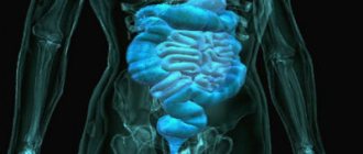
Modern computed tomography allows using a 3D model to display the loops of the large intestine.
Compared to endoscopic colonoscopy, this procedure is painless and is performed without anesthesia. CT scan of the intestine requires moderate distention of the intestinal loops, so the patient may experience minor discomfort and the urge to defecate. Virtual colonoscopy is indispensable for Crohn's disease, a disease in which there is a high probability of damage to the intestinal wall by the endoscope. The main advantages of the virtual colonoscopy technique are speed and accessibility, detailed 3D visualization of the entire large intestine with high-quality display of its loops and other organs of the abdominal cavity and retroperitoneal space (liver and bile ducts, kidneys and adrenal glands, pancreas, spleen), vessels and bones on level of research, layer-by-layer viewing of structures of interest in all planes.
Indications for virtual colonoscopy
- Gastrointestinal bleeding;
- colon polyps;
- colon cancer;
- suspicion of nonspecific ulcerative colitis, Crohn's disease;
- abnormalities in the development of colon loops;
- injuries;
- unsatisfactory or questionable data from other diagnostic methods;
- planning surgery on the large intestine;
- monitoring the effectiveness of treatment.
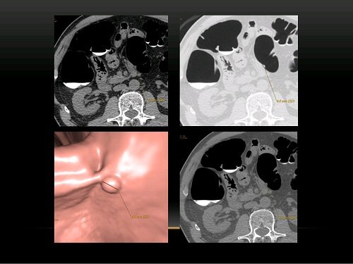
Broad-based intestinal polyp identified on virtual (CT) colonoscopy. The study was performed by M.A. Petrova, a radiation diagnostics doctor at the KGNY.
What is CT colonoscopy?
Computed tomography (CT) is a non-invasive test that allows doctors to look “inside” a patient’s body.
A tomograph (a special X-ray machine) receives a large number of images (so-called slices), which are processed by a computer and displayed on a monitor screen or printer.
Computed tomography provides high-quality images of many internal organs, bones and blood vessels.
Virtual colonoscopy is a special type of computed tomography that examines the large intestine (colon - in Latin). Conventional colonoscopy (not virtual) is an endoscopic diagnostic method in which the endoscope is inserted directly into the colon.
Preparing for the study
Carrying out a virtual colonoscopy requires careful preparation; it is best to do it in the morning. On the eve of the study, the last meal is allowed at 14:00; starting from 16:00, take the laxative Fortrans or Lavacol (each sachet for 1 liter of water, for body weight > 70 kg, take 3 sachets, for
Before the study, you must follow a slag-free diet for 3-4 days: exclude legumes, brown bread, chocolate, fresh milk, fresh and pickled vegetables and fruits.
During the period of preparation for the study, take medications prescribed by your attending physician according to the usual (prescribed) regimen.
To improve the quality of the conduct and description of the study, you should provide the results of ultrasound, CT or MRI, which the patient underwent previously, as well as, if available, other medical documents and extracts.
If it is necessary to administer a contrast agent, blood should be donated for biochemical analysis to determine the level of urea and creatinine. This can be done immediately before the test in our hospital ( express tests ) or you can bring the results of a biochemical blood test no more than 2 weeks old.
To obtain the most accurate result, before the procedure it is necessary to remove jewelry, clothing with metal fittings and/or thread, belts and other items containing metal from the abdominal area.
Bowel preparation schedule
| Step | Appointed time | lead time | Medicine(s) to take |
| 1 | Until 11:00 |
| |
| 2 | AT 16:00 |
| |
| 3 | AT 18:00 |
| |
| 4 | AT 20:00 |
|
to come back to the beginning
Alternative methods for diagnosing the large intestine
- Plain radiography of the abdominal organs - determination of colonic obstruction.
- X-ray irrigoscopy - diagnosis of tumors and inflammation, polyps, abnormalities of the large intestine. During the procedure, a mbarium-containing substance is used.
- Ultrasound of the abdominal cavity and pelvis are common screening methods. CT is more informative.
- An MRI of the abdominal organs is performed to clarify the diagnosis and requires special bowel preparation.
- Traditional endoscopic colonoscopy also examines loops of the colon, but requires anesthesia and risks damaging the intestinal wall. Unlike virtual colonoscopy, it allows you to evaluate the appearance of its mucous membrane and take tissue for morphological examination under a microscope.
You can see prices for services
Preparing for a CT colonoscopy
| Diet for 3 days | Exclude:
| You can eat:
|
| Diet the day before | Exclude:
| You can drink:
|
| Keep in mind |
| |
Schemes of drug preparation for CT colonoscopy/FCS
| Dosage | Does not depend on mass:
| |
| Breeding | Each pair of sachets (sachet A+B) is diluted in one liter of still water | |
| Chair markings | Important! Add 20 ml of any of the following drugs to the last liter of solution: Urografin, Trazograph, Omnipaque, Ultravist, Xenetics or Optirey (sold in the pharmacy chain) | |
| Reception scheme |
| |
| Time of receipt | On the eve of the study from 16.00 to 20.00 - take Moviprep solution, liquid food is allowed | |
| Effects | Painless, loose stools appear 1-2 hours after the start of treatment. The stool will turn into a clear or slightly colored liquid 2-3 hours after taking the last portion. | |
Computed tomography of the colon - virtual colonoscopy from the patient's point of view
The patient lies down on a diagnostic couch, which slides inside the “donut” of the tomograph.
A small tube is inserted into the patient's rectum to a depth of about 5 cm. A small amount of air is supplied through this tube into the rectum - this is necessary to improve the quality of the image and straighten the intestine.
After this, the couch is moved into the scanner. The patient is asked to hold his breath from time to time. Usually, the patient first lies on his stomach, and then he is asked to roll over onto his back, sometimes vice versa.
The whole procedure takes about 15 minutes.
Virtual colonoscopy does not cause pain; there may be slight discomfort from distending the rectum with air.
As a rule, description of images takes about 30 minutes.
MR imaging of the intestine for diagnosing Crohn's disease
Crohn's disease is a fairly common, severe disease that damages the small intestine. It is extremely difficult to reach it with an endoscope without worsening the patient’s condition. Ultrasound also does not provide an understanding of the complete clinical picture. Therefore, MRI is more often used for diagnosis. The sensitivity of this method is more than 95%, the procedure is well tolerated by patients and does not cause complications.
In case of Crohn's disease, it is necessary to conduct regular examinations to monitor the patient's current condition and adjust the treatment regimen. The most effective and safest method is MRI, which allows you to obtain detailed visualization of the entire intestine, detect the inflammatory process, thickening or dissection of the walls, abscesses and fistulas
As we said earlier, colonoscopy is strictly contraindicated for many patients with this disease, and CT gives a high dose of radiation and cannot be performed frequently.
Advantages and disadvantages of the method
Compared to standard video colonoscopy, virtual colonoscopy has a number of advantages and disadvantages. The advantages of this method include:
- less traumatic, lower risk of complications compared to endoscopic examination;
- no need to use sedatives or anesthetics - after the study, the patient can return to normal life;
- the ability to examine all parts of the intestine even in the presence of obstruction.
In addition to all of the above, the method of introducing air or a contrast agent into the intestines is also considered an advantage. This is done using a very thin and short tube that is inserted into the rectum and not into other parts of the intestine.
Among the disadvantages of virtual colonoscopy are:
- difficulties in diagnosing polyps less than 10 mm in diameter;
- inability to perform tissue biopsy during the study.
- the need for endoscopic colonoscopy after detection of tumors in the intestine by this method for the purpose of biopsy from a tumor or polyp and subsequent histological examination;
In addition, virtual colonoscopy is one of the less common procedures, so it is not available in all clinics.
MSCT of the intestine
Many thanks to doctor Artyom Valerievich Gorinov.
On June 14, he did an MSCT scan of my abdominal cavity. I was in a difficult situation after previous studies - I had to not just do the next one, but decide which one (or series) was best to do in my situation to clarify the diagnosis. The attending physicians did not have a clear opinion. I went to different Moscow clinics directly to CT and MRI doctors to understand where to do expensive tests and who to trust.
In Medsi on Belorusskaya, Roman Valerievich took a break from his work and talked to me, listening to my doubts and looking at my medical records. He spoke in detail about the research - he described the procedure itself, what exactly the images will show, and what we will not see or only indirectly. What contrast is introduced during the procedure. Then he opened my card (all this was not at the reception - I asked the administrator to connect me with the MRI doctor and consult), explained that I needed additional consultation with an endocrinologist regarding the iodine administered during the MSCT procedure. And if I decide to do it with them, then I will definitely need to take kidney function tests. I named them. Gave me a reminder on how to prepare for the study. At this stage I decided that I would make an appointment with this doctor. We signed up in a week. During the week, I heard positive reviews about this professional from doctors at appointments twice more. I also came across people who took pictures and a difficult procedure for the patient (preparation and process) called Virtual Colonoscopy with this doctor. They said that the doctor acts very clearly and delicately. He does everything himself, even what assistants usually do during the procedure. So on June 14 everything went smoothly. The doctor again paid attention and answered all questions before starting.
He clearly explained how best to behave afterwards in order to quickly remove the contrast from the body and recover. After receiving the results, I received detailed explanations from him. Although, as I understand it, this is no longer his responsibility, but that of the attending physicians. But we still need to get to them, and here I, as a patient, had the opportunity to get answers to the most important things. I also thank the entire MRI department shift that day for their participation. The administrator, the nurse, the laboratory assistant who helped the doctor and me as a patient during the procedure. Thanks to the professionalism and human qualities of Roman Valerievich. It would be great if the clinic’s management has an incentive system specifically for such specialists.
Best regards, Angela Moore. I have been buying a MEDSI insurance policy for about 10 years.
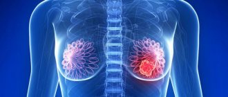
MRI for diagnosing rectal cancer
Colorectal cancer is one of the most serious problems of modern oncology. Patients usually “grow” their tumors for years without even knowing they have the disease. Therefore, it is important to screen for colorectal cancer.
MRI combines the advantages of other non-invasive methods (CT and ultrasound), but does not have their disadvantages (does not produce radiation and does not cause the risk of tissue damage). The study allows you to see adjacent lymph nodes, which often affect metastases, and evaluate the physiological parameters of the tumor (thanks to visualization of blood flow during dynamic MRI with contrast).
The method makes it possible to detect cancer at an early stage, assess the extent of the tumor, choose a treatment method and monitor the process.