Goals
During the procedure, the doctor may find in the patient:
- Polyps;
- Ulcers;
- Inflammation;
- Tumors.
In addition, by resorting to manipulation, the doctor can take a biopsy sample for examination, as well as perform therapeutic actions.
There is a recommendation that a colonoscopy should be performed at age 50. Everything else is individual.
Medical practice shows that most colon tumors are detected at mature stages, since diagnosis is carried out too late.
Early diagnosis allows you to identify precancerous diseases, which means you can start treatment on time. This is where colonoscopy comes to the rescue.
Is it possible to do a colonoscopy during pregnancy - indications and features
Basic "colonoscopy" Can a woman have a colonoscopy during pregnancy - and in what cases?
April 27, 2021 Colonoscopy
Pregnancy is a state of the body in which taking certain medications and performing certain medical procedures is restricted or prohibited. Let's find out whether it is possible to perform a colonoscopy during pregnancy - i.e. examination of the intestines using an endoscope with an optical device inserted into the body through the anus.

Negative properties of the procedure
As a rule, colonoscopy during pregnancy is performed only in the early stages and in emergency cases due to the danger to the life of the mother, if such an examination cannot find an equivalent informative value and a safer alternative for both the mother and the fetus. There are several reasons why it is not advisable to have a colonoscopy:
- Negative effects of painkillers on the fetus. In this context, colonoscopy under general anesthesia is completely prohibited for pregnant women; pain relief is carried out only on site. This restriction applies not only in the very early stages of pregnancy (up to 3 weeks), when the embryo is still on its way to the uterus and is not attached to it, i.e. practically does not interact with the mother’s body.
- Long lying position on your back during the procedure and tension in the abdominal muscles. This can lead to: increased uterine tone (tension of the muscles of the uterus), which can lead to embryo rejection, early miscarriage or premature birth at a later stage; dilatation of the cervix; disturbances in the blood supply to the fetus and, as a consequence, lack of oxygen due to the narrowed inferior vena cava supplying the uterus - hypoxia is associated with miscarriage and various pathologies of fetal development, including growth arrest.
In women already breastfeeding, it is recommended, depending on the medications used during the examination, to interrupt feeding for several hours to 24 hours and express the first portion of milk.
Use during menstruation
Another female-specific period—menstruation—colonoscopy should also be postponed if possible.
The reason for this is physical inconvenience during the procedure, as well as a possible increase in menstrual bleeding due to preparatory measures (especially enemas), increased intestinal motility during the examination itself, as well as medications used during the examination. However, all these restrictions are not as serious as during pregnancy, so a colonoscopy is still performed if necessary.
Indications
The procedure is performed for the following tasks:
- dysphagia or loneliness;
- prolonged gastrointestinal bleeding;
- severe diarrhea of unknown cause;
- strong suspicion of a colon tumor or adhesion process;
- cholecystopancreatitis, cholangitis or choledocholithiasis;
- excruciating abdominal pain with nausea and vomiting and other severe symptoms.

Alternatives to Colonoscopy
Unfortunately, colonoscopy is the most effective method for examining internal organs in a number of cases - and doing something equivalent will be problematic.
It combines the possibility of physical presence, for example, to take the necessary sample, with the quality and objectivity of examination of internal organs.
Therefore, it will be problematic to find a complete replacement for this procedure.
However, there are additional laboratory tests that can at least partially replace this procedure. These include x-rays, tomography (MRI) and ultrasound. They are non-invasive, i.e. do not require direct intervention in the woman’s body, i.e. cannot cause particularly serious damage.
If only basic information needs to be collected, then they are used, although, for example, CT examination during pregnancy is also strongly discouraged, although it is the most appropriate examination, for example in the case of intestinal adhesions. But in some situations, unfortunately, it is difficult to find a complete replacement.

Let's sum it up
In principle, colonoscopy is one of the most common and effective methods for examining internal organs. But is it even possible to have a colonoscopy during pregnancy? This is very undesirable, in many cases this possibility is extremely undesirable.
You may be interested in: Placental polyp after a medical abortion: first symptoms and treatment without surgery
But if necessary, if there are no procedures that could completely replace this type of examination, then, at the discretion of the doctor, colonoscopy is also acceptable.
The doctor will weigh the risks, assess whether it is more dangerous to conduct the study or not to conduct it, and decide what to do in a particular case.
Preparation
Preparation usually takes two to three days. The doctor gives recommendations to the patient, including following a diet, taking laxatives, and minimally eating the day before.
Diet plays an important role during the preparation period.
The patient needs to switch to a low-fiber diet, reduce or abandon fresh vegetables and fruits to a minimum, and forget about dried fruits, nuts, and whole grains.
A couple of days before the procedure, you need to give up solid food. There are broths, clear fruit juices, tea and coffee, jelly. The day before you need to drink more fluid.
Four hours before the procedure, you need to completely abstain from food and water, since the procedure is done on an empty stomach.
Plan of tests and examinations during pregnancy by week
Pregnancy check-ups per week is a schedule developed by doctors for conducting tests and visiting close specialists. It is based on the recommendations of the Ministry of Health and Social Development of the Russian Federation and is aimed at maintaining health and well-being during pregnancy.
It is important that the expectant mother visits a doctor, follows the doctor's recommendations and undergoes routine examinations. A gynecologist monitors the progress of pregnancy, the risk of possible complications, the development of the fetus and the condition of the pregnant woman herself.
studies and a series of tests help to identify possible problems in a timely manner and provide the necessary assistance.
In a normal pregnancy, examination of pregnant women in the first trimester includes visiting a gynecologist once a month in the first trimester, in the second trimester - from 36 weeks onwards once every 2-3 weeks, and before giving birth, the gynecologist examines the pregnant woman every week.

Examination plan for a pregnant woman in the antenatal clinic
To receive legal assistance in overcoming pregnancy, you must register with the Prenatal Clinic. It is better to do this at 6-8 weeks of pregnancy (be sure to take your passport and insurance policy with you).
Examinations in the first trimester (from 1 to 13 weeks)
In the first trimester, the gynecologist will refer the pregnant woman to a highly specialized doctor for a mandatory examination:
- ENT - examines for chronic or infectious diseases of the respiratory and hearing organs, smears of the mucous membrane;
- Dentist - examines the oral cavity, teeth;
- Ophthalmologist - gives recommendations on the method of delivery (in the case of high intraocular pressure, a cesarean section may be recommended);
- Cardiologist - examines the heart;
- radiologist - all family members with whom the pregnant woman lives must do fluorography and transfer this data to the therapist; The pregnant woman herself is prohibited from having x-rays or fluorography;
- therapist - gives an opinion on the results of the examination.
The gynecologist measures blood pressure, height, weight and pelvic size and enters this data into the pregnant woman’s medical record. Gives recommendations on taking vitamins, proper nutrition, daily routine and prescribes directions for testing:
- general urine analysis. The analysis allows you to quickly assess the general health of a pregnant woman and how her kidneys are functioning. For analysis, morning urine is collected (at night the kidneys work more actively, the urine becomes more concentrated, so this diagnosis is more accurate);
- smear through the flora. A smear on the flora reveals possible infections and inflammations. In case of anomalies, the gynecologist prescribes additional examinations. A test for sexually transmitted diseases (STDs) is often ordered if the smear test is poor. Some infections are dangerous for the normal development of the fetus. In this case, they are treated taking into account the duration of pregnancy and the quality of the drug (the choice of drug is not general, but local);
- general blood analysis. A blood test during pregnancy is carried out at registration and repeated in each trimester. Thus, the doctor has the opportunity to monitor the patient’s condition. Based on the results of a clinical blood test during pregnancy, determine the number of leukocytes, platelets, hemoglobin, assessed by SOE and other indicators; blood chemistry. The analysis helps to identify failure of important organs: the liver and pancreas. High concentrations of creatinine and urea are found in renal failure. High bilirubin levels associated with liver problems. The functioning of the pancreas is monitored by glucose levels so as not to miss the possible development of gestational diabetes;
- blood clotting test. If there are no deviations, the test is carried out once per trimester;
- blood type and Rh factor. Even if you know your blood type, you should still do this test. In the event of possible major blood loss or unplanned surgery, this information can save the life of mother and child. If a woman has a negative Rh factor, and the father of the child is positive, then a Rh conflict may occur. This means that the mother's body perceives the baby as a foreign body and produces antibodies to eliminate it. This reaction can lead to anemia, miscarriage, or death of the fetus in the womb. If the mother's Rh factor is negative, then the blood is donated by the future father. If the Rh factor is positive, a pregnant woman undergoes an antibody test once a month until the 32nd week of pregnancy. After the 32nd week of pregnancy and twice a month until the end of pregnancy. If this is your first pregnancy and there are no antibodies before the 28th week of pregnancy, doctors suggest administering a serum that subsequently blocks the production of antibodies;
- tests for HIV, syphilis, hepatitis. These tests are carried out at the beginning of pregnancy and at 30-35 weeks (due to the long incubation period of diseases);
- TORCH infection. The TORCH test shows the expectant mother’s immunity to toxoplasma, rubella, cytomegalovirus, herpes and some other infections. They are very dangerous for the intrauterine development of a child. If antibodies are not detected, the pregnant woman should take all preventive measures to avoid getting sick while carrying a child;
- double test. Includes ultrasound and blood tests for levels of human chorionic gonadotropin (hCG) and plasma produced protein (PARP-A). These tests show whether a child is at risk of developing a chromosomal abnormality (Down syndrome).
Examinations in the second trimester (from 14 to 27 weeks)
In the second trimester, the gynecologist begins to measure and record the height of the uterine fundus and the volume of the abdominal cavity. A pregnant woman visits a gynecologist more often: once every two to three weeks. If the condition of the expectant mother is normal, the doctor prescribes what examinations the pregnant woman should undergo:
- at 18-21 weeks. At 18-21 weeks, she undergoes a second screening, the “triple test”, with the help of which doctors more accurately determine the risk of possible pathology of the child;
- at 18-21 weeks they do a second routine ultrasound, assess the condition of the placenta and amniotic fluid, observe how the baby develops, by this time you can already know the gender;
- do a general urine test;
- They do a second examination for syphilis.
The doctor may order additional repeat blood, urine, and other tests if necessary to What tests are given to a pregnant woman in the third trimester?
- In the period from 30 to 34 weeks, do a third ultrasound examination, determine the size and approximate weight of the baby, observe its position in the uterus, examine the condition of the placenta, whether there is an umbilical cord, and monitor the quantity and quality of amniotic fluid.
- Within 32-35 weeks, do a CTG (cardiotocography), study the functioning of the embryo’s cardiovascular system, and observe its motor activity. This test helps determine how the child is feeling and whether he or she has developmental problems.
- From 36 weeks until delivery, a gynecologist examines the pregnant woman every week.
- At week 30, the doctor gives a referral for a general urine test, a general blood test, a biochemical blood test, an HIV and hepatitis test, a third test for syphilis and a smear for cytological examination.
What is an exchange card
At the end of the second trimester, between the 22nd and 23rd weeks of pregnancy, the gynecologist gives the expectant mother a smear.
You might be interested in: Wen behind the ear: what it looks like, causes and treatment
This is a medical document in which the doctor records the course of pregnancy and the woman’s health status.
A pregnant woman should carry it with her passport and OMS guidelines if she is in urgent need of medical attention. This becomes especially important in the last trimester.
The exchange card must consist of three mandatory parts:
- The first part contains information entered by the gynecologist at the prenatal clinic. Here you can find out the personal data of a pregnant woman, information about chronic and past diseases, previous pregnancies and births, all measurement data during a medical examination, copies of ultrasound, CTG, examination results, recommendations after examination by other specialists.
- The second part contains information entered by the maternity hospital doctor. Before discharge, it is written down how the birth went, the prenatal period, if there were complications, and if necessary, recommendations for treatment are written down. This information is necessary for the gynecologist at the maternity hospital when a woman comes for her first examination after childbirth.
- The third part contains information about the born child. Enter height, weight, degrees on the Apgar scale. This information is important for the pediatrician, who examines the baby and copies all the data about the child, starting with the exchange card and ending with the medical record of the little patient.
Brief table of examinations during pregnancy by timing:
Duration of pregnancy (weeks) Type of examination and tests15
| 5-7 | First visit to the gynecologist, confirmation of pregnancy. |
| 7-11 | Visit to the gynecologist, registration at the prenatal clinic. Measuring pressure, pulse, temperature, pelvic size, weight and height of a pregnant woman. What tests need to be done: - Coagulogram; - general urine analysis; - general blood analysis; — chemical blood test; — blood test for TORCH infection; — blood group analysis and Rh factor analysis; - blood test for HIV, hepatitis B and C, syphilis; - cytological smear. |
| 2 weeks after registration | Visiting a doctor: - dentist; - ophthalmologist; - ENT specialist; - cardiologist; - therapist. |
| 11-14 | First scheduled ultrasound, First examination (“double test”) |
| 16 | Examination by a gynecologist |
| 16-18 (allowed up to 20 weeks) | Second routine Ultrasound examination and second screening (“triple test”) |
| 20 | — gynecological examination; - general urine analysis; - second study of syphilis. |
| 22 | Gynecologist |
| 24 | — Gynecological examination; — Examination of the legs for varicose veins; - Analysis of urine. |
| 26 | Gynecologist |
| 28 | - Gynecologist; - Examination of legs for varicose veins; - Urine test. |
| 30 | - gynecologist; - Analysis of urine; - blood analysis; - blood analysis; - third test for syphilis; - HIV test; - flower tampon. Third planned ultrasound |
| 33-34 | Fetal cardiography (FCC) |
| 35 | — Gynecologist — examination for varicose veins — urine test — fetal CTG. |
| 37 (once a week until delivery) | Gynecologist |
- 22330
Methodology
Diagnostic manipulation usually takes place faster than therapeutic procedure.
Sedation is used for patient comfort.
He is laid on his left side, his legs are bent, his knees are brought to his stomach.
The colonoscope is lubricated with Vaseline and carefully inserted through the anus, moving slowly.
To ensure a good view, the intestines are inflated with gas.
The image is shown on the monitor screen, the process itself is recorded.
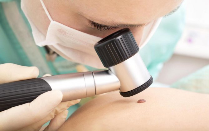
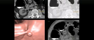
Dermatoscopy
After examining the intestines, the colonoscope is removed.
Recovery
Usually the procedure is performed in an outpatient setting. Therefore, the patient can leave the clinic as soon as the anesthesia wears off.
There are certain recommendations regarding recovery and more:
- You cannot drive or drink alcohol for 24 hours;
- Immediately after the procedure, it is recommended to walk around so that the gas leaves the intestines faster;
- It is recommended to rest during the day;
The doctor gives more specific recommendations, taking into account your condition and the nature of the procedure.
In case of fever, bleeding, or abdominal pain, you should consult a doctor.
Popular publications:
- Black feces Why? Causes? — 01/07/2015 05:06 — Read 45157 times
- Video capsule endoscopy: frequently asked questions - 11/12/2013 17:02 - Read 44864 times
- Capsule endoscopy. Description, reviews, advantages and disadvantages - 03/24/2013 17:15 - Read 41818 times
- Abdominal syndrome. Abdominal pain - 01/10/2015 05:02 - Read 23458 times
- Capsule colonoscopy - indications, contraindications, price - 10/30/2015 07:20 - Read 18897 times
Key words: Colonoscopy, intestinal colonoscopy, colorectal cancer, colon cancer, polyps, colonoscopy indications, diet during colonoscopy
- < Back
- Forward >