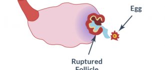
The fertilized egg is the embryo along with the embryonic membranes. The period of formation of the fertilized egg is the first stage in the development of pregnancy.
After the fusion of germ cells, the egg is at the stage of active division. First it is divided in half, then into four and so on. The number of cells and the size of the embryo gradually increases. It moves along the fallopian tube into the uterus to attach there. Implantation occurs approximately seven days after fertilization. Until this time, the embryo receives nutrients from the egg, and after attachment - from the mucous membrane of the uterus. This feeding continues until the placenta is formed.
The placenta is formed from the outer layers of the fertilized egg, densely covered with villi. They are the ones who remove the mucous uterine layer at the implantation site and are introduced into the prepared area.
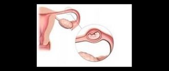
The presence of a fertilized egg is the first sign of a normally started pregnancy. It can be seen on an ultrasound monitor 14 days after the delay, and the embryo can be seen only after five weeks.
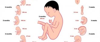
The size of the fertilized egg according to weeks
If the fertilized egg is only four millimeters in size, this indicates a very early stage of pregnancy - up to six weeks. Most often, this size of the fertilized egg indicates a period of four weeks. At five weeks it is equal to six millimeters, and at five weeks and three days it is seven.
We can say with certainty that the fertilized egg grows by one millimeter every day for up to 15-17 weeks. Then it increases by two and a half millimeters daily.
It happens that an ultrasound of a pregnant woman gives an inaccurate gestational age. This often happens, since in the first weeks it is difficult to establish a specific date based on the size of the fertilized egg. In the future, it is recommended to repeat the ultrasound examination, starting from the sixth or eighth week of pregnancy.
Deformations of the fertilized egg
A small fertilized egg in the early stages is not a reason to worry, since usually everything returns to normal after some time. It is important to initially monitor the process using ultrasound. It’s another matter when the fertilized egg has irregular outlines.
This phenomenon is a pathology and most often indicates abnormal development of the fetus. Normally, the egg should have an oval or round shape. If it is flattened on the sides, then this is the first sign of uterine hypertonicity. The patient needs to be under the supervision of a doctor for some time. If no dangerous symptoms develop in the future, then the threat is eliminated.
If a pregnant woman experiences pain or vaginal discharge appears, immediate action must be taken. Bed rest and absolute rest are prescribed. Medications to reduce uterine tone and hormonal medications are required. It is possible that the woman will have to spend the entire remaining period before giving birth in the hospital.
Abnormal development of the fertilized egg
In addition to deformation of the fertilized egg, the development of any anomalies is possible. Many of them can cause miscarriage or fetal death. The most common anomalies are presented below.
1. If there are two embryos in the fertilized egg, then this is not an anomaly, but rather the norm for the development of twins. The sizes of embryos may differ from each other. A difference of several millimeters is most often characteristic of monochorionic twins. However, at an early stage it is impossible to accurately determine whether the children will be twins or twins.
2. Small fertilized egg. This pathology is diagnosed by comparing the gestational age and the size of the ovum. Such studies help determine the correct development of the fetus in the uterus. The gestation period is also determined by the size of the fertilized egg. It is necessary to carefully monitor the development of the fertilized egg; too slow growth may indicate fetal fading. In this case, it is advisable to prescribe the patient a blood test for hormone levels.
3. If the fertilized egg is larger than the embryo, then the pregnancy may be frozen. Pregnancy can also be anembryonic. All signs of pregnancy are characteristic, the membranes of the fetus are formed, but the embryo is missing. Anomalies can only be diagnosed by ultrasound examination of the fetal egg no later than 6-7 weeks after conception. In this case, it is very important to establish the exact duration of pregnancy.
4. Elongated shape of the fertilized egg. Normally, the fertilized egg should be round in shape. Elongated contours may signal the possible loss of a child. The reason most often lies in uterine hypertonicity. It is important to diagnose the deformity in time and take measures to reduce tone with the help of antispasmodics. The fetus can be saved if it continues to develop and a heartbeat can be heard. The patient needs to stop sexual activity, minimize physical activity and avoid emotional turmoil. If this pathology is present, complete bed rest is prescribed. Ultrasounds are performed every few days to monitor changes in the gestational sac.
5. Abnormal location of the fertilized egg. Usually the egg is implanted at the bottom or center of the uterus, but sometimes it occurs in the area of the internal os.
6. Death of the fetus inside the womb. Diagnosed by listening to the heartbeat using ultrasound. The cause may be inflammatory processes in the mother's body, the presence of infections, late toxicosis, placenta previa or abruption, diabetes mellitus, pyelonephritis, pneumonia, influenza, polyhydramnios or oligohydramnios, intoxication with harmful substances.
Such diagnoses are usually made in the very early stages of pregnancy, which makes it possible to control the process. Thus, there are many chances to maintain the pregnancy and give birth to a child.
Detachment of the ovum
Detachment of the ovum is the early rejection of the egg from the wall of the uterus. If spontaneous abortion is diagnosed in time, there is a good chance of maintaining the pregnancy.
Detachment of the ovum manifests itself in aching pain in the lower abdomen and lower back, bleeding or dark red discharge.
Causes of pathology:
- disturbances in the functioning of the ovaries;
- inflammatory processes;
- exacerbation of infectious processes;
- presence of tumors;
- underdevelopment of the genital organs;
- strong physical activity;
- severe toxicosis;
- stress.
If you notice the first signs of detachment, you should immediately call emergency help. Before the specialists arrive, the woman needs to lie on her back and raise her legs up, placing them on some kind of support (on the wall, the back of the sofa).
Signs of an incipient miscarriage
In the initial stage of spontaneous abortion, aching, sometimes cramping pain in the lower abdomen or lumbar region is observed. When the placenta separates from the wall of the uterus, bloody discharge appears from the genital tract. As the detachment process progresses, the bleeding intensifies. Often, heavy bleeding may occur, leading to severe anemia. Together with the blood, the fertilized egg comes out of the uterus. After this, the uterus begins to contract and the bleeding stops. If parts of the membranes and placenta remain in the uterus, it will not contract and bleeding will continue. Heavy uterine discharge can become life-threatening. The cervix remains slightly open, which favors the development of inflammatory diseases due to the entry of pathogenic microorganisms there.
Sometimes the elements of the fertilized egg retained in the uterus are very small, and bleeding may stop, but subsequently polyps are formed from these elements, preventing the healing of the surface of the uterus. They can lead to prolonged bleeding from the genital tract.
Determining whether the fetus is developing in the womb or not can be determined by determining the level of human chorionic gonadotropin (hCG) over time. An increase in the amount of this hormone in two days indicates a normal pregnancy. If the level of human chorionic gonadotropin remains the same or decreases, it means that the development of the fetus has stopped.
Absence of embryo in fertilized egg
In the earliest stages of pregnancy, the embryo in the fertilized egg is not yet visible, and this is the norm. However, starting from the fifth week, it should already be noticeable on the device monitor. If it is not possible to visualize the embryo, then a repeat ultrasound examination is scheduled after two weeks.
If upon repeated examination of the embryo and no heartbeat is observed, then an anembryonic pregnancy is diagnosed. In this case, curettage is performed.
It is important to know that despite the absence of an embryo, pregnancy is confirmed during testing. Pregnancy tests also show a positive result. The fact is that in the female body all the processes corresponding to bearing a child have already been launched and certain hormones are being produced.
It is impossible to name the exact causes of this pathology. Most often, its occurrence is associated with genetic factors. In addition, anembryonia can be caused by the use of certain medications that are prohibited for use during pregnancy.
Anembryony does not indicate the impossibility of successfully conceiving a child in the future. It is quite possible that the next pregnancy will be favorable. However, planning should begin no earlier than six months later.
It is important for the patient to provide not only physical rehabilitation, but also psychological one. The loss of an unformed child is a difficult ordeal.
large fertilized egg and small embryo
2012, after two ZBs, my husband and I were examined for infections and so on, the doctor allowed me to get pregnant after a course of COCs. And here it is, the long-awaited month and the long-awaited 2 stripes, everything worked out the first time, at the very beginning it was a little smeared, I was in storage, and at 16 weeks bleeding began, no matter how hard I tried, I couldn’t control it, but it flowed like a river. They stopped the bleeding, sent me for an ultrasound, but it was beating! My little heart, and the baby is fine. After this incident, my pregnancy was perfect! I led a very active lifestyle and at 41 weeks I gave birth to my beautiful daughter, 4 kg without any tearing. I fed her until she was 1.5 years old. They really wanted the second one, but for now the helmsmen decided to take precautions. In 2015 we started planning, passed all the main tests, everything was normal. We went to the sea, just to be sure) and move on. It turned out in 2021. I find out about pregnancy very early, a week before my period, because my breasts are filled and hurt not as usual. Also a small daub at the beginning, preservation and ultrasound at 9 weeks. Spontaneous abortion. Urgent anesthesia, curettage, antibiotics. Doctors shrug their shoulders. I myself find information about the need to consult a hematologist, go, get tested for mutations, and they find some defects. I take vitamins, my husband and I take all the tests again and after 6 months we try again, but pregnancy occurs only in 2021. This time everything is different, I feel good, there is no spotting, I am in touch with the hematologist, my blood tests are perfect, I don’t need Clexane, I ask you to at least prescribe Actovegin into a vein. I lay in storage while they injected Actovegin, did physio, and I was sure that everything would work out. At 7 weeks, ultrasound, heart beating, everything is fine, discharged. At 10 weeks there is a strange spotting, I go for an ultrasound scan at 8 weeks. Vacuum aspiration, I take a karyotype test, the result is monosomy. After half a year, more examinations, hysteria, hormones and now in 2021 2 stripes! There are no limits to happiness! But at 6 weeks of blood, I run to an ultrasound, where, as the doctor said, “porridge”, he puts on a hematometer and assumes that the embryo has come out. Of course, I don’t donate anything, no blood tests, but a day later I understand that my periods have stopped. I run to take hCG, and then the result is 7 weeks old, I sign up for an ultrasound, I go to the same doctor, and she says with such confidence, hCG is not an indicator, it can grow even after a miscarriage. But I insist on an ultrasound, she inserts the sensor and her eyes widen, when asked what’s wrong, she says everything like that, I just can’t believe it and turns the monitor to show how the heart is beating. Miracle! And here I am sure that the baby has come to live and everything will definitely be fine.
At 10 weeks my morning sickness disappears and I go to the doctor. Upon examination, the uterus corresponds to the term, we go for an ultrasound, but there is no embryo in the fertilized egg. Why? For what?
They give me a medical interruption, give me antibiotics, and discharge her after a week. After another two, I go for an ultrasound, they see the remains of the fertilized egg and the accumulation of blood in the tube, as the pharynx is closed, the doctor says take antibiotics and see you in a week. But I understand that this is not a joke and in a week it may be too late, but I still want children. I go to another doctor, she admits me to the hospital. Anesthesia, hystera, antibiotics, the remainder was very large and adhered very strongly to the uterus, they could barely be removed. The next day we do an ultrasound, preparing for discharge, and there is another placental polyp. God! Again anesthesia, hystera, everything was removed. The anesthesia was severe, I couldn’t take the standard dose anymore, they barely put me to sleep.
Here's the story. Despite all these trials, I want to get pregnant again, to bear and give birth to a healthy baby, to kiss, hug, give him my love, breastfeed and sing lullabies.
Don't give up! Everything will be fine!