Requirements for the placement of an X-ray room
According to the technical specifications of a medical organization, design documentation for an X-ray room is developed by a person who has the appropriate license. Acceptance of the office for operation is carried out by a commission consisting of representatives:
- medical organization;
- construction organization;
- the region’s X-ray and radiological department (RRO), the functions of which, in Moscow, for example, are carried out by the City X-ray and radiological department on the basis of the State Budgetary Institution “Scientific and Practical Clinical Center for Diagnostics and Telemedicine Technologies of the Department of Health”;
- sanitary-epidemiological service.
Documentation required for acceptance of the cabinet into operation:
- license for medical activities;
- sanitary and epidemiological conclusions:
- for an X-ray machine (having a registration certificate from the Russian Ministry of Health and operational documentation);
- for an x-ray room project (having a technological design agreed with the RRO, a technical passport);
- for hidden work;
- ventilation efficiency checks;
- testing of the protective grounding device indicating the current spreading resistance of the main grounding conductors;
- checking the condition of the grounding network of medical equipment and electrical installations;
- dosimetric measurements;
- monitoring the operational parameters of the device;
- testing of individual and mobile radiation protection equipment;
- dosimetric measurements for planning radiotherapy;
- measuring the insulation resistance of wires and cables.
- control and technical equipment for the X-ray machine;
- registration of workplace training;
- on the classification of working persons as personnel of groups A and B;
- on the appointment of persons responsible for radiation safety, accounting and storage of X-ray machines, industrial radiation control;
- on training of personnel on radiation safety;
- confirming the accounting of individual radiation doses:
- patients (sheet for recording patient dose loads during X-ray examinations);
- personnel (card for recording individual radiation doses of personnel);
Based on the results of acceptance into operation, a sanitary and epidemiological conclusion is issued for the right to operate the X-ray room. The selection of premises for the X-ray room (department) is carried out taking into account a number of requirements.
| X-ray room placement options | |
| valid: | invalid: |
|
|
The design of the X-ray room determines the conditions of its operation and the composition of the equipment and studies performed, and changing these parameters requires obtaining a new sanitary and epidemiological conclusion.
The composition and area of premises for X-ray examinations are regulated by SanPiN depending on the type of X-ray machine (composition of a set of stands, etc.), the presence or absence of digital image processing capabilities, diagnostic methods (tomography, mammography) and diagnosed diseases (heart and blood vessels, gastrointestinal tract). -intestinal tract, lungs), as well as the need to import a gurney. For example, a fluorography room for mass examinations consists of a treatment room (at least 14 m2), a dressing room, a waiting room (6 m2 each), a staff room (9 m2), and a darkroom (if the device is not digital).
As a rule, the ceiling height in the treatment room should be at least 3 meters, the doorway should be 1.2 meters wide and 2.0 meters high. An automatically illuminated “Do Not Enter” light sign is placed above the door. The cabinet floors are made of electrical insulating materials, waterproof, antistatic and spark-free. Other surfaces, like the floor, must be washable and disinfectable, but also be matte and smooth. Finishing materials must have a sanitary and epidemiological certificate.
The X-ray machine is placed so that the primary radiation beam is directed towards the main wall. Direct sunlight into the X-ray room is undesirable, so it is preferable to orient the windows to the north-west, and in the darkroom they should be protected by light-proof curtains. In addition, protective shielding is also provided for offices on the lower floors and near other buildings.
Attention is also paid to temperature conditions (usually 18-20°C depending on the type of room) and ventilation (in new offices it should be autonomous).
Changes in the structure of the clinic
First of all, we inform you that major changes have been made in the structure of the clinic
, namely, the list of units that are recommended to be included in the structure of the clinic has been adjusted (clauses 8 and 9 of the Rules).
In particular, the following were excluded from the list of such units:
- dental department (office) ( note:
in this case, the dental office will remain within the structure of the Center (Department) of General Medical Practice (Family Medicine)); - trust office;
- room for crisis conditions and medical and psychological relief;
- medical aid room for smoking cessation.
In addition, the changes affected departments (offices) of primary specialized health care, offices of medical specialists and diagnostic departments (offices) of a polyclinic
.
Thus, it was previously recommended to have:
- department (office) of primary specialized health care (there are no more such departments (office));
- departments of primary specialized health care (trauma and orthopedic, surgical, therapeutic, otorhinolaryngological, ophthalmological, neurological departments, etc.) (i.e. the list of these departments was actually open
); - offices of medical specialists (however, the list of such offices was not specified in any way);
- diagnostic departments (offices) and laboratories (the presence of the following departments was necessary without any exceptions or clarifications): fluorography room;
- department (office) of functional diagnostics;
- department (office) of radiation diagnostics;
- clinical laboratory;
- biochemical laboratory;
- microbiological laboratory.
Now, in the structure of the clinic, specific departments (offices) of primary specialized health care and diagnostic departments (offices) are recommended.
, which
are determined depending on the size of the population attached to the clinic
. The indicated recommended departments (offices) are as follows:
| Name of department (office) | Up to 30 thousand people assigned to the clinic | 30 - 50 thousand people assigned to the clinic | Over 50 thousand people assigned to the clinic |
| Departments (offices) of primary specialized health care: | |||
| Cardiology room | + | + | + |
| Urological office | + | + | + |
| Otorhinolaryngological office | + | + | + |
| Ophthalmological office | + | + | + |
| Phthisiatric room | + | + | + |
| Endocrinologist's office | + | + | + |
| Surgeon's office | + | + | + |
| Neurologist's office | + | + | + |
| Office of Traumatology and Orthopedics | + | + | + |
| Emergency Traumatology and Orthopedics Room | + | + | + |
| Department (office) of infectious diseases | + | + | + |
| Department (office) of medical prevention for adults | + | + | + |
| Primary oncology office | + | + | |
| Rheumatologist's office | + | ||
| Gastroenterologist's office | + | ||
| Pulmonologist's office | + | ||
| Office of a nephrologist (nephrology office) | + | ||
| Coloproctology office | + | ||
| Geriatric department (office) | + | ||
| Department (office) of medical rehabilitation | + | ||
| Palliative care room for adults | + | ||
| Department of visiting palliative medical care for adults | + | ||
| Diagnostic departments (offices): | |||
| Clinical diagnostic laboratory (provided for in the absence of the opportunity to carry out research in other medical organizations licensed to perform work and services for clinical laboratory diagnostics) | + | + | + |
| Biochemical laboratory (a microbiological laboratory is provided if it is not possible to carry out research in other medical organizations licensed to perform work and services for clinical laboratory diagnostics) | + | + | + |
| Department (office) of functional diagnostics | + | + | + |
| Department (office) of ultrasound diagnostics | + | + | + |
| Radiology Department, which may include an X-ray room | + | + | + |
| X-ray room for lung radiography (fluorography) | + | + | + |
| X-ray mammography room | + | + | + |
| Department (office) of endoscopic diagnostics | + | + | |
| X-ray computed tomography room and (or) magnetic resonance imaging room | + | ||
Note:
Please note that despite the fact that in this table there is no
therapist’s office
, it,
as before, will be present in the structure of the clinic
(the only thing is that it is now allocated separately, and not as part of the list of departments (offices) for the provision of primary specialized medical care sanitary, as was done earlier).
We believe that, on the one hand, clarifying the list of departments (offices) for the provision of primary specialized health care is a positive change that guarantees at least some minimum of medical specialties for which patients will be able to receive care in clinics. In addition, the introduction of such diagnostic rooms as an X-ray mammography room, an endoscopic diagnostic department (office) and an X-ray CT room and (or) an MRI room is positive.
On the other hand, linking the departments (offices) of a clinic to the size of the population attached to it, in our opinion, is unfair from a social and ethical point of view. So, in fact, it turns out that the health of citizens of sparsely populated regions will become less important than the health of residents of large cities, since the latter’s clinics always have a larger population, which means there will be more offices and specialists.
Although, of course, it is clear that budget funds for financing primary care are not unlimited and it is impossible to ideally “staff” all clinics.
Regarding the structure of the clinic, we note that another innovation is the vaccination room
, which is now recommended to be provided in the clinic.
X-ray room equipment requirements
When equipping an X-ray room, you should take into account Order of the Ministry of Health and Social Development of Russia dated May 15, 2012 No. 543n “On approval of the Regulations on the organization of primary health care for the adult population,” which sets the standard for equipping an X-ray room. Order No. 543n provides for the presence of an X-ray machine itself, a printer, a developing machine or a computer radiography system, a set of protective equipment, a negatoscope, a server, an automated workstation with a computer and a package of application programs, and a rack. Order of the Ministry of Health of Russia dated 03/07/2018 No. 92n “On approval of the Regulations on the organization of primary health care for children” additionally provides for the presence of a bactericidal air irradiator of a recirculating type, a container for disinfection, and waste collection. Order of the Ministry of Health and Social Development of the Russian Federation dated December 7, 2011 No. 1496n “On approval of the Procedure for providing medical care to the adult population for dental diseases” regulates the standard of equipment for the X-ray department (office) of a dental clinic. At a minimum, the following items should be present during X-ray dental examinations:
- dental x-ray diagnostic apparatus;
- dental chair;
- disposable products (masks, gloves, napkins, towels);
- bactericidal lamps (irradiators) for indoors and mobile;
- developing machine for dental films;
- wall-mounted negatoscope;
- scissors;
- dental radiovisiograph;
- individual protection means;
- means and containers for disinfecting instruments;
- tables and chairs for doctors and laboratory assistants;
- cabinets (separately for chemical reagents, medicines, drying room for X-ray films, for medical clothing).
SanPin requires the presence of a sink with a supply of hot and cold water, and for urological examinations - a viduara (a dish-type toilet with a widened panel and a tank for a disinfectant solution, a metal grill and a foot pedal).
The organization that received the medical X-ray machine must notify the sanitary and epidemiological supervision authority about this within 10 days. X-ray machines are accounted for in the receipt and expenditure journal, and the possibility of their loss and uncontrolled use must be excluded. Safety requirements for X-ray machines are established by Resolution of the Chief State Sanitary Doctor of the Russian Federation dated July 7, 2011 No. 91 “On approval of SanPiN 2.6.1.2891-11 “Radiation safety requirements during the production, operation and decommissioning (disposal) of medical equipment containing sources of ionizing radiation” . Medical installations intended for X-ray examinations and patient therapy cannot be disposed of in a general landfill, but must be disposed of by an appropriately licensed organization as radioactive waste.
Rules for organizing the activities of the X-ray fluorography room
Page 1 of 3Next ⇒
These rules determine the procedure for organizing the activities of the fluorographic office.
A fluorography room is created as an independent structural unit of a medical organization or as a structural unit of the X-ray diagnostics department of a medical organization.
In the fluorography room, all types of fluorographic studies can be performed for the purpose of diagnosis or prevention in accordance with the technological capabilities of the installed equipment.
The management of the fluorography room is carried out by a radiologist, who is appointed and dismissed by the head of the medical organization within which he was created.
A specialist who meets the qualification requirements for specialists with higher and postgraduate medical and pharmaceutical education in the field of healthcare, approved by order of the Ministry of Health and Social Development of Russia dated July 7, 2009 No. 415n, is appointed to the position of radiologist in the fluorography room (registered by the Ministry of Justice of Russia on July 9, 2009, No. 14292 ), as amended by order of the Ministry of Health and Social Development of Russia dated December 26, 2011 No. 1644n (registered by the Ministry of Justice of Russia on April 18, 2012, No. 23879), in the specialty “radiology”.
The staffing level of the fluorography room is established by the head of the medical organization in which it was created, based on the volume of diagnostic and treatment work, the size of the population served and the recommended staffing standards established by Appendix No. 10 to the Procedure for the provision of medical care in the field of “radiology”, approved by this order .
Specialists who meet the Qualification Characteristics of Positions of Workers in the Healthcare Sector, approved by Order of the Ministry of Health and Social Development of Russia dated July 23, 2010 N 541n (registered by the Ministry of Justice of Russia on August 25, 2010 N 18247) in the specialty “radiology” are appointed to the positions of nursing staff in the fluorography room.
The set of premises for a fluorography room is determined on the basis of industry standards and sanitary norms and operating rules for fluorography rooms, taking into account the recommendations of companies that produce this type of equipment, and is agreed upon in the prescribed manner.
The fluorography room is equipped with special equipment and diagnostic equipment in accordance with Appendix 12 to this Procedure.
The fluorography room can be equipped with an analogue (film) x-ray fluorograph or a digital x-ray fluorograph (digital x-ray machine for radiography of the chest cavity). In cases of reconstruction of an existing office or construction of a new fluorography room, a digital fluorography machine (digital X-ray machine for chest examination) is installed in it.
Employees working in an X-ray fluorography room are entitled to benefits provided by law when working with sources of ionizing radiation.
The main tasks of the fluorography room personnel are:
conducting screening (preventive) fluorographic examinations of the thoracic organs of the attached population;
retrospective study of fluorograms in order to increase the efficiency of fluorographic examinations of the population;
organization of the second reading of fluorograms during verification (preventive) fluorography;
referral for an in-depth examination of persons who need to determine the presence or absence of pathology or clarify the nature of the identified pathology after fluorographic examination;
transfer of data on patients with suspected respiratory tuberculosis and tumors of the chest organs to specialized health care institutions and obtaining updated diagnoses from them;
organization of diagnostic fluorographic examinations of the chest organs in the presence of clinical indications and upon the direction of a doctor;
maintaining office accounting and reporting documentation in accordance with established standards;
annual analysis of the volume, quality and effectiveness of research conducted;
introduction into practice of new effective methods of fluorographic research and progressive forms of organizing work in fluorographic rooms;
implementation of measures to ensure the quality of the research carried out and the correct functioning of diagnostic equipment;
ensuring radiation safety and safety precautions for patients and personnel when conducting fluorographic studies in accordance with current sanitary rules and radiation safety and safety regulations.
The load on the X-ray fluorography room is set in accordance with the current temporary standards for conducting fluorography studies, taking into account the technological capabilities of the equipment installed in the room.
The main criterion limiting the amount of work performed by personnel is the effective dose limit - 20 mSv per year on average for any consecutive 5 years, but not more than 50 mSv per year. Calculation of radiation exposure of personnel is carried out through individual dosimetry.
| Appendix No. 5 to the Procedure for the provision of medical care in the “radiology” profile, approved by order of the Ministry of Health of the Russian Federation dated “___”________ 201___ No. ____ |
1Next ⇒
Recommended pages:
Use the site search:
Radiation protection equipment for the X-ray room: stationary
In accordance with OSPORB-99/2010, the use of technical radiation protection equipment (stationary, mobile and individual) is mandatory when conducting x-ray procedures. Parts of the patient's body outside the radiation field must be protected by lead rubber personal protective equipment.
Stationary radiation protection means are the walls, floor, ceiling, doors, and windows of the X-ray room. The design documentation for the X-ray room must provide for the attenuation of X-ray radiation to a level at which the dose limit for the relevant categories of exposed persons will not be exceeded. The operating load of the X-ray machine, the distance from the focus of the X-ray tube to the calculation point and the direction of radiation are taken into account. Values for calculations can be obtained from the technical documentation for the X-ray emitter or calculated based on the regulated SanPiN values. For building materials such as concrete, brick, plasterboard, lead equivalents have been established - an indicator of the protective effectiveness of the material, equal to the thickness of the lead plate in millimeters, which attenuates the X-ray dose rate the same number of times as the given material (Decision of the Customs Union Commission dated 12/09/2011 No. 878 “On the adoption of the technical regulations of the Customs Union “On the safety of personal protective equipment”). Based on calculations of the reduction factor of the absorbed dose rate of X-ray radiation in the air at a given point in the absence of protection to the value of the permissible absorbed dose rate, the values of lead equivalents of stationary protection elements are determined.
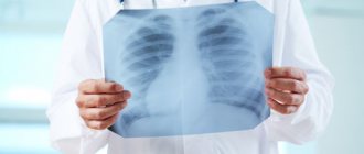
Which specialist conducts the examination?
It is possible to undergo fluorography in any public clinic upon the referral of a family doctor or a specialized specialist.
Mobile X-ray machines are sent to remote rural areas and places of work for examination as part of medical examination.
What is the name of the specialized doctor who does fluorography and interprets the results?
The diagnostic event is carried out by a laboratory assistant who has undergone special training. The decoding is carried out by a radiologist - a specialized specialist in radiation diagnostics.
What are the responsibilities of a radiologist?
The responsibilities of a radiologist include the following:
- carrying out a diagnostic procedure based on the standards of medical care;
- entering information into medical documentation;
- participation in meetings and consulting with specialists from other fields of medicine, assistance in making a diagnosis;
- providing information to patients about radiation exposure at the time of diagnosis.
Note! A practicing radiologist does not prescribe further treatment for patients, but makes a presumptive diagnosis based on the examination results.

Required Skills
The radiologist should know:
- aspects of radiation safety, standard indicators;
- rules for conducting x-ray examination;
- pathogenesis and clinical picture of pathologies for which fluorography is performed;
- basic information about radiation studies of diseases.
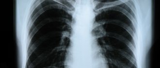
Fluorography has long become a routine diagnostic examination that detects tuberculosis, atypical formations and other pathological changes.
The need for annual diagnostic testing is regulated by the relevant law, the purpose of which is to prevent the spread of tuberculosis in the Russian Federation.
The study is not prescribed for women during pregnancy, lactation and children under 15 years of age.
Diagnostics are quick and easy to access and can be assessed in a short time by experienced radiologists.
In a state clinic, upon referral from a doctor, research is provided on a budgetary basis. The only drawback is radiation, but the dose is very low.
Radiation protection equipment: mobile and individual
Radiation protection equipment for personnel and patients is classified into:
| mobile | individual |
|
For children: |
X-ray rooms for various purposes must have a mandatory set of mobile and personal radiation protection equipment. For example, urography and angiography rooms must have a full set of radiation protection equipment, and in a fluorography room, a large screen, collar and apron or skirt is sufficient. The minimum values of the protective effectiveness of mobile and personal radiation protection equipment for personnel and patients, expressed in lead equivalent values, are established by SanPiN.
Protective materials and radiation protection equipment must have sanitary and epidemiological reports, technical documentation, and markings. Monitoring of the operational parameters of radiation protection equipment is carried out by accredited organizations at least once every two years.
How to undergo fluorography without a referral correctly
In order not to waste time visiting a medical facility again, you need to follow a few simple rules that will allow you to receive a free service without a referral or not pay for it twice. These rules include preparation for the procedure and the availability of the necessary documents.
Is advance preparation required?
Before the FLG examination, you do not need to follow any diets, take special medications or carry out preparatory procedures. When choosing a study at a clinic at your place of residence, you need to know what documents are needed to undergo fluorography:
- passport with registration,
- compulsory health insurance policy (CHI).
If you have these documents, you can go straight to the office and receive the service. Most often it is carried out even without prior registration.
Before fluorography, you should not wear clothes with large metal buttons, zippers, rivets or other dense parts, otherwise the doctor will ask you to undress. Neck jewelry or a brooch will also interfere with exploration. If the patient wants to undergo the procedure without undressing, then they should come in a thin turtleneck or T-shirt. The woman will have to unfasten her bra.
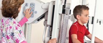
What should be done in the fluorography room?
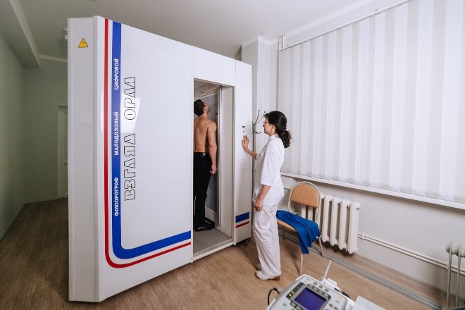
If necessary, the specialist will ask you to remove outer clothing and jewelry that may distort the photo. When the patient is prepared, he will be asked to approach the device screen and take a special position: move his shoulders slightly forward.
The requirement to breathe and not breathe is mandatory. The immobility of the organ will allow you to get a clear image on the first try. You will only have to hold your breath for a few seconds. After this, the doctor will tell you when you can come for the result.
Is it possible to take a fluorogram on the same day as other procedures?
When undergoing a medical examination, you have to save time and undergo several procedures at once. If an FLG study has been completed, then X-ray diagnostics or CT scanning are not recommended. These types of studies involve exposure of the body to hard radiation, and the total dose can be quite high.
After the fluorograph, you can take any tests and visit the offices of any specialists. The study does not in any way affect the patient’s blood or urine, his vision, nervous system or heart activity.
Production control
The administration of a medical organization is responsible for organizing production control according to a program agreed upon with the state sanitary and epidemiological inspection body.
Production control includes, inter alia, technical improvement, control over the professional training of persons working with x-ray radiation, radiation control and control of the operational parameters of x-ray equipment.
| Radiation control | ||
| planned | an object | periodicity, order |
| radiation dose rate at personnel workplaces, in rooms and areas adjacent to the X-ray room procedure room | during technical certification of the X-ray room, obtaining a sanitary and epidemiological report | |
| technical condition and protective effectiveness of mobile and personal radiation protection equipment | 1 time every 2 years | |
| Group A personnel (as well as persons periodically participating in special x-ray examinations) | constantly with registration of measurement results once a quarter, storage period - 50 years after dismissal, annually statistics are transferred to the territorial center of state sanitary and epidemiological supervision | |
| patients | for each study, information is provided to the health authorities of the constituent entities of the Russian Federation | |
| unscheduled | operating conditions of the X-ray room | changing the purpose of the office and/or adjacent rooms, replacing the X-ray tube, protective equipment, in emergency situations |
Monitoring of the operational parameters of medical equipment is classified into periodic and current. Control of operational parameters is carried out by an accredited organization, the results are recorded in a protocol. For X-ray equipment with a service life of more than 10 years, control is carried out at least once every two years to assess the possibility of its further operation.
Features of X-ray dental examinations
During X-ray dental examinations, a limited set of organs is exposed to irradiation: the brain, salivary glands, thyroid gland, part of the red bone marrow (MR 2.6.0098-15. 2.6.1. Ionizing radiation, radiation safety. Assessment of radiation risk in patients during X-ray radiological examinations. Methodological recommendations approved by the Chief State Sanitary Doctor of the Russian Federation on 04/06/2015). Most dental studies fall into the negligible and minimal risk categories.
The specifics of ensuring radiation safety during X-ray dental examinations are determined by the type of X-ray equipment used.
| X-ray machine | Acceptable placement option | Composition, room area, sq. m (not less)* |
| Dental apparatus working with conventional film without reinforcing shielding | Only in the X-ray room of a medical organization | Procedural – 8; Photo laboratory – 6; |
| Dental apparatus and pantomograph working with a highly sensitive film and/or digital image receiver, including a visiograph (without a darkroom) | In the premises of a dental institution located in a residential building, including in adjacent residential premises | Procedural – 6; |
| Panoramic apparatus, pantomograph | Only in the X-ray room of a medical organization | Procedural – 8; Control room – 6; Photo laboratory – 8. |
| * plus 4m2 for each additional device. | ||
The operation of X-ray dental equipment is permitted if there is a registration certificate from the Ministry of Health of Russia and a sanitary-epidemiological conclusion. A set of mobile and personal protective equipment for personnel and patients in an X-ray diagnostic room for dental examinations consists of a large screen (or curtain for devices with highly sensitive image receivers), a one-sided lung apron and a collar for staff and a dental apron or cape and apron to protect the patient's gonads.
Requirements for protection from non-radiation factors
Safe conditions for conducting x-ray examinations must be ensured by fire-fighting and anti-epidemic measures, measures to protect against exposure to electricity and lead.
Electrical safety is ensured by the following means. The possibility of contact with open current-carrying parts of electrical purposes is excluded; heating radiators must be covered with insulating shields, sockets must have a grounding contact, and in newly built offices there must also be a residual current device located at least 1.2 meters from the floor. Cables and wiring are laid in underground channels, floor and wall boxes; exit hatches must be sealed.
Metal parts of stationary X-ray equipment and equipment are connected to the ground bus with a copper wire. The X-ray equipment is connected to the network by a switching device (switch), which, when opened, de-energizes all parts of the equipment. The distance between the switching device and the control panel of the X-ray machine should be no more than 1.5 m at a height of at least 1.6 m from the floor level.
The presence of exposed lead or lead-containing surfaces in the X-ray room is not permitted. The noise level from technical equipment should not exceed 60 dBA (comparable to the noise in the first line of residential development in large cities).
Movable devices must remain stable, and the ceiling mounting of the elements must have a tenfold load reserve. Parts of the equipment that the patient touches with the body should be treated with a solution of chloramine and ethanol.
In addition, the X-ray room should have carbon dioxide fire extinguishers. The values of parameters of non-radiation factors in the X-ray room are determined by accredited and licensed organizations at least once every two years.