When is ophthalmoscopy prescribed?
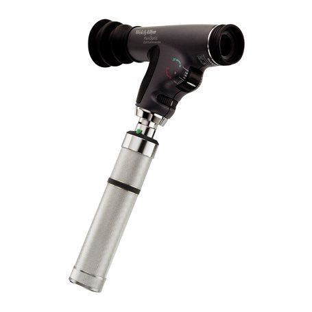
This research method is the most common in ophthalmology. It is carried out using an ophthalmoscope - a special device that allows you to examine in detail the internal structures of the eye: the retina, vitreous body, optic disc, blood vessels, as well as the peripheral parts of the eyeball. Thanks to ophthalmoscopy, it is possible to identify severe pathologies of the visual organs at the very beginning of development. Among them:
- cataract;
- neoplasms;
- damage to the optic nerve;
- retinal detachment;
- glaucoma;
- macular degeneration;
- melanoma;
- cytomegalovirus retinitis;
- pathology of the blood vessels of the eyes.
Moreover, during an examination with an ophthalmoscope, not only ophthalmological diseases can be detected, but also diseases of a systemic nature, including diabetes mellitus, hypertension, renal failure, and tuberculosis.
Ophthalmoscopy is prescribed for every visual examination. It can be considered a standard procedure that is included in any examination by an ophthalmologist. A referral for an ophthalmoscopy can be given not only by an ophthalmologist, but also by a cardiologist, infectious diseases specialist, gynecologist, and therapist. This diagnostic method is very important for pregnant women. During pregnancy, patients with myopia have an increased risk of retinal detachment. With the help of an ophthalmoscope, this dangerous disease can be detected at a very early stage and laser coagulation can be prescribed.
The following diseases and symptoms may also be indications for ophthalmoscopy:
- color blindness;
- eyeball injuries;
- myopia;
- inflammatory processes in the eye;
- traumatic brain injury;
- headache;
- epilepsy;
- violation of movement coordination.
There are also a number of limitations to the purpose of this research method. It is not performed in case of increased photosensitivity, lacrimation, clouding of the optical media of the eye, miosis and other pathologies of the pupils. Under these conditions, it will be impossible to examine the fundus in detail. In addition, symptoms such as tearing and photophobia may increase. Ophthalmoscopy is not always prescribed for angle-closure glaucoma. There is a risk of increased intraocular pressure. The method is not used in the presence of cardiovascular diseases. Contraindications may include various infectious and inflammatory diseases of the anterior parts of the eye.
Examination price
If the patient applied to a free medical institution under the policy, the cost of the doctor’s services and the procedure performed will be free. A person will only need to buy medications that dilate the pupil.
If the patient goes to a private clinic, the cost of the procedure is included in the services of an ophthalmologist. The price of the clinic depends on the qualifications of the specialist. Most often, the price varies from 500 to 1500.
The ophthalmoscopy procedure is one of the main examinations in ophthalmology. Using this method, you can look at the fundus of the eye and identify its deviation from the normal parameters of the organ of vision. It is possible to identify systemic diseases of the cardiovascular system that affect microcirculation vessels. In this case, the ophthalmologist is able to promptly refer the patient to specialists and prevent the development of irreparable complications.
Types of ophthalmoscopes
Such devices are monocular and binocular. With the first type of ophthalmoscope, the doctor examines one eye. The binocular is designed in such a way that it makes it possible to examine the patient with both eyes, which provides a clearer picture. There are other types of ophthalmoscopes:
- Helmholtz device. It is monocular and allows direct examination of the fundus.
- Goldman lens. With its help, you can enlarge the image several times, which makes it possible to examine the fundus and periphery in the smallest detail. This device is often used when examining patients with myopia. With this refractive error, various pathological processes can occur in the eye structures.
- The Spencer head mirror is a binocular ophthalmoscope that allows for more accurate diagnosis in both eyes.
- Slit lamp. Ophthalmoscopy using it is called biomicroscopy, which studies the condition of the retina and vitreous body. Modern slit lamps are equipped with devices that take photographs.
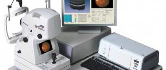
- Laser ophthalmoscope. It also allows you to take photographs during the inspection. Some laser-type devices are equipped with a video camera. It displays the image on a computer monitor.
- Electronic ophthalmoscope. It can be used for any diagnostic method.
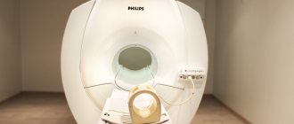
In addition, these devices are divided into stationary, hand-held (portable) and head-mounted.
Preparation for ophthalmoscopy
For a better view, before the examination, the doctor instills mydriatic drops into the patient’s eyes. Under their influence, the pupil dilates. The effect of mydriatics lasts for several hours, during which time photophobia and poor near vision may occur. When going for an ophthalmoscopy, take sunglasses with you. If you are allergic to certain medications, inform your ophthalmologist before instilling the drops. It is also recommended to talk about the medications you are taking and any existing diseases. Women need to remove makeup before the procedure. Actually, no special preparation is required. It lasts no more than 15 minutes, depending on which ophthalmoscopy technique is chosen. There are several of them: direct and indirect (reverse), contact and non-contact, monocular and binocular. Let's take a closer look at them.
Preparatory stage
Ophthalmoscopy does not require special preparation. Pupil dilation is achieved using a drug called Tropicamide. Instillation is carried out only in the absence of allergies. Typically, the drug composition is administered 15 minutes before the main procedure. A diagnostic kit for direct ophthalmoscopy is prepared in advance. The doctor must be notified of the presence of ophthalmic diseases in the family history.
The patient is warned about the appearance of unpleasant sensations after the procedure. Sometimes there are problems focusing your eyes. Therefore, the patient should not come to a medical facility unaccompanied. You may also need sunglasses after an ophthalmoscopy.
This will avoid disorientation in bright light. The discomfort will disappear a few hours after the examination, and the risk of complications is almost completely absent. If a negative reaction occurs during ophthalmoscopy, the patient must immediately tell the doctor about it. Otherwise, the likelihood of developing adverse reactions will increase.
Direct ophthalmoscopy
It is carried out using a directed beam of light. The ophthalmologist sits opposite the patient and places an ophthalmoscope on his eye, which illuminates the fundus of the eye. The image can be zoomed in and out. The distance between the device and the eye of the subject is 4 cm. The technique is designed based on the fact that the eye is an optical system capable of magnifying the image by 14-16 times. Thanks to this, it is possible to detect even minor changes in the fundus. The method also allows you to determine refractive errors. Direct ophthalmoscopy also has disadvantages:
- Small viewing angle. It is not possible to examine the fundus in full.
- Close contact with the patient.
- Lack of stereoscopic picture.
Performing ophthalmoscopy on young patients
The fundus examination in children performed at the Optic Center clinic has a number of its own specific features. Since the fundus of the eye tends to change very quickly at an early age, this diagnostic technique should be performed more often than in adult patients. The number of examinations per year can be 3-4 visits to an ophthalmologist.
The procedure itself is no different in principle from adult ophthalmoscopy. You just need to prepare your child in advance for a visit to a specialist and tell him about the upcoming study.
Indirect ophthalmoscopy
Reverse ophthalmoscopy is performed using a conventional lamp, which is located next to the patient, to the left and slightly behind his back. As a result of this arrangement, the subject finds himself in the shadow. There is no direct contact between the subject and the device. This is a non-contact ophthalmoscopy. The ophthalmologist sits opposite him at arm's length, brings the ophthalmoscope to his eye and directs a beam of light from the lamp, reflected from the mirror of the device, into the patient's eye. At this moment, you can notice that the pupil turns red. Next, the doctor brings a biconvex lens (magnifying glass) to the patient’s eye. He holds it from the patient’s face at a distance of 7-8 cm. The optical product magnifies the image and makes it possible to examine the fundus of the eye.
Indirect ophthalmoscopy can be performed using various lenses and mirrors. The picture turns out to be stereoscopic. The doctor sees the image upside down. The examination is especially effective in diagnosing cataracts in the maturation stage. Indirect binocular ophthalmoscopy has other advantages:
- wide overview, the ability to examine peripheral parts;
- speed of implementation;
- lack of patient contact with the device;
- Possibility of use even in poor lighting.
If we talk about the disadvantages, the following fact should be mentioned: reverse ophthalmoscopy does not make it possible to greatly enlarge the image, as in the direct method. In this case, the doctor can prescribe both types of studies. The reverse one helps to quickly examine the fundus of the eye and identify pathology, and the direct one allows you to study in detail the areas of the fundus affected by the disease.
In addition to direct and indirect ophthalmoscopy, other methods are also used. One of the most famous is biomicroscopy. Let's find out what its features are.
Biomicroscopy
This diagnostic method is also called ophthalmoscopy using a slit lamp. With its help, it is possible to examine in detail all areas of the fundus. Contact and non-contact lenses with different optical powers are used in the examination. They allow you to study the state of the internal structures of the fundus as accurately as possible.
Non-contact binocular ophthalmoscopy - examination using an aspheric lens equipped with strong diopters (+60.00, +90.00, +78.00D). It provides a clear reverse image of the fundus with a field of view of 70-90 degrees. The distance between the patient's cornea and the binocular non-contact ophthalmoscope is only 1.5-3 cm. The device is located perpendicular to the eye. The slit lamp is retracted to the maximum distance, after which it is slowly brought closer to the subject. The doctor examines the center and peripheral part of the fundus of the eye in a wide view, and the device does not come into contact with the patient’s eye.
Contact binocular ophthalmoscopy using a slit lamp and contact optics requires instillation of anesthetic drops into the patient's eyes. After their instillation, a disposable contact lens (diagnostic) is installed on the cornea. The patient places his chin on the rest, pressing his forehead against the bar of the slit lamp. You need to look straight ahead. After this, an examination of the fundus begins. A variety of types of ophthalmoscopes with different numbers of lenses and mirrors can be used. The width of the view depends on their number. Using non-contact binocular ophthalmoscopy, the doctor can obtain a high-quality image in which all parts of the retina will be visible.
Side effects
Ophthalmoscopy is a safe and painless procedure. However, it may cause discomfort. Some patients experience diplopia and blurred vision at close range. Mydriatic drops have such side effects. There may be other short-term unpleasant symptoms:
- redness of the skin around the eyes;
- nausea and vomiting;
- dry mouth;
- dizziness;
- increased intraocular pressure.
This is extremely rare and can occur if there is an allergy to the eye drops that the patient was not aware of. All of the above symptoms go away quickly. If necessary, the doctor will prescribe an appropriate drug to eliminate them.
Ophthalmoscopy results
The reliability of the method is 90-95%. Moreover, it is considered one of the simplest and most accessible in ophthalmology. In this regard, it is used in almost any ophthalmological examination. Normal results include healthy blood vessels and no damage to the retina or optic disc. The ophthalmologist pays attention to the size and color of the disc, the shape and sharpness of its edges, the presence or absence of hemorrhages, blood clots, and symptoms of inflammation. Ophthalmoscopy allows you to identify the early stages of eye diseases. The procedure only takes a few minutes. An annual examination is a good prevention of ophthalmopathologies, and if they are present, it is possible to start treatment on time.
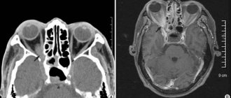
Why is a fundus examination necessary?
This procedure is called ophthalmoscopy. It allows you to assess the condition of the retina and its individual structures: the macula, the optic nerve, the choroid. Before the test begins, special drugs are injected into the eye to dilate the pupil - drops “Irifrin”, “Atropine” and others. No other special training is required. To do this, you need to remove contact lenses if you use them. Also, do not forget to warn about increased intraocular pressure if there is such a problem.