For a long time, doctors could diagnose the disease only when obvious symptoms developed, relying only on experience and clinical methods. By the 30s of the last century, a non-invasive diagnostic technique was developed, which became the successor to classical radiography - computed tomography (CT). The level of development of the technical base increases every year, modern hardware devices for radiation diagnostics are constantly being improved, becoming more informative and safe.
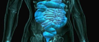
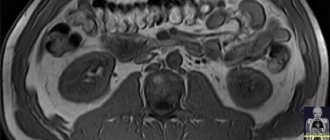
Beam scanning is a universal method, as it allows you to study any internal organ of the body without instrumental penetration by visualizing the processes occurring inside the body. Abdominal organs such as the kidneys and adjacent systems and structures are clearly visualized. To enhance image clarity, a scanning mode such as kidney CT with contrast is used.
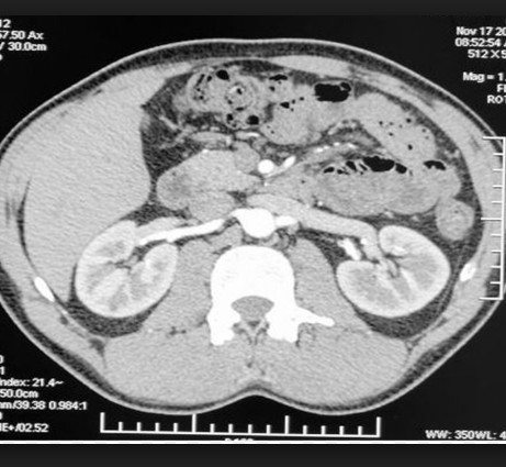
The operation of scanning machines is based on the production of ionizing radiation, which, penetrating into biological tissues, is partially absorbed by them. The denser the fiber structure, the more X-rays it absorbs. Hollow and liquid substances, on the contrary, transmit rays almost unhindered. Based on the analysis of the difference in such absorption, the equipment processes the signals and transmits them in the form of images to the computer screen.
Features of CT scan of the kidneys with contrast
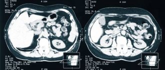
Radiation screening is carried out step by step on a special tomograph. The machine produces multiple scanning sections in a longitudinal position at any given depth. This allows you not only to obtain many static images of the studied area, but also to combine photos into a multidimensional projection to view the area from all sides, rotating it at any angle and precisely zooming in on anomalous zones. The study allows you to quickly determine the source of the disease, since in most cases the foci of the anomaly are visible immediately, at the first diagnosis. If the pathology is microscopic in size or the cause of the inflammation is not clear, specialists prescribe a CT scan of the kidneys with contrast.
This procedure differs little from the standard screening protocol. The main feature is the use of a special coloring amplifier - contrast - in the process. The solution is a “dye” that has a ferromagnetic base and shows up well in photographs, making them more contrasty and clear. Images are displayed on the monitor in black and white, denser formations appear light, and hollow and liquid structures appear dark. Some pathologies, such as cancerous lesions, may be similar in density to adjacent tissues and are more difficult to distinguish in photographs. Staining is used to identify them.
There is no point in being afraid of this mode of procedure. In the absence of individual contraindications, dye preparations are absolutely harmless and do not leave any side effects. When studying this area, an intravenous solution is administered. Distributed throughout the bloodstream, the substance accumulates in soft tissues, tinting them, especially vascular channels and capillaries. CT with staining helps to clearly see even microscopic pathologies in detail.
The entire process of scanning and intravenous administration of the amplifier is supervised by experienced physicians. At the slightest deviation from the norm, the procedure is stopped; all actions of doctors are aimed at avoiding complications. To determine whether the patient has a negative reaction to the drug, drug testing is performed in advance. If the immune system has a positive response (expressed in an allergic reaction - itching, redness at the injection site), enhanced screening is not prescribed; alternative diagnostic methods are proposed.
Stages of the kidney CT procedure
Screening is carried out in several stages:
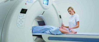
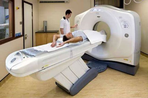
- Preparation and instruction. The client is escorted to a separate room where he can change clothes, leave all foreign metal objects, and check his pockets. The medical worker talks about how to behave during the session, escorts him to the equipment, places him on the retractable part of the tomograph, and fixes the patient in a stationary position.
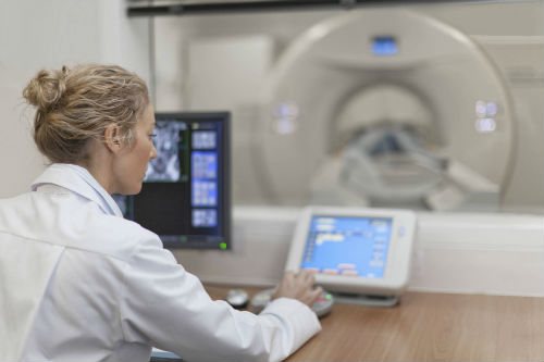
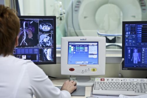
- Scanning. In the enhancement mode, contrast is introduced before shooting, which spreads throughout the body over the course of 5-10 minutes and accumulates in the tissues. In some cases, the patient is given a catheter with an automatic injector so that the contrast is introduced into the blood gradually. At the main stage, the ring part of the installation moves along the human body and produces layer-by-layer scans. The images are displayed on the computer of the radiologist, who monitors the examination from an adjacent room.
- Results. After the session is completed, the patient is escorted to the waiting room. Preparing the final description of the images will take about an hour.
There is no rehabilitation required after the CT scan, so you can plan your daily activities on the day of the scan. It is also not necessary to wait for the conclusion at the clinic; it is enough to leave an email address to which the diagnostic documentation will be sent.
When is a kidney CT scan with contrast prescribed?
Screening using a hardware installation is classified as an expensive procedure, so it is not prescribed during a routine preventive examination. If a specialist decides to refer a patient for such a study, then there are significant reasons for this, and one should not refuse diagnosis.
Most often, screening is indicated in controversial situations when other methods have not given an effective result. Scanning is a more informative method if the cause of the disease has not been discovered, if the results of an ultrasound examination or urography turned out to be controversial, and also if there are suspicions about the course of oncological processes in this area.
Contrast is also used only when absolutely necessary. The treating doctor will definitely explain and justify the choice of this diagnostic regimen. Kidney CT scan with contrast is performed for the following indications:
- displacement, change in location of organs;
- cystic formations on tissues;
- infection of unknown etiology;
- congenital or acquired pathologies of the configuration of vascular canals;
- disturbance in the processing of protein compounds;
- acute or chronic diseases of the body’s natural “filters”;
- post-traumatic pathologies;
- monitoring the effectiveness of treatment;
- control of the rate of formation of thrombotic barriers in blood vessels;
- study of adrenal formations;
- disturbance in organ development;
- preoperative period, assessment of the patient’s operability;
- stagnation of fluid, leading to stretching of the walls of organs;
- visual control during tissue sampling (biopsy);
- determination of the nature and nature of neoplasms.
CT does not involve penetration into biological tissues, therefore it is absolutely painless and does not require anesthesia in the standard mode. Suspicions of improper operation of filtrates arise when deviations from the norm in laboratory tests are detected; such methods cannot detect the root cause of the pathology. Computer scanning is designed to cope with this task.
Indications for CT scan of the kidneys
Computed tomography of the kidneys is an effective way to identify pathologies and study the structure of organs in real time. The results of the study are saved electronically and printed in the form of several images, which are analyzed by specialists.
Kidney CT is performed for patients in the following clinical cases:
- chronic, acute kidney diseases;
- congenital kidney pathologies;
- urolithiasis disease;
- kidney damage and trauma, abscesses;
- benign and malignant tumors;
- inflammation of the lymph nodes located in the retroperitoneal space;
- pathological changes in the adrenal glands;
- preparation for surgery;
- monitoring the effectiveness of chemotherapy.
Patients at the Yusupov Hospital with symptoms of kidney disease undergo a comprehensive examination, which makes it possible to determine the location of the kidneys, their features, and identify the condition of nearby organs. Specialists use high-precision European equipment, so the study is completed in the shortest possible time.
Are there any prohibitions on performing a CT scan of the kidneys with contrast?
Like any medical procedure, hardware screening has a list of contraindications. Contrast also belongs to the medicinal group, which has its own prohibitions on use. The most important contraindications are:
- The period of pregnancy at any stage of the course. Regardless of a woman’s confidence that she is not pregnant, she will definitely have to take an analysis or a rapid pregnancy test. This is necessary in order to eliminate any possibility of harm to the intrauterine fetus. The study is carried out under the influence of X-rays, which aggressively affect any actively growing tissue, which can negatively affect the health of the unborn baby. Enhancing staining is also contraindicated during pregnancy, since the drug will penetrate through the mother’s blood into the child’s organs, which can lead to intoxication of his body. Dyes are also not used during breastfeeding, as they easily penetrate into any liquid substance, including milk.
- Children's age up to 12-14 years. During this period, the young patient actively develops. A child should not be exposed to harmful radiation.
- Urinary dysfunction. With reduced urinary function, the dye can remain in the body for a long time, which will negatively affect the functioning of organs and can lead to poisoning.
- Allergy to medications. CT scan of the kidneys with contrast is contraindicated in case of allergic rejection to the components of the drug. During the procedure, mild nausea, an acidic taste in the mouth, hot flashes as the solution spreads throughout the body, and a slight burning sensation at the injection site are considered normal. You should not be afraid of such manifestations; they will pass quickly. But if the patient feels difficulty breathing, gagging, or swelling, the procedure must be stopped immediately.
- Severe anxiety, mental or neurological disorders, fear, excessive pain, shock. Such factors will not allow the patient to remain motionless for a long time, which is required to obtain the correct result. In these situations, a person’s motor activity cannot be controlled. The solution may be to use sedatives or complete anesthesia.
- Excess body weight by more than 120-200 kg. It is difficult for people of a large weight category to undergo a CT scan, since tomographs are not designed for such weight. An obese person will not fit inside the installation. In this case, it is recommended to resort to the use of special equipment of an open configuration, which has no weight restrictions.
Patients are often afraid to undergo radiation examination, worrying that they may receive a high dose of radiation. There is no point in worrying about this, since any ionizing effect is recorded in the patient’s medical record, and doctors carefully monitor the annual level received by the person. The X-radiation in the device is so low that even with repeated access to diagnostic scanning, it is very difficult to exceed the annual radiation exposure rate.
How to prepare for a CT scan of the urinary system
Proper preparation for any diagnostic procedure is the key to its quality and effectiveness. A CT scan of the urinary system is performed on a full bladder - this is necessary for a reliable assessment of the condition of the urinary tract. To achieve tight filling, it is enough to drink a glass of water 1 – 2 hours before the test and not go to the toilet. Another important point is to fast for at least 4 hours before the tomography.
In cases where contrast administration is required, renal function should first be assessed - a blood test for creatinine. The result must be fresh, made no more than 10 days ago. Creatinine numbers allow you to calculate the glomerular filtration rate - the main indicator of kidney function and the presence of renal failure.
Are preparatory measures necessary for a CT scan of the kidneys with contrast?
Since the area being examined is at the level of the peritoneum, it is important that the gastrointestinal tract is in a “quiet” state at the time of the examination. To do this, you should stick to a simple diet for several days: give up flour, dairy, legumes, fresh plant foods, carbonated liquids, and alcohol. A few hours before the test, stop eating altogether and come to the procedure on an empty stomach.
A kidney CT scan with contrast is prescribed only by a doctor based on the patient’s medical history. The subtleties and features of the procedure, important recommendations and training instructions will be provided by both the doctor and the radiologist on duty at the diagnostic center.
To eliminate the risks of critical complications, blood tests for hormonal levels are mandatory. This is necessary in order to determine the possibility of using contrast.
As general recommendations for preparing for the procedure, it is necessary to prepare a package of documents, including a medical record, a referral from the treating specialist, the results of other examinations and previous images if CT was performed more than once. It is necessary to take care of comfortable clothing during the screening and remove metal objects from the body. Once the session is over, the person can return to their normal lifestyle. A rehabilitation period is not required, since the procedure does not affect the patient’s well-being.
CT scan of the kidneys with contrast: preparation for the study
The process of administering contrast often frightens patients. However, if a person has no contraindications to this procedure, there is nothing to be afraid of.
When preparing for a kidney CT scan with contrast, it is recommended to abstain from eating at least 4 hours before the start of the study. In the same way as with a CT scan of the kidneys without the use of contrast, it is necessary:
- do not consume food and drinks that increase the formation of gases in the body;
- do not wear tight and uncomfortable clothing when preparing for a CT scan, and if necessary, change into special clothing issued by the staff for the procedure;
- remove all metal jewelry, glasses or dentures with metal inserts.
The contrast agent can be injected or orally into the body.
If intravenous contrast is needed, it is administered using an automatic injector. The substance begins to act on average after twenty seconds. In this case, the patient may feel a false urge to go to the toilet, but this condition passes quite quickly and a CT scan can be started. You should not be alarmed if you experience mild nausea and a metallic taste in your mouth - this is a normal reaction to the administration of contrast.
The oral method of administration means that the patient drinks a special solution, having previously done a CT scan without contrast. After which, after some time, a computed tomography scan is done with a contrast agent. A few hours before a CT scan using contrast, you must refrain from eating not only food, but also drinks.
General recommendations for computer scanning:
- During the procedure, you must remain absolutely calm and not make any movements;
- 24 hours before the test you need to give up not only alcoholic beverages, but also drinks containing caffeine;
- It is important to inform the doctor in advance if the patient had a magnetic resonance imaging, x-ray or ultrasound scan shortly before the CT scan: the CT scan may have to be postponed;
- do not wear clothes and jewelry with metal elements;
- You should definitely notify your doctor or nurse if you are allergic to iodine or have any contraindications.
How is a kidney CT with contrast performed?
Screening is carried out according to the standard protocol for this diagnostic mode. At the preparatory stage, the client is registered and questioned on issues related to the disease. Next, detailed instructions are provided. A person is invited into a room with equipment, placed on the moving part of the tomograph, and sensors are attached to the area being studied. If necessary, the limbs are secured with straps built into the unit. This is necessary to maintain stillness throughout the session.
In a kidney CT scan with contrast, a healthcare professional inserts a catheter connected to an auto-injector into a patient's vein. Through which saline solution is injected into the patient’s blood. Next, the conveyor with the human body is located inside the installation, contrast is added to the saline solution, which spreads throughout the body within half a minute. The installation begins a step-by-step scan. The dye enters the blood throughout the entire session, which lasts about half an hour.
When the examination is completed, the patient is helped up, the sensors are removed, the catheter is removed, and he is asked to go to the waiting room and wait for the CT results. This may also take 30-50 minutes, please be patient as correct interpretation of the results is worth the time spent.
CT scan of the kidneys: preparation for the study
To conduct a computed tomography scan of the kidneys with contrast in St. Petersburg, the patient will need to undergo a biochemical blood test for creatinine, which will reveal the presence or absence of renal failure. Some centers offer on-site evaluation of this analysis using a special express system.
Kidney tissue can be examined using three different examination zones, depending on what disease the attending physician prescribes MSCT suspects. Computed tomography of the kidneys is included in the scanning area “abdominal organs and retroperitoneal space”. This area is used to obtain information about the condition of all internal organs or when searching for neoplasms and their metastatic screenings. In the vast majority of cases (more than 80%), a CT scan of the kidneys with contrast is performed in such situations. Preparation consists of fasting for at least 4 hours before the test. This is necessary for better visualization of organs located in the abdominal cavity.
When diagnosing the cause of high blood pressure or identifying tumor formations after ultrasound examination (ultrasound), doctors may prescribe tomography of the kidneys and adrenal glands. You do not need to be fasting to complete this study. If tumor formations are detected, intravenous contrast will also be required.
If examination of the entire urinary system and assessment of urodynamics is required, diagnostics of the urinary system is used, which includes examination of the kidneys, ureters and bladder. It is also better to fast before this test. Contrast will be needed in situations where there is a disturbance in the outflow of urine from the kidney due to external compression of the ureter. To exclude a tumor in such a situation, it is advisable to administer a contrast agent.
To summarize the above, preparation for a kidney CT scan with contrast consists of taking a blood test for creatinine and observing fasting in certain scanning areas.
You can find out what kind of CT scan of the kidneys to do and how to prepare for it from our employees by calling 8 (812) 317-00-37 or leaving a request on our website.
What are the pros and cons of a kidney CT scan with contrast?
The medical measure has both pros and cons, which you should be aware of in advance. So the advantages of CT include:
- No pain during the procedure.
- There is no injury to the skin or internal organs.
- obtaining a quick conclusion on which the diagnosis will be based.
- A visual picture of the processes occurring inside the body, clear photographs.
- Determination of pathological zones with high resolution of the device.
- The study is possible even with metal implants implanted into the body.
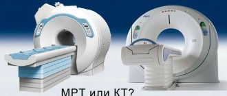
- Relatively cheap (more financially advantageous than MRI).
The disadvantages of beam scanning include:
- Increased dose of radiation.
- Proportion of probability of developing allergic rejection to a medicinal dye.
- Impossibility of studying pathologies in children and pregnant women.
- Lower quality of photographic images than similar MRI.
What will a multislice CT scan of the kidneys show?
The study can be carried out to find out the causes of pain in the lumbar region, increased body temperature in combination with pain in the lower back, urination problems, the presence of blood in the urine, poor results of a urine test, and also in cases where alternative research methods cannot identify this or that other violation and provide dubious information.
MSCT urography is performed to diagnose the following disorders:
- consequences of injuries to the lower abdomen or back;
- infectious disorders;
- anomalies of kidney development, in particular, hypoplasia, polycystic disease, aplasia, duplication, pelvic, cross, thoracic and other dystopias, rudimentary kidney, accessory, horseshoe-shaped kidney and others;
- necrotic and dystrophic processes;
- stones;
- abscesses;
- pyelonephritis, other inflammations;
- benign and malignant tumors, as well as metastases from other organs;
- cystic formations.
A comprehensive CT scan of the kidneys and bladder is often performed, which also shows bladder development abnormalities, cystitis, squamous cell carcinoma, bladder adenocarcinoma, bladder neck obstruction, metastatic lesions and other pathologies.
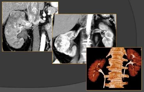
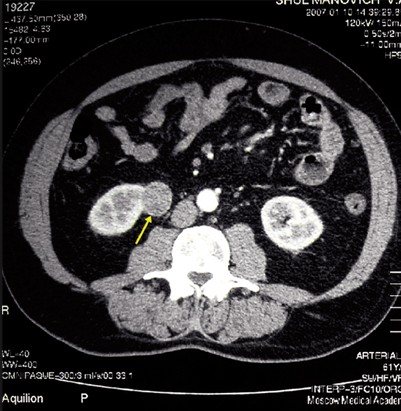
For differential diagnosis of cysts, tumors, inflammations, a CT scan of the kidneys with a contrast agent is performed. Differentiation of such formations is based on the property of malignant formations to accumulate a radiocontrast agent, while the density of the tumor increases by more than 5-15 units on the Hounsfield scale. Computed tomography without contrast is not performed when diagnosing tumors; contrast enhancement is required to obtain the most detailed information.
The main signs of benign cysts on CT are a round shape of the formation with a smooth wall, clear contours, as well as homogeneous content with a density of less than 20 units. An exception is hyperdense cysts, the density of which can be more than 20 units due to the content of a large amount of protein or hemorrhagic components. Also, CT signs of cysts are the presence of nodular seals in the wall, internal septa and multi-chamber formation. After administration of a contrast agent, the density, shape and size of a simple cyst do not change, while the density of the renal parenchyma increases.
In modern practice, the main method for verifying calcifications is CT. The presence of calcifications in the cyst may indicate a possible malignancy of the process. However, small linear calcifications can occur in the septum or wall of benign cysts.
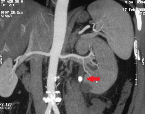
The contrast procedure also makes it possible to determine areas of growth of secondary tumors from the pelvic organs, structures of the abdominal cavity and chest, to diagnose stones in the kidneys and ureters, as well as vascular disorders. For example, with angiomyolipoma, an unenhanced CT photo shows areas of adipose tissue with reduced density. On post-contrast images in the cortical phase, a hyperdense formation is visualized, the degree of which depends on the size and vascularization of the tumor.
For a comprehensive assessment of the extent of the pathological process, a CT scan of the kidneys and abdominal cavity is often performed, which shows the mutual relationships of the structures of a given anatomical region, the condition of the blood vessels and the lymphatic system.
Why do CT angiography of the kidneys?
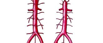
A CT examination of renal vessels is carried out to identify pathologies of the renal arteries, veins, as well as oncological processes that affect the blood vessels. Angiography makes it possible to detect abnormalities in the development of renal arteries, stenoses, vasorenal conflicts, arteriovenous malformations, and the degree of compression of vessels by germinating tumors.
Diagnosis is carried out with contrast enhancement, since without the administration of the drug such a scan will not be informative enough.
Preparing for a CT scan of the kidneys with contrast
The study requires compliance with simple preparation measures. So, the patient should not eat 3-4 hours before screening. Diagnosis of the bladder is carried out in an average or full degree of fullness; for this, the patient needs to drink 1 glass of clean water half an hour before the examination and not urinate. This is necessary in order to smooth out the folds of the bladder as much as possible.
If possible, the patient should take with him a doctor’s referral, data from previous diagnostics, hospital extracts or medical records and other documents to the study.
The contrast procedure requires the patient to undergo a blood test to determine creatinine and urea levels. If these parameters deviate from the norm, the decision about the possibility of conducting a contrast study will be made by the attending physician or radiologist.
How to do a CT scan of the kidneys
During the procedure, the patient is placed horizontally on his back on the moving table of the tomograph. If contrast is necessary, the laboratory assistant installs a catheter into the antecubital vein, through which a contrast solution will be supplied in the future.
After this, the table with the patient moves into the ring of the device, while the abdominal area is located at the level of this ring. During screening, the ring will perform rotational movements around the patient, and the tabletop will move progressively, while the speed of its movement depends on the diagnostic purposes.
A CT scan of the kidneys without contrast usually takes 15 minutes, with contrast it lasts up to 25-30 minutes, taking into account the patient’s positioning and other preparatory measures.
After screening, the patient receives a series of black-and-white images, printed on film or recorded onto a medium, and later receives a written description from the radiologist.
What are the consequences after a CT scan of the kidneys with contrast?
Consequences may be associated with insufficient preparation for the study. If doctors do not detect an allergic reaction to contrast in time, this can lead to a severe rejection of the immune system to the components of the drug, up to Quincke's edema and a severe drop in blood pressure.
Such cases are extremely rare, as doctors carefully monitor the progress of the procedure. At the slightest suspicion of deviations from the norm, prompt measures are taken to prevent complications.