MRI of the pituitary gland is a modern type of diagnosis performed using a tomograph. Using magnetic radiation, the device scans the patient's tissues in different planes. Many of the resulting layer-by-layer images are transmitted to a computer screen, which allows for a detailed study of the organ or anatomical area being viewed.
Many patients are frightened when they are recommended to undergo MRI diagnostics of the pituitary gland, since in the vast majority of cases this study is prescribed for suspected adenoma.
But do not panic if you have been prescribed an MRI of the pituitary gland.
Adenoma is a really common and quite serious disease that can affect your health and require medical or even surgical medical care. However, this disease is not fatal, and malignant tumors of the pituitary gland are extremely rare.
Pituitary adenoma, especially if diagnosed in a timely manner, responds well to treatment, and accurate diagnosis using magnetic resonance imaging will make it possible to identify the disease even in the early stages.
What is MRI of the pituitary gland?
The pituitary gland is an endocrine gland located deep in the brain in an area called the sella turcica.
Despite its small size (on average 7-10 mm), the pituitary gland is a very important organ that produces a huge amount of hormones. This important endocrine gland produces substances that affect other glands, causing them to produce their own hormones. The pituitary gland can be compared to the command post for controlling the entire body.
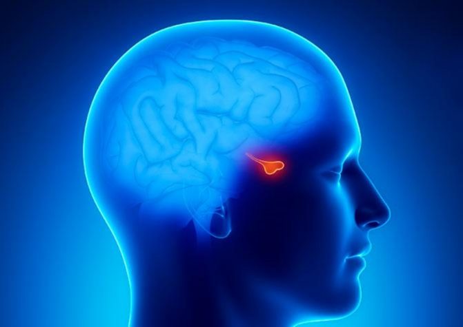
Location of the pituitary gland
Indications for MRI of the pituitary gland
A magnetic resonance imaging study of the pituitary gland is carried out if there is a suspicion of abnormalities in its functioning, which can manifest itself in symptoms and conditions such as:
- hormonal imbalances;
- regular headaches;
- visual disturbances, especially limitation of visual fields;
- discharge from the nipples in women and men;
- a sharp increase in body weight;
- decreased libido, erectile dysfunction;
- infertility.
MR examination is currently the only diagnostic method that allows obtaining high-quality visualization of the pituitary gland.
Due to its small size and location deep at the base of the skull, the pituitary gland is difficult to visualize with other diagnostic methods, such as ultrasound and CT.
If there are symptoms such as persistent headaches of varying intensity and location and vision problems, magnetic resonance imaging of the brain may be initially prescribed. The resulting images can reveal pathological processes in the area of the pituitary gland. In this situation, the patient will be prescribed targeted MRI diagnostics aimed at visualizing the structures of the pituitary gland and the sella turcica.
MRI of the pituitary gland: what the study will show
The vast majority of MRI studies of the pituitary gland are prescribed when the following diseases and conditions are suspected:
Pituitary adenoma
This tumor is formed from glandular cells. As an adenoma develops, the pituitary tissue grows and can put pressure on the optic nerves, leading to visual impairment. The tumor can produce certain hormones, depending on the location in a certain lobe of the pituitary gland, which causes the target glands to work harder, releasing more substances into the blood than the body needs to maintain certain functions.
Pituitary adenomas are divided into:
- Hormone-secreting. Can provoke the development of endocrine diseases and conditions, for example: hyperprolactinemia, acromegaly, etc.
- Hormonally inactive.
Pituitary adenomas are also classified by size:
- microadenomas (less than 10 mm);
- macroadenomas (more than 10 mm);
- giant adenomas (more than 40 mm).
An MRI examination of the pituitary gland can produce sections about 1 mm thick, which can even detect microadenomas up to 2 mm in size.
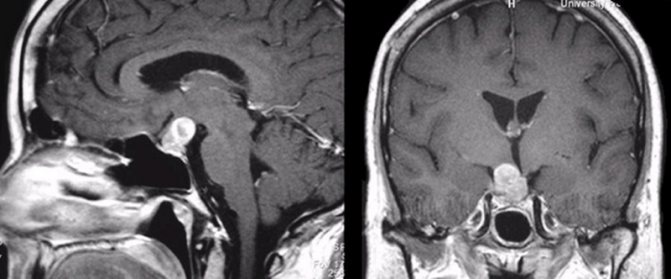
Pituitary adenoma detected during MRI with contrast
Malignant pituitary adenomas are extremely rare. If the course of the disease is malignant, MRI images will show how the tumor grows into other brain tissues.
Metastases to the pituitary gland from other organs are an even more rare occurrence, which is accompanied by severe symptoms in the form of visual impairment, even loss of vision, and severe headaches.
Problems with conception caused by increased levels of the hormone prolactin
MRI of the pituitary gland may be prescribed to women who have problems conceiving or who are about to enter into an IVF protocol.
An excess of the hormone prolactin, produced by the anterior pituitary gland, can lead to amenorrhea, infertility, and impaired libido.
Examination of the pituitary gland is necessary for differential diagnosis, since an increased amount of prolactin in the blood does not always indicate a malfunction of the gland.
Empty sella syndrome
This is the name for a pathology in which the meninges fill the cavity of the skull called the sella turcica, which leads to compression of the pituitary gland.
An empty sella turcica can be either a congenital structural feature or an acquired pathology and is most often asymptomatic. In some cases, the disease may be accompanied by severe symptoms.
The patient may experience:
- intense persistent headaches;
- autonomic disorders;
- mood swings;
- visual disturbances.
If the presence of the syndrome is suspected, an MRI of the area of the sella turcica and pituitary gland is prescribed in order to exclude or confirm the suspected diagnosis.
Other, less common endocrine diseases
Using magnetic resonance imaging of the pituitary gland, the following can be detected:
- Cushing's syndrome;
- acromegaly;
- diabetes insipidus;
- gigantism.
These diseases and conditions occur because the pituitary gland produces either too much or too little hormones, which in turn causes other endocrine glands to release too much or too little hormone into the blood.
MRI with contrast: preparation for the study
There are no strict rules for preparing for the diagnosis of most organs. There are certain recommendations:
- It is recommended to come to the procedure with contrast on an empty stomach, limiting food intake two to three hours before the appointed time, this helps prevent the development of nausea;
- if intravenous sedation is planned, the patient must comply with the requirements of the anesthesiologist;
- before examining the pelvic and abdominal organs, it is necessary to adhere to a diet excluding gas-forming products;
- on the eve of diagnosis of the genitourinary and digestive systems, bowel preparation is required (a laxative or enema is used); The last meal should take place 6 hours before the visit to the clinic, liquids - 4 hours.
Before an MRI with contrast, you should refrain from eating and drinking
The contrast agent penetrates not only into the blood, but also into breast milk. During lactation, breastfeeding can be resumed no earlier than 24 hours after the injection, so you should prepare in advance and stock up on food for the baby. Milk that is unfit for consumption is expressed and discarded.
Is contrast necessary for MRI of the pituitary gland?
Contrast during MRI of the pituitary gland is used in the vast majority of cases.
Due to the small size of the organ, conventional MRI images are not very informative. The only exception is an MRI of the pituitary gland with an already established diagnosis of macroadenoma, when it is necessary to evaluate the dynamics.
Contrast enhancement makes it possible to see all the structures of the pituitary gland, and in the presence of adenomas, to consider their size, location and possible growth into neighboring brain tissue.
For contrast, gadolinium-based drugs are used, which are well tolerated and harmless to the patient.
MRI of the pituitary gland with and without contrast - what is the difference?
When a patient comes to the MRI center for examination of the pituitary gland, depending on the diagnostic purposes, he is offered an MRI with or without contrast enhancement. It should be said right away that tomography of the pituitary gland in the vast majority of cases must be carried out with the introduction of a contrast agent. It is contrast that makes it possible to detect all, even very small pituitary tumors. For example, using non-contrast, native MRI, it is impossible to find a pituitary microadenoma due to its small size. It is the introduction of contrast that allows doctors to:
- assess the distribution, boundaries and relationship of pathological processes;
- identify all neoplasms, their size, location and degree of fusion with surrounding tissues;
- identify and evaluate inflammatory processes and the level of tissue necrosis;
- assess the degree of vascularization, that is, the rate of accumulation and release of the contrast agent, for differential diagnosis. This characteristic allows specialists to judge the benignity or malignancy of the neoplasm.
Contraindications for MRI of the pituitary gland
Although MRI diagnostics is a progressive and safe technique, there are several absolute contraindications to its implementation.
MRI is not performed if:
- presence of a pacemaker;
- the presence of any metal devices in the body: prostheses, vascular clips. The exception is devices made of titanium, since this metal is inert to the effects of a magnetic field.
MRI of the pituitary gland cannot be performed in the following categories of patients:
- women in the 1st trimester of pregnancy;
- children under 5 years old;
- very plump people whose weight exceeds 130 kg.
Contraindications
Contraindications to tomography are divided into relative and absolute. If a patient has absolute contraindications, then MRI will be prohibited for him, regardless of whether there are compelling reasons for this examination or not. These include:
- metal implants, insulin pumps, pacemakers present in the body.
- the presence in the body of the subject of metal objects received as a result of injury.
Relative or temporary bans on scanning include:
- first trimester of pregnancy. In exceptional cases, diagnostics can still be carried out on expectant mothers if the expected benefit from the procedure for the mother exceeds the potential risk to the fetus;
- the patient is in extremely serious condition;
- epilepsy;
- contrast intolerance;
- disorders of the kidneys;
- fear of closed spaces;
- involuntary movements of the limbs.
If the patient has any doubts or concerns before undergoing a tomography, he can always ask all the questions that interest him to the radiologist.
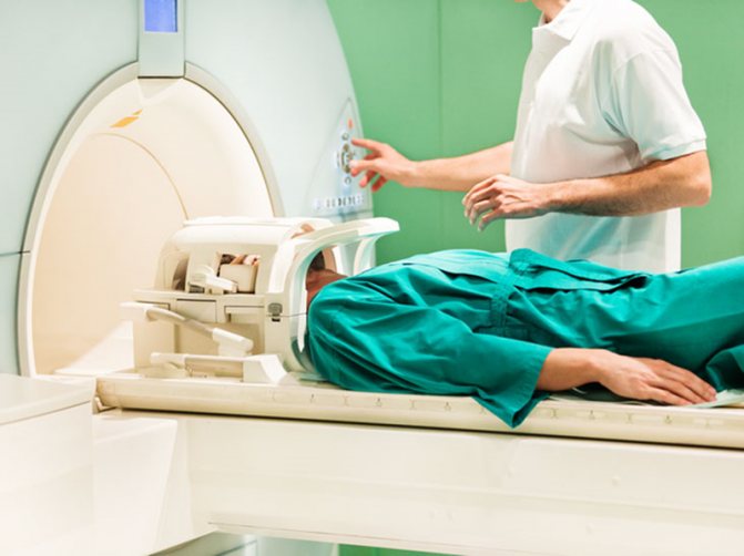
Preparing for MRI of the pituitary gland of the brain
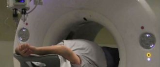
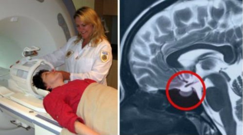
Before the examination, the patient will be asked to remove all metal-containing objects. The radiologist will ask the necessary questions and also tell you how the procedure will go. The patient lies down on a couch, which is then placed in the tomograph tube.
MRI procedure of the pituitary gland
A contrast agent will be injected at some point during the procedure. As a rule, no unpleasant sensations arise. Some patients note that when the drug is administered, they feel warm or slightly dizzy.
Interpretation of MRI results of the pituitary gland
The procedure for MRI diagnostics of the pituitary gland with contrast lasts about 30 minutes.
The examination results recorded on disk and a written conclusion based on the diagnostic results are given to the patient 20-30 minutes after completion of the procedure.
MRI diagnostics of the pituitary gland is the only technology today that allows us to identify pituitary microadenomas and determine their exact location. The procedure is quick and painless for the patient. Within 30 minutes after diagnosis, the patient can receive the examination results and transfer them to the attending physician to draw up a further treatment plan.
| What does an MRI show? |
| How is an MRI of the brain done with contrast? |
| MRI of the brain: what does tomography show? |
| MRI of the brain with contrast |
| MRI of the sella turcica and pituitary gland in Rostov-on-Don |
| MRI of the cerebellum of the brain |
Indications and contraindications for diagnosis

The specialist can refer the patient for a tomography after conducting an initial examination and passing all the necessary tests. In this case, an MRI is prescribed if, based on the results of other examinations, it was not possible to identify the cause of the deterioration in health. Indications for scanning may include:
- metabolic problems;
- decreased visual acuity;
- in women - menstrual irregularities;
- in men - impotence;
- excessively high levels of TSH in the blood;
- headache.
As for contraindications to undergoing MRI of the pituitary gland, they are usually divided into permanent and temporary. If there are permanent contraindications, examination is strictly prohibited:
- People with pacemakers. During operation of the scanner, they can not only fail, but also begin to move, which will cause injuries.
- Patients with metal implants, including dental ones.
- People with tattoos with metal connections.
Temporary contraindications may include:
1. The first 3 months of bearing a child. To date, there has not been enough research into the negative effects on the fetus, so scanning is prohibited in the first 3 months of pregnancy. However, an MRI in early pregnancy can only be performed if the woman's life and health are at risk.
2. The patient's serious condition. If the patient is unconscious after injury or under a ventilator. In this case, it is not possible to carry out the examination.
3. Mental illness. Another contraindication to scanning is the presence of mental illness in the patient. In some cases, for example, if the situation is urgent, MRI of the pituitary gland can be performed in such patients under anesthesia.
4. Uncontrollable tremors of the limbs. It simply does not make sense to conduct examinations of such patients during exacerbations for the reason that the quality of the images will be unsatisfactory.
A separate list of contraindications includes MRI of the pituitary gland with contrast:
- liver pathologies;
- kidney diseases;
- diabetes;
- contrast intolerance.
Before the scan, each patient must inform the radiologist about the presence of dental implants, dentures, metal objects in the body, tattoos, chronic diseases, regardless of whether an MRI of the pituitary gland is planned with or without contrast. This will avoid a large number of complications and make the diagnosis as safe as possible.