- MRI of the kidneys with contrast (contrast)
- Price
- doctor
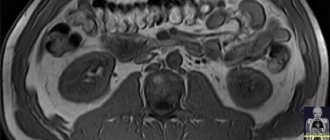
Magnetic resonance imaging, or MRI of the kidneys and renal arteries, is a diagnostic method that allows you to assess the condition of the kidneys and abnormalities in their structure. This method is one of the most effective and safe today and can be prescribed to children.
Briefly about
Address: Moscow, metro st. 1905, no. 7, building 1
Working hours: seven days a week, 24 hours a day
Equipment: Powerful Philips 1.5 Tesla
Specialists: All employees have at least 5 years of experience
MRI of the kidneys - features of conducting with contrast, detection of diseases
The magnetic resonance imaging method has wide diagnostic capabilities.
It is based on the effect of a magnetic field and radio frequency waves on the human body, which help to obtain accurate multi-layer images of any organ. Using them it is easy to determine the presence of pathology and its localization. The kidney function is to filter the blood and is vital for humans. If the functioning of an organ is disrupted, the entire body suffers. According to statistics, such diseases are quite common. Sometimes they are asymptomatic, sometimes with pain: burning, tingling in the lower back, difficulty urinating. Complications can be serious, including death. An MRI of the kidneys will help to conduct a detailed diagnosis and make the correct diagnosis.
Preparing for the study
Preparation for an MRI study of the kidneys does not imply any serious restrictions. 2 – 3 hours before the scan you must refrain from eating food, water, or medications. It is worth remembering that during the examination the presence of any metal jewelry, hairpins, watches, or glasses on the body is prohibited. Other contraindications to the study:
- presence of a pacemaker in the body;
- fixed dental, orthopedic prostheses, hearing aids;
- vascular clips;
- pins, spokes, fixing bolts, etc.
It is not recommended to apply decorative cosmetics that may contain metal microparticles. You must leave your mobile phone, any electronic gadgets, removable dentures, and hearing aids outside the doors of the diagnostic room.
If the patient suffers from claustrophobia and is terrified of closed spaces, on the eve of the study you can take light sedatives, such as Novo-Passit, tincture of valerian, etc. More serious drugs can only be taken as prescribed by a doctor.
This completes the preparation for MRI of the kidneys without contrast. If contrast enhancement is expected, an allergy test is performed immediately before the procedure, which will help assess the body’s reaction to the injected substance.
MRI examinations (general preparation rules)
Kidney MRI scan with contrast
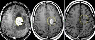
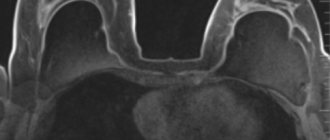
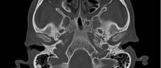
There are two types of magnetic resonance imaging used in modern medicine: simple and with contrast. The latter is prescribed for oncological processes. This type of study allows you to get a more informative picture of the disease present. The patient is injected intravenously through a catheter with a special substance that enhances tissue contrast. It will help you see not only tumors and metastases in the pictures, but even determine their type and stage of the process. In terms of its capabilities, the MRI method with contrast for examining the kidneys is comparable to a biopsy, in which tissue is taken for histology. The contrast agent accumulates differently in tumors, metastases and cysts, which makes it possible to distinguish them from each other and determine the degree of malignancy. After the procedure, the drug will be excreted from the body through urine and feces.
Kidney MRI with contrast
In general, MRI of the kidneys is a fairly informative diagnostic method, but in some situations the attending physician needs to obtain additional data that a standard examination cannot provide. In such cases, an MRI of the kidneys with contrast is prescribed. The study allows us to obtain a clearer structure of tissues, identify neoplasms, and determine the characteristics of their blood supply. To obtain this data, the patient is injected with a contrast agent.
Currently, drugs based on gadolinium salts are used. The drugs are quite safe, but extremely rarely, they can lead to the development of complications, for example, allergic reactions. In addition, this type of study requires more time to conduct and is much more expensive due to the high cost of the drug. For this reason, MRI of the kidneys with contrast is performed only if indicated.
What kidney and urinary tract diseases will an MRI show?
The range of disorders in the body detected by this method is wide. MRI examination of the kidneys shows pathologies both in tissues and blood vessels. It allows you to see:
- organ sizes;
- tissue structure and changes in them;
- structure of the bladder;
- neoplasms;
- vascular condition;
Among the kidney and urinary tract diseases diagnosed on MRI:
- aneurysm and vasoconstriction;
- renal artery thrombosis;
- abnormalities in the structure of blood vessels;
- renal infarction;
- abscesses, carbuncles and hematomas;
- inflammatory processes (pyelonephritis, glomerulonephritis);
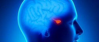
- adrenal oncology;
- presence of parasites.
When is an MRI of the kidneys necessary?
It is carried out in the following cases:
- renal symptoms of unknown etiology when the diagnosis has been completed (the patient’s face and limbs are swollen, pain in the lower back is observed, if this is accompanied by high body temperature, headache, vomiting and weakness);
- control of organs after surgery;
- there are traumatic injuries to the lower body;
- poor blood and urine tests if no pathologies are found during ultrasound examination;
- the presence of malignant processes and you need to find out their location and stage;
- high blood pressure;
- urography with contrast is contraindicated;
- burning and pain in the lower back;
- difficulty and pain when urinating;
- suspicion of an anomaly (organ enlargement, prolapse, congenital polycystic disease).
What to choose: checking the kidneys and adrenal glands with an MRI or ultrasound
To confirm the diagnosis, if there is a clear picture of a specific disease, an ultrasound may be prescribed. This method identifies stones and tumors. When the symptoms of the disease are smoothed out or the medical specialist is unable to determine its cause using ultrasound, the patient is sent for a tomography, which will show what exactly is happening in the human body: the picture of the disorders, the dynamics of their development. However, the same urinary stones can be suspected on tomography only by indirect signs. The method of examination should be determined by the attending physician.
Which is better: MRI or CT scan of the kidneys?
Today, doctors do not have a clear opinion about which is better - MRI of the renal system or CT. This is due to the fact that these methods are different in themselves and, accordingly, provide different information. With CT, you can find out the physiological data of the tumor, and with MRI, you can classify it in detail.
CT is considered a more harmful method, which has more contraindications. This is explained by the participation of X-rays during scanning. MRI in this regard is a safer diagnostic method, but at the same time more expensive.
Doctors sometimes recommend that their patients undergo both an MRI and a CT scan in order to obtain the maximum amount of data and make an accurate diagnosis.
Advantages and disadvantages of kidney MRI examination
It is safe for humans, so it can be prescribed to children. Unlike X-rays and computed tomography, the person being examined does not receive radiation. During the examination, several images are obtained at once in different planes, when this is impossible to obtain with an X-ray machine. You will have to be irradiated over and over again.
The downside is the price of a kidney MRI. As well as the duration of the procedure, during which you need to lie still. There are a certain number of contraindications. The tomograph is prohibited for people:
- with metal implants;
- cardio and neurostimulants;
- defibrillators;
- ferromagnetic implants or prostheses;
- vascular stents and clips.
The magnetic field of the device can be deadly for them. It has the property of attracting metal objects to itself and moving them in the human body. Those, in turn, can damage surrounding tissues and structures.
Pregnancy is also a contraindication. It is not suitable for people with high body weight (over 120 kg), as well as those suffering from fear of closed spaces (claustrophobia), epilepsy and hyperkinesis.
There are also prohibiting factors in which diagnosis is not carried out, but is possible in extreme cases. Among them is the installation of non-magnetic implants. The taboo is valid for 6 weeks after surgery. During this period, the device will be fixed. These include:
- insulin pumps and other drug infusion devices;
- joint prostheses;
- artificial heart valves;
- bone plates and pins.
Contrast during MRI of the kidneys is not recommended for those suffering from diseases of the urinary system and those who are intolerant to the contrast agent.
How is an MRI of the kidneys and urinary tract done with contrast?
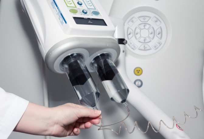
Contrast during MRI can be supplied automatically using an injector
After choosing a clinic and making a preliminary appointment, you need to show up for the procedure. Be sure to tell your doctor about possible pregnancy, allergies to contrast, or metal parts in the body.
If you have diabetes mellitus or persistent hypertension, you may not be aware of concomitant renal failure, and then it is better to first donate blood for creatinine. But contrast-induced nephropathy is an incidentally rare complication, so this measure is not mandatory. Gadolinium and similar paramagnetic drugs practically do not cause significant side effects, unlike iodine-containing agents, therefore MRI is preferable to excretory urography not only in terms of information content, but also in safety. Contrast can be bolus: the substance enters the vein automatically using an injector during certain phases of the study.
For unforeseen situations, there is a button at hand to contact the staff; the procedure is monitored from the adjacent room. After the diagnosis is completed, you can lead your usual lifestyle; there are no restrictions on driving a car or performing precision work. In order for the contrast agent to leave the body faster, it is recommended to increase the drinking regime.
The results will be ready in an hour, all questions can be asked to the radiologist. In 99% of cases, after deciphering the images, the diagnosis is known, but final verification is only legitimate by the attending physician, most often after histology. In the most difficult cases, they resort to collegial evaluation of tomograms.
How the kidneys and urinary tract are examined during an MRI
No special preparation is needed. Approximately 6 hours before the scheduled time of the study, you should stop eating and drinking. Meals should be light, not heavy. You should also not smoke. A few days before the procedure, try to eat right, exclude heavy fatty and protein foods, as well as foods rich in fiber from your diet. Try to drink more clean water, avoid drinking alcohol and soda. Eliminate stress and nervous tension.
Wear loose clothing for the examination (some medical centers offer special shirts). Do not forget to remove all metal objects from your pockets and remove metal jewelry. It’s best to leave all this at home so you don’t accidentally forget to take it off. On the day of the examination, it is better for girls and women not to use cosmetics with metal particles, even very small ones. When exposed to radiation, they can burn the skin.
It is better if you are referred by a doctor. He will monitor your condition in advance using tests and make a decision on the use of contrast.
Find out at your medical facility how tomography is performed on children. Due to their age, children cannot remain motionless for a long time and will be offered sedation or anesthesia. It is better to prepare the child in advance, explain how and what will happen in the process.
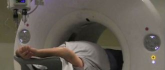
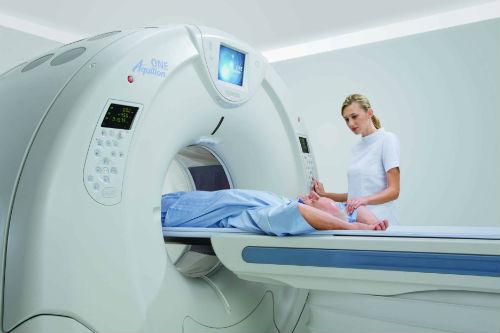
MRI examinations of the adrenal glands, kidneys and bladder are performed in the supine position. If this is your first time on it, then you need to know what it is:
- The medical center will ask you to fill out a questionnaire where you need to indicate your physiological data, medical history and health characteristics. This is necessary for additional control of your safety and security.
- Afterwards you will be taken to the locker room, where you need to leave your things and remove metal objects. Put on clothes for tomography there.
- When the time comes, the doctor will invite you into the office with the device.
- You will be given earplugs or headphones. They are needed to avoid strain on the ears, since the design makes loud noise.
- If contrast injection is expected, a catheter will be installed.
- They will explain where the panic button is located. Press it if something goes wrong or if you feel unwell.
The procedure will last about 20 minutes. All this time you will be under the supervision of a specialist located in the next room.
While lying in the machine, try not to move or cough to ensure high-quality results.
How to prepare for an MRI of the kidneys and urinary tract
No special preparation is required before a routine MRI.
No special preparation is required before a routine MRI. If the examination is planned with a contrast method, then the patient’s last meal should be approximately 6 hours before the start of the procedure. Preparation for an MRI of the kidneys should be carried out in accordance with all doctor’s instructions to obtain a reliable and high-quality examination result. To do this, the patient needs:
- Be dressed in underwear made of natural material;
- Immediately before starting the procedure, remove all metal-containing items such as: jewelry, hairpins, watches, clothes with metal fittings;
- Place your mobile phone, hearing aid, keys and other metal objects in a special safe.
Some clinics use sedatives to reduce feelings of fear and anxiety in children. For high-quality prevention of gas formation, several days before the start of the MRI procedure itself, it is recommended to take white or black coal orally, at the rate of 1 tablet per 10 kg of weight, 3 times a day after meals.
Where to get an MRI of the kidneys in Moscow
There are 157 diagnostic centers in the capital where magnetic resonance examinations are carried out. Call us at 8(495)221-63-87 and we will help you choose an institution in Moscow where you can have your kidneys checked for MRI. Our consultants will introduce you to discounts and bonus programs and select a center with the optimal price for a kidney MRI.
The cost of the procedure depends on the type of device and the qualifications of the medical personnel.
There are two types of tomographs: open and closed. The first is suitable for small children and those who are afraid of narrow, enclosed spaces. Has restrictions on body volume and weight of people. It is less noisy and powerful, which is why the cost of MRI of the kidneys is lower. How much and where exactly – our specialists will tell you. In the open, complex examinations such as intestines and magnetic resonance angiography are not possible.
The closed tomograph is a classic. It has a higher magnetic field strength, therefore the quality of the resulting images is also better. It's noticeably noisier.