Causes of kidney cancer
Kidney oncology is the proliferation of a malignant neoplasm of an organ. The tumor may appear on one kidney or both. Metastases tend to be detected in other organs. Experts say that the occurrence of kidney cancer is associated with many provoking factors.
- Heredity – affects the genetic predisposition of the body to the development of the disease. If the parents suffered from cancer, then the children are more likely to develop malignant neoplasms.
- Often cancerous tumors appear due to exposure to radiation or frequent contact with harmful chemicals.
- Smoking promotes the penetration of a large number of carcinogens into the body. Most patients diagnosed with kidney cancer are addicted to this addiction.
- Obesity promotes the appearance of tumors in the kidney tissue at an early stage of the appearance of excess weight. Addiction to fatty foods increases the risk of cancer.
- Diabetes mellitus and hypertension can also become a provoking factor in the development of kidney cancer.
- Any harsh mechanical impact on the kidneys can provoke disease. Falls, injuries, and blows to the kidney area can cause cancer.
- Excessive drug use and uncontrolled use of drugs increases the risk of kidney cancer.
- Chronic diseases of viral etiology and problems associated with their treatment provoke the appearance of malignant tumors.
No one has yet established a clear cause for the occurrence of malignant neoplasms. There are many points of view on this issue. Given the undoubted success in the treatment of kidney cancer in Moscow, it is still too early to talk about a complete, 100% victory over cancer. Scientists will be able to defeat cancer only after the universal mechanisms of carcinogenesis and ways to prevent the development of malignant tumors are discovered.
Classification
Currently, in Russia and in most countries of the world, the TNM classification (T-tumor, N-node, M-metastasis) is used.
- T—primary tumor TX —primary tumor cannot be assessed
- T0 - no data on the primary tumor
- T1 - tumor no more than 7 cm in greatest dimension, limited to the kidney
- T1a - tumor up to 4 cm
- T1b - tumor 4-7 cm
- T2 - tumor more than 7 cm in greatest dimension, limited to the kidney
- T3 - the tumor spreads into large veins, or invades the adrenal gland or surrounding tissues, but does not extend beyond the Gerota's fascia
- T3a - tumor invasion of the adrenal gland or perirenal tissue within Gerota's fascia
- T3b - Tumor extends into the renal vein or inferior vena cava below the diaphragm
- T3c - the tumor extends into the inferior vena cava above the diaphragm or invades its wall.
- T4 - Tumor extends beyond Gerota's fascia
- N - regional lymph nodes NX - regional lymph nodes cannot be assessed
- N0 - no metastases in regional lymph nodes
- N1 - metastasis in one regional lymph node
- N2 - metastases in more than one regional lymph node
- M - distant metastases MX - distant metastases cannot be assessed
- M0 - no distant metastases
- M1 - distant metastases
- G - histopathological grading GX - degree of differentiation cannot be assessed
- G1 - well differentiated tumor
- G2 - moderately differentiated tumor
- G3-4 - poorly differentiated/undifferentiated tumor
Grouping by stages
| Stage I | T1 | N0 | M0 |
| Stage II | T2 | N0 | M0 |
| Stage III | T1 | N1 | M0 |
| T2 | N1 | M0 | |
| T3 | N0, N1 | M0 | |
| Stage IV | T4 | N0, N1 | M0 |
| any T | N2 | M0 | |
| any T | any N | M1 |
Kidney cancer: symptoms
The main problem of early detection of the disease is the asymptomatic course of the disease. In the early stages, there are no signs of kidney cancer, appearing only in the later stages.
- Sharp or aching pain in the lower back appears.
- Blood appears in the urine, possibly with clots.
- A tumor-like formation is palpated in the projection of the kidney.
- Increased blood pressure, red blood cell levels, calcium levels.
- Hyperthermia is a constant increase in body temperature.
In the later stages of the disease, other characteristic symptoms occur: appetite decreases, the person loses weight sharply, anemia causes pale skin, the erythrocyte sedimentation rate increases, the patient experiences general weakness, and quickly gets tired.
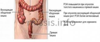
Nature of metastases
- When metastases appear in the lungs, coughing and hemoptysis appear.
- When metastases appear in the bones, severe pain occurs in the projection of the affected area, pathological bone fractures, and compression of the spinal cord. Upon palpation, a new formation on the bone is clearly identified.
- With metastases in the brain, rapidly progressive neurological manifestations occur.
- When metastases occur in the liver, jaundice occurs, the abdomen enlarges due to accumulation in the abdominal cavity, that is, ascites.
What to do in the postoperative period?
On average, a patient stays in the hospital for about a week after laparoscopic partial nephrectomy. Drinking and eating are usually allowed the next day after surgery, and walking is allowed in the evening of the same day. After the operation, as before, a broad-spectrum antibacterial drug is administered.
During the postoperative period, you will be advised to:
- drink 1-2 liters of water per day;
- do not lift weights exceeding 5 kg;
- do not undergo heavy physical activity.
You should immediately consult a doctor if you have:
- blood appeared in the urine;
- body temperature increased;
- severe abdominal pain occurred.
After surgery, your doctor will schedule consultations for you to undergo examinations, blood and urine tests, ultrasound examinations, and computed tomography scans to evaluate the effectiveness of treatment. After 5 years, if there is no evidence of tumor progression, the patient is removed from the register.
Make an appointment at the urology clinic of the First Moscow State Medical University named after. Sechenov, you can see a urologist, oncologist, Doctor of Medical Sciences G. N. Akopyan by phone or through the interactive Appointment form on our website.
September 11, 2019
Hakobyan Gagik Nersesovich - urologist, oncologist, MD, doctor of the highest category, professor
All clinical observations...
Diagnostics
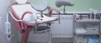
Oncology is a serious problem, but despite the severity of the disease, kidney cancer is treatable, especially if diagnosed early. If you have pain in the kidney area, you should contact a urologist. A specialist doctor will conduct a preliminary examination, palpation, collect anamnesis and prescribe the necessary tests. To make the most accurate diagnosis, additional studies are prescribed, which are carried out in the laboratory using radioisotope, ultrasound, and x-ray methods.
All patients, without exception, donate blood for biochemistry and do a routine clinical blood test. A general urine test is performed. The presence of a tumor is diagnosed, its location is clarified, the exact dimensions and relationships with the internal structures of the kidney and nearby organs and vessels are determined.
- The X-ray diagnostic method provides the most complete information when using multislice computed tomography with contrast and three-dimensional image reconstruction or MSCT.
- In some cases, excellent results can be obtained using magnetic resonance imaging or MRI.
- X-ray or computed tomography of the chest is used to diagnose lung metastases.
- Radioisotope scanning of skeletal bones or bone scintigraphy helps diagnose bone metastases.
- Radionuclide testing or nephroscintigraphy helps determine the functions of both kidneys.
The puncture allows you to obtain tumor tissue samples, which are then examined in detail in the laboratory. The condition of the lymph nodes, bones, bronchi and lungs, and organs located in the abdominal cavity is subject to mandatory checks. Additional studies are designed to determine the presence of metastases present in the body.
Computer 3D modeling of the tumor and the operation itself is used when planning the operation.
Indications for surgery
Endoscopic removal of part of the kidney is performed in the presence of a malignant neoplasm. In this case, the tumor must be located within the organ, without spreading to surrounding structures.
In addition to the above, surgery is prescribed in the following cases:
- neoplasm of a single kidney;
- bilateral malignant kidney disease;
- a tumor of the one remaining functioning kidney, without which the patient will be forced to constantly undergo dialysis;
- diseases of the kidney that are not affected by the tumor, which can lead to loss of its function (kidney infarction, partial kidney damage as a result of injury or tuberculosis).
Contraindications for laparoscopic partial nephrectomy include the following:
- acute inflammatory and infectious diseases;
- severe concomitant pathologies;
- diseases of the circulatory system associated with blood clotting disorders in the stage of decompensation;
- extreme obesity;
- late stages of pregnancy.
Acute inflammatory processes are a contraindication for surgery due to the possible risk of infection of the abdominal cavity. The presence of pronounced scars in the abdominal cavity leads to technical difficulties in isolating the kidney from the surrounding tissues, which makes laparoscopic resection of the kidney impossible.
Kidney cancer: treatment
Patients diagnosed with a serious illness are most interested in the question: can kidney cancer be treated or are all efforts futile, including surgery? The experience of our clinic clearly confirms that timely treatment and removal of a kidney tumor (cancer) in Moscow often demonstrates good results. With early diagnosis and radical treatment, success is achieved in 90% of cases. To help patients, urologists use several constructive techniques: immunotherapy, targeted therapy, surgery.
- Laparoscopic partial kidney resection with tumor is used more often than classic open surgery.
- If resection is technically impossible, kidney nephrectomy, open or laparoscopic removal of the organ along with the tumor is performed.
- Ultrasound-guided tumor cryoablation.
- Chemoembolization.
- For inoperable cancer and metastasis, targeted and symptomatic therapy is used.
We treat kidney cancer, and the prognosis is favorable in the vast majority of cases if treatment is started at an early stage. If the disease is detected and treatment is started at a later date, the prognosis will be less favorable. To exclude risk factors, it is necessary to conduct a preventive ultrasound of the kidneys annually. Early detection of cancer, when there are no obvious symptoms of the disease, gives a high probability of a positive result.
The best result is achieved by surgical intervention. Most often, operations are performed to resection or completely remove an organ affected by a malignant tumor.
- Resection is the removal of part of an organ with a detected and precisely localized tumor.
- Kidney nephrectomy is the complete removal of an organ affected by cancer.
The type of surgery is selected depending on the stage of the disease, size and location of the tumor. Resection is used for stage 1 or 2 kidney cancer. In later stages or a large tumor size, it is advisable to remove the entire kidney. The stage of the disease is determined based on the results of a complete medical history, examination, and histology. The method is chosen taking into account the patient’s age and concomitant diseases.
Doctors at our clinic have the opportunity to treat the patient as carefully as possible, choosing gentle treatment methods. Nowadays, the best results are achieved by minimally invasive surgeries to remove the tumor while preserving the organ. At the same time, doctors do not perform open intervention or make deep incisions to gain access to the diseased organ. Laparoscopic or robot-assisted kidney resections facilitate the subsequent rehabilitation process. Additional therapy is prescribed according to indications. If metastasis does not occur, then the patient has every chance of a healthy life.
Laparoscopic partial nephrectomy
Symptoms
Currently, as described earlier, due to the developed diagnostic system, kidney tumors are detected at an early asymptomatic stage. However, some of the symptoms characteristic of a kidney tumor are described below.
- Pain. In most cases, it is local in the lumbar region of the affected side. Irradiation of pain to nearby areas usually does not occur. The type of pain in the vast majority of cases is aching, dull and, in some cases, pulling. The pain can be either constant or intermittent.
- Macrohematuria. Blood in the urine is the most serious symptom of urological diseases. If this symptom is present, diagnostic measures are necessary to detect the source of bleeding. The admixture of blood in the urine can also be invisible to the eye, but can only be determined in laboratory conditions, the so-called microhematuria. Both of these conditions require immediate consultation with a urologist.
- Palpable formation. When the formation reaches a large size, a kidney tumor can be determined by conducting a general palpation examination of the patient by a urologist. The tumor can be felt when the doctor's palm is placed on the anterolateral surface of the abdomen. This symptom is a sign of an advanced oncological process and requires immediate investigation and determination of the correct diagnostic and treatment tactics.
Contraindications to laparoscopic partial nephrectomy
Today, as in any area of laparoscopic surgery, there is a list of contraindications in which surgical treatment is associated with a high risk of complications such as bleeding, or surgical treatment will be difficult.
Contraindications:
- impaired functioning of the blood coagulation system;
- infection;
- late stages of pregnancy.
- acute inflammatory processes;
- the presence of scars in the abdominal cavity from previous operations entails significant difficulties in isolating the kidney and surrounding tissues.
Preparation for surgical treatment
Before the operation itself, bowel preparation is carried out and broad-spectrum antibiotic therapy is prescribed to prevent infectious complications.
Stages of laparoscopic kidney resection (operation progress)
The operation is performed under general anesthesia (putting you to sleep during the entire operation). The patient is placed on the operating table in a special position - on the healthy side, while the table is extended, thereby allowing the kidney to be brought closer to the anterior abdominal wall. Access to the kidney is performed using 3-4 mini-holes of about 1 cm on the anterior abdominal wall; a laparoscope (video camera) is passed through one hole, and other necessary surgical instruments are passed through the rest. Subsequently, one of the holes is slightly extended to remove a special package with a tumor from the abdominal wall. Visibility with the help of a laparoscope increases several times, which allows the surgeon to work more accurately and accurately. This is especially necessary during resection, since unlike kidney removal (nephrectomy), resection is a technically more complex manipulation that requires more precise actions. Carbon dioxide is injected into the abdominal cavity, which allows you to increase the working space; after the operation, the gas is completely removed.
After creating optimal conditions, the surgeon begins to isolate the kidney; to do this, he takes the organs to the side and dissects the tissue. The stage of isolating the vessels of the kidney follows after isolating the kidney itself and examining it. Depending on the complexity of the tumor, its location and size, the surgeon decides on the method of temporary hemostasis (stopping the blood supply to the kidney to ensure minimal blood loss during surgery). The following are the three most commonly performed methods of temporary hemostasis:
Clamping the main trunk (applying a bulldog clamp). This method allows you to completely block the flow of blood to the kidney, thereby ensuring a “dry” surgical field. This method of hemostasis has a time limit of no more than 40 minutes. If hemostasis lasts more than 40 minutes, based on literature data, kidney death occurs. In our clinic, the maximum time for this type of hemostasis is 15 minutes.
Selective segmental ischemia (applying a bulldog clamp to the artery that supplies blood to the segment of the kidney in which the tumor is located). The method allows you to avoid clamping the main trunk of the renal artery.
ZERO ischemia. A method in which the kidney vessels are not compressed. According to domestic and foreign literature, compression of the renal blood flow entails some changes in the functional ability of the kidney. Considering the above, our clinic has accumulated experience in performing laparoscopic resection using ZERO ischemia.
After this, the most crucial moment begins - the resection stage. After removing the area of the kidney with the tumor, the edges of the kidney are sutured. Next, the abdominal cavity and the bed of the removed tumor are examined for bleeding. After making sure that there is no bleeding, the tumor is placed in a special bag and removed from the abdominal cavity. The surgeon then removes the instruments and stitches the tissues together. The duration of the surgical procedure varies from 1.5 to 3 hours, depending on the complexity.
The immediate postoperative period in the ICU
After surgical treatment, the patient is transferred to the anesthesiology and intensive care unit (ICU) under the supervision of a resuscitator, where dynamic monitoring of vital functions is carried out (control of blood pressure, heart rate, respiratory function, amount of urine excreted, etc.), in addition The drainage discharge (a special silicone tube installed in the wound cavity), urine color, temperature and general condition of the patient are assessed.
The most common complaints in the immediate postoperative period:
- pain in the area of postoperative wounds, often not requiring additional drug pain relief, which differs from pain during open operations, when analgesic therapy is required;
- moderate nausea resulting from the administration of various drugs necessary for anesthesia;
- the presence of a urethral catheter is necessary for dynamic control of the amount and color of urine. It is deleted the next day.
Postoperative period in the department
The next day after the operation, the patient, in agreement with the resuscitator, is transferred to the urology department under the supervision of the attending physician.
In the department, an examination will be carried out by the attending physician, the operating surgeon and the head of the department. Antibacterial, anti-inflammatory, infusion and symptomatic therapy was prescribed and prescribed. The urethral catheter and safety drainage are removed in the department on the first or second day.
The hospitalization period ranges from 5 to 7 days. Upon discharge, your attending physician will prescribe therapy and dates for consultations and examinations. After 5 years, if there is no evidence of a relapse, the patient is removed from the oncology register.
The cost of laparoscopic kidney resection for a tumor is approximately 200 - 250 thousand rubles.
Prices vary based on the volume of operational assistance. This price includes all costs for the operation itself, anesthesia, disposable consumables, postoperative stay in intensive care, as well as the administration of medications, stay in the ward, food, necessary postoperative examination and treatment.
Urologist Mamedkasimov Nariman Akitovich has extensive experience in performing laparoscopic operations for kidney tumors. Patients operated on by him do not remain unattended even years after the operation, due to the work of a quality control system for operations performed and monitoring of overall relapse-free survival.