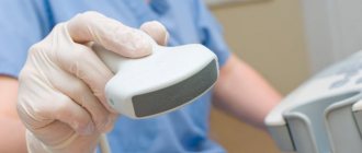
Cystoscopy – examination of the inner surface of the bladder using an endoscopic system.
Cystoscopy is one of the main methods used to diagnose neoplasms and other diseases of the bladder. The study is carried out using a cystoscope, which is inserted into the urethra and then advanced into the bladder.
In medical practice, 2 types of cystoscopes are used: rigid and movable.
The rigid cystoscope has practically been replaced by flexible equipment, but it is used for retrograde catheterization of the ureters and transurethral resection of the prostate gland.
Using a movable cystoscope, you can observe the internal structure of the bladder on the monitor screen. A movable cystoscope consists of a thin, hollow metal tube 30 cm long, equipped with a light source and an optical system.
The equipment allows the doctor, using the magnification function, to examine the condition of the mucous membrane of the urethra and bladder. The cystoscope is equipped with a large number of different optical fibers and lenses that transmit a complete image of these organs from the inside
In addition, the cystoscope has special channels that allow delivery directly into the bladder of the instruments necessary for the manipulation (biopsy forceps, loops for removing polyps, needles for aspiration, etc.).
1
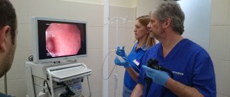
Ureterorenofibroscope Pentax FUR-9RBS
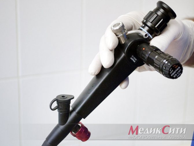
2 Pentax FUR-9RBS ureterorenofibroscope
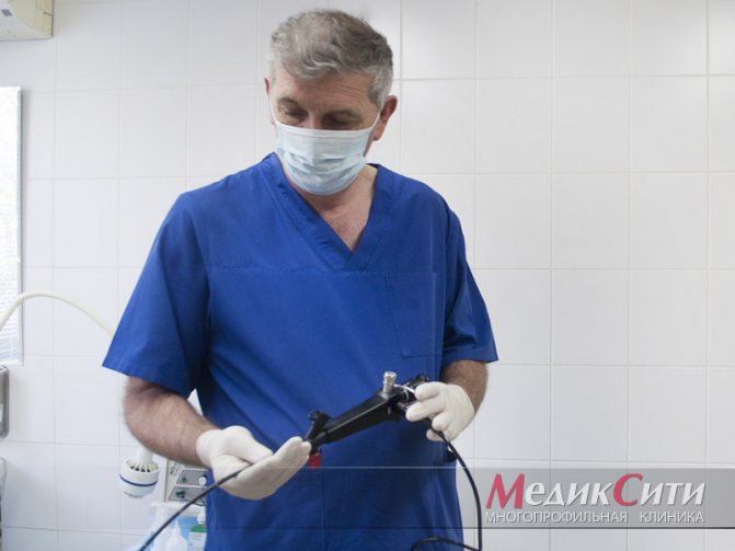
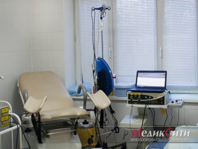
3 Cystoscopy in MedicCity
More about the disease
Strictures (narrowing) of the urethra are diagnosed in 1–2% of urological patients.
Men are more susceptible to pathology due to the anatomy of the genitourinary organs. Narrowing of the urinary canal can be congenital or acquired, short (up to 2 cm) and long (more than 2 cm). As a result of the negative impact of traumatic or infectious factors on the mucous membrane of the urinary canal, dead cells begin to be replaced by scar-connective tissue, which causes a narrowing of the lumen. In many cases, not only the mucous membrane, but also deeper tissues are involved in the pathological process. As the disease progresses, more and more problems arise with urination - difficulty in passing, incomplete emptying of the bladder, stagnation of urine, etc., up to acute urinary retention due to obliteration (complete closure of the lumen) of the urethral duct.
How is an ultrasound of the urethra done in men?
Preparations include normal hygiene procedures. An important feature of the examination for men is that the patient must undergo it with a full bladder. A couple of hours before visiting the office you need to drink about a liter of water. Due to the anatomical features, before the examination, 10 to 25 ml of a special solution is injected into the urethra. The contrast agent displaces air from the lumen and allows you to get a clear image on the screen. The patient sits on the couch, lies on his back and takes the “frog” position (legs apart and bent at the knees and hip joints).
Decoding the results
An ultrasound diagnostic specialist, based on the received image, records the data and draws conclusions. There are three possible results, which are described below.
Normal indicators
During the study, no foreign body, narrowing of the lumen or swelling of the canal walls was detected. In this case, the cause of the patient’s complaints must be sought in another organ of the genitourinary system.
Inflammatory process
The main symptom in this case is an increase in the thickness of the walls of the urethra, which caused a decrease in the lumen.
Why surgery is needed
Scar tissue changes are irreversible. Stricture disease cannot be treated with medication, and methods such as bougienage of the urethra have a temporary effect. The only way to permanently eliminate pathological changes and restore normal function of the lower urinary tract is through surgery. To effectively treat stricture disease of the urethra, a wide range of plastic and reconstructive surgeries are used, aimed at replacing the damaged area of the urinary canal with healthy tissue.
Benefits of contacting us
The surgical center of the GMS clinic provides treatment for urethral strictures of any length, location and complexity, including previously unsuccessfully treated or operated ones. Surgical tactics are selected by our specialists individually in each case. The intervention option depends on the root cause that caused the narrowing of the urinary canal, the severity of the disease, and the volume of intervention.
At the surgery center at GMS Hospital, operations for stricture disease are performed by experienced specialists with many years of practice. Mostly, plastic and reconstructive interventions are performed using gentle microsurgical techniques, which provides the following advantages:
- minimal surgical trauma;
- low risk of surgical complications;
- short recovery period;
- good therapeutic and aesthetic result;
- rapid healing and return to a healthy life.
There is no universal operation that would be suitable for all clinical cases. Each stricture is unique, and accordingly, the success of the intervention is ensured only in the case of a qualified selection of the most effective surgical technique. Our specialists have a thorough knowledge of all the arsenal of plastic and reconstructive surgeries on the urethra and their variations.
Cost of treatment for urethral strictures
The prices indicated in the price list may differ from the actual prices. Please check the current cost by calling +7 495 104 8605 (24 hours a day) or at the GMS Hospital clinic at the address: Moscow, st. Kalanchevskaya, 45.
| Name | Price |
| Balloon dilatation/cutting of ureteral stricture | RUB 107,520 |
| Laser incision/bougienage of ureteral stricture | RUB 90,244 |
| Plastic surgery of the lower third of the ureter (Boari operation) | RUB 178,563 |
| Endoscopic balloon dilatation of urinary tract strictures | 126,000 rub. |
Dear Clients! Each case is individual and the final cost of your treatment can only be found out after an in-person visit to a GMS Hospital doctor. Prices for the most popular services are indicated with a 30% discount, which is valid when paying in cash or by credit card. You can be served under a VHI policy, pay separately for each visit, sign an agreement for an annual medical program, or make a deposit and receive services at a discount. On weekends and holidays, the clinic reserves the right to charge additional payments according to the current price list. Services are provided on the basis of a concluded contract.
Plastic cards MasterCard, VISA, Maestro, MIR are accepted for payment. Contactless payment with Apple Pay, Google Pay and Android Pay cards is also available.
Western standards of treatment (evidence-based medicine)
Continuous staff development
Regular interaction with leading Russian and foreign medical institutions
Modern medical equipment and advanced diagnostic and treatment methods
Unified standard of service
We work around the clock 24/7/365
Causes of the disease
Congenital urethral strictures account for only 2% of all cases. Much more often, urologists encounter acquired narrowing of the urinary canal caused by the following reasons:
- traumatic injuries to the urethra;
- inflammatory and infectious diseases of the genitourinary system;
- iatrogenic - arising as a result of urological diagnostic and therapeutic procedures;
- nonspecific dystrophic-degenerative diseases (balanitis obliterans, lichen sclerosus).
Signs and symptoms
Symptoms of the disease are expressed in a violation of the outflow of urine from the bladder and is manifested by the following signs:

- thin, sluggish stream of urine;
- difficulty urinating;
- the need to strain when urinating;
- incomplete emptying of the bladder;
- intermittent or splashing stream of urine;
- leakage of urine after urination;
- discomfort and pain when urinating;
- lower abdominal pain;
- discharge from the urethral canal and relapses of its inflammation.
With severe strictures, urine is released in rare drops, and when the lumen of the canal is obliterated, acute urinary retention develops - an emergency condition requiring urgent medical intervention.
After cystoscopy
After cystoscopy, the following unpleasant sensations may be observed (which disappear over time):
- pain and burning in the bladder (to reduce these symptoms, it is recommended to drink more fluids);
- frequent urination;
- urinary retention;
- lower back pain;
- chills, fever;
- bloody discharge in the urine (should disappear after a couple of days).
If the discharge does not disappear within 2 days after cystoscopy, then you should urgently seek advice from a urologist.
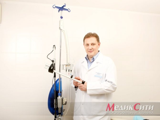
1 Consultation with a urologist after cystoscopy
2 Consultation with a urologist after cystoscopy
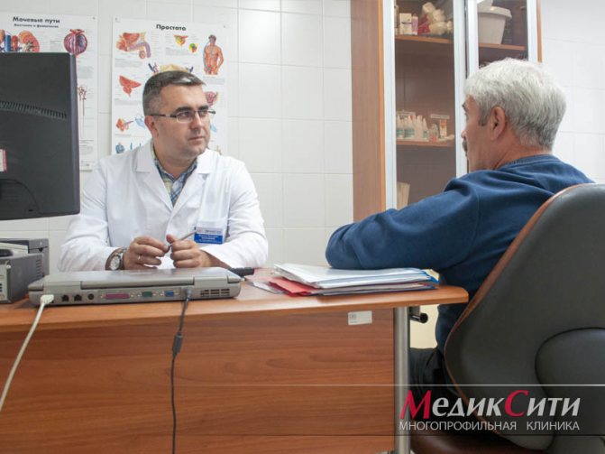
3 Consultation with a urologist after cystoscopy
Diagnostics
Since the clinical signs of stricture disease are similar to many other diseases of the urinary system, differential diagnosis is carried out, including:
- examination by a urologist;
- urofluorometry;
- cystometry;
- retrograde urethrocystography;
- cystourethroscopy;
- Ultrasound of the urethra and bladder.
Additionally, CT and MRI of the urethra may be prescribed to assess the degree of damage to the urethral meatus and the involvement of nearby tissues in the scarring process. If a malignant process is suspected, a biopsy of the affected areas is performed.
Indications for the procedure
Cystoscopy is prescribed to diagnose diseases or clarify their severity and course. The procedure allows you to differentiate pathologies that have similar symptoms. With its help, neoplasms are excluded, and if they are present, the type of tumor is determined, which can be benign or malignant.
Cystoscopy is indicated for the following diseases and symptoms:
- Cystitis. It manifests itself as painful urination, pain in the sacrum or lower back. There is increased frequency of urination, a decrease in the urine produced per micturition, which becomes cloudy and foul-smelling.
- Tumor. The symptoms are similar to cystitis, but analysis of a smear from the urethra shows the presence of atypical cells.
- Prostate hyperplasia or adenocarcinoma, prostatitis. They are manifested by a frequent desire to urinate, incomplete emptying of the bladder, and urinary retention. The desire to defecate usually occurs at night.
- Male infertility. It is carried out to study the condition of the seminal tubercle.
- Enuresis, if there are no mental disorders or neurological abnormalities.
Endoscopy is also performed to identify the causes of pyuria or hematuria, conditions in which pus and blood are found in the urine excreted, respectively. With its help, you can exclude or confirm urolithiasis with the location of stones in the bladder. It allows you to evaluate the effectiveness of the treatment selected by the doctor and, if necessary, adjust it.
What treatments are used
Treatment tactics are selected based on the results of a comprehensive examination, depending on the location, severity and extent of scar-sclerotic changes. If the cause of the pathology is an infectious-inflammatory process, drug treatment is prescribed - antibiotics, anti-inflammatory drugs, etc. Surgical treatment is aimed at restoring the anatomical shape, integrity and functionality of the urinary canal.
To restore urethral patency, the following types of interventions are used:
- anastomotic urethroplasty - excision of the stricture of the urinary canal and restoration of its integrity by forming an anastomosis (the healthy ends of the urethra are sutured). It is performed for scar lesions of the posterior parts of the urethral canal (prostatic, membranous, bulbar zone) with small structures, no more than 2 cm.
- replacement (augmentation) urethroplasty - pathologically altered areas are excised and replaced with an allograft, which is used as a fragment of the mucous membrane of the cheek, gums (buccal plastic surgery) or the foreskin of the patient's penis. This technique has many modifications; the operation option is selected depending on the degree of tissue damage and the causes of the pathological process.
- intraurethral stenting is an endoscopic procedure that involves installing a special stent into the lumen of the canal, which acts as a supporting frame and does not allow the lumen to narrow.
- internal urethrotomy is a minimally invasive operation aimed at dissecting the stenotic area. It is carried out for scar changes no more than 0.5 cm in length.
At the GMS Surgery Center, urethral stricture is treated using advanced, minimally invasive methods. Our surgeons thoroughly master all variants and modifications of surgical techniques aimed at restoring the patency, anatomy and functionality of the urethra and perform operations at the highest level.
How is an ultrasound urological examination performed?
In order to conduct the highest quality examination and receive effective and high-quality treatment, you must carefully prepare for the procedure. A few days before the procedure, it is necessary to prepare the intestines for this purpose; be sure to exclude from the diet all foods that contribute to increased gas formation. Such products include cabbage, buckwheat, legumes, brown bread, and carbonated drinks.
Before the examination, you can take enzymatic preparations and drugs that help reduce the formation of gases in the intestines. For constipation, you can take activated charcoal and laxatives or plant decoctions. In some cases, the examination is performed using a rectal probe. Before such an examination, it is necessary to do a cleansing enema. Ultrasound examination is carried out strictly on an empty stomach. It is not recommended to eat food 12 hours before it starts.
Disease prevention
Prevention of the formation of scar-sclerotic changes consists in the prevention and timely treatment of sexually transmitted infections, inflammatory processes, and avoidance of injuries to the genitourinary system. Remember - self-medication is unacceptable, and all medical procedures must be performed by qualified specialists!
Prevention of relapse of the disease lies in choosing an adequate treatment method. You can get maximum information about the pathology and existing treatment options at a consultation with a urologist at GMS Hospital. You can make an appointment with a specialist around the clock, using the feedback form on the website or by phone.
Reception is strictly by appointment only!!!
Doctor Plus LLC License No. LO-77-01-004801
Registration at the clinic by phone
Make an appointment at the clinic
- +7 (495) 125-49-50
- Addresses of clinics in Moscow
- Daily
- call me back
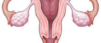
Urological ultrasound for women
allows you to timely identify the main causes leading to the development of diseases and prescribe modern and comprehensive treatment. Using this technique, you can easily and quickly identify the most common urological diseases in women.
Very often, women mistakenly consult a gynecologist when urological problems arise. To understand the causes and determine an accurate diagnosis, you need to consult a urologist. It is convenient to undergo an examination and get a consultation with a urologist by contacting our clinic.
You can make an appointment for ultrasound diagnostics with our consultants by phone
Prices for ultrasound Clinic addresses Ultrasound at home Cardiac echo Urology center Calling a urologist to your home