Ultrasound sensor is the key to your ultrasound's performance. In this blog, we will explain the different types of ultrasound transducers and identify the types of studies you can use them for.
In the end, we will offer you some good points that you should keep in mind when purchasing inverters.
But first of all – what is an ultrasonic sensor and what does it do?
An ultrasound transducer, also called a probe, is a transducer that produces sound waves that bounce off the tissues of your body and cause an ECHO.
The transducer also receives echoes and sends them to a personal computer, which uses them to generate an image called a sonogram.
Additionally, an essential component of every ultrasound transducer is the piezoelectric crystal.
It serves both to generate and receive ultrasonic waves. Unfortunately, the medical imaging industry has been using the same piezoelectric material for over 40 years. Up until a few years ago. Then a new type of crystalline material and ultrasonic probe technology appeared.
Types of Ultrasound Sensors
You can find ultrasonic transducers in different shapes, sizes and with different functions. This is because you need different specifications to maintain image quality in different parts of the body.
Transducers can either be passed through the surface of the body - external transducers - or inserted into an opening, such as the rectum or vagina - these are internal transducers.
Are there any other differences? Yes! Ultrasonic transducers vary in design based on:
- Frequency;
- Piezoelectric crystal;
- Aperture.
Below we list three more common types of ultrasonic transducers—linear, convex (standard or microconvex), and phased array. In addition, we have included other sensors that are available on the market and can be found in medical equipment warehouses.
Structure of an ultrasound machine
The structure of an ultrasound machine is complex and consists of the following basic elements:
- monitor;
- control panel: keyboard, trackball, knobs;
- computer part: Back-End: Motherboard, memory, expansion cards and ultrasonic hard drive;
- peripheral devices: thermal printer, cardiograph, etc.;
- Front-End: a set of ultrasound boards and modules (for ultrasound control, imaging, etc.);
- sensors
- power supplies: main, auxiliary, computer power supply;
- built-in protection, UPS.
All elements of the ultrasound system are closely related to each other:
- The software controls the operating modes of the sensors, power supply, and boards.
- The sensors interact via feedback with the power supply and ultrasound generation and control boards.
- The operation of the sensors, image quality, and the operation of the entire device depend on the operation of the system’s power supply.
Therefore, in order for the ultrasound scanner to work properly and uninterruptedly, it is important to constantly monitor all elements and properly maintain the equipment.
If you want to buy an ultrasound machine, be sure to pay attention to our catalogs:
- Ultrasound scanners and ultrasound machines
- Ultrasound machines for general imaging
- Veterinary ultrasound machines
- Ultrasound machines for gynecology and obstetrics
- Portable ultrasound machines and scanners
- Ultrasound machines for cardiology
- Comprehensive IT solutions for ultrasound machines and ultrasound scanners
Linear ultrasound sensor
So, what are the specific features of using a linear converter (eg GE 9L)?
First, the piezoelectric crystal placement is linear, the beam shape is rectangular, and the near-field resolution is of good quality.
Second, the size, frequency, and application area of a linear ultrasound transducer depend on whether the product is designed for 2D or 3D imaging.
Linear ultrasound probe for 2D imaging
In addition, the linear ultrasound transducer for 2D imaging has a wide footprint, and its center frequency is 2.5 MHz-12 MHz.
You can use this sensor for various applications, such as:
- Vascular examination.
- Venipuncture, visualization of blood vessels.
- Breast.
- Thyroid.
- Tendons, arthrogenic.
- Intraoperative laparoscopy.
- Determination of the thickness of fat deposits as well as muscles for daily health checks and locomotive syndrome testing.
- Photoacoustic imaging, ultrasonic imaging of velocity changes.
Linear ultrasound probe for 3D imaging
The linear ultrasound transducer for 3D imaging has a wide footprint and a center frequency of 7.5 MHz-11 MHz.
What can you use this converter for?
- Breast;
- Thyroid;
- Arterial carotid artery for vascular use.
Possibilities
- Selecting a Probe.
- Rectangular Beam Shape.
Convex ultrasound probe
A convex ultrasonic transducer (eg GE C1-6) is also called a curved transducer because the piezo crystal arrangement is curved.
In addition, the beam type is convex, and the sensor is good for in-depth studies.
Even though image resolution decreases as depth increases.
The amount, frequency and location of use also depend on whether the product is used for 2D or 3D imaging purposes.
Convex ultrasound transducer for 2D imaging
Finally, the convex 2D imaging sensor has a wide footprint and a center frequency of 2.5 – 7.5 MHz.
You can use it for:
- Abdominal examinations.
- Transvaginal and transrectal examinations.
- Organ examination.
Convex ultrasound transducer for 3D imaging
The convex ultrasound transducer for 3D imaging has a wide field of view and a center frequency of 3.5 MHz-6.5 MHz.
You can use it to examine the abdominal cavity.
In addition to convex converters, there is a subtype called micro-convex.
It has a much smaller footprint and generally doctors will use it in neonatal and pediatric applications.
Appropriateness of sensors for clinical use
Now that sensor types and properties have been linked to imaging and acoustic windows, they can help guide the selection of sensors for specific clinical applications. The suitability of certain sensors for specific applications has evolved historically. The main considerations are the target area of interest and its extent, as well as the available acoustic windows required for access.
Abdominal Imaging
When transducer arrays were first commercialized for abdominal imaging (including obstetrics and gynecology) in the 1970s, they were of the linear type (type A). In most cases, patient contact area was not a critical issue, and some of these linear arrays were long enough (e.g., 8 cm *they were called irons) to cover, say, the head of a third-trimester fetus. However, it soon became clear that large coverage could be achieved by using curved or convex arrays (Type C) without the risk of handling rather cumbersome linear sensors.
Convex probes are the probe of choice for most common 2D imaging areas involving the abdomen. The form factor, related to ergonomic factors and suitability of the sensor shape and FOV for the application, for abdominal 3D is still evolving. The three key descriptors for these arrays are the contact surface (total aperture size), FOV, and radius of curvature. The contact surface is usually in the shape of a rectangle, circle or ellipse.
For mechanical 3D sensors, the current preferred form factor is the mechanically scanned convex array; Fully electronic 2D arrays are now becoming available. In these cases, two FOVs are specified for orthogonal scanning directions. Alternatively, phased arrays, due to their small contact surface and wide FOV, are also used for the abdominal cavity. 2D matrix arrays are becoming increasingly common due to their superior image quality, resolution and ease of use.
Intercostal imaging
It is used more often for scanning the heart and liver. Due to restrictive anatomy and limited acoustic windows caused by ribs and air-filled lungs, transducer choice here is limited to phased arrays. Even in this area, initial attempts were made to use line arrays; however, they were quickly discarded due to shading of the ribs and the superiority of the phased array sensor format (*depending on what purpose and depending on who! Linear sensors are still used for intercostal scanning, especially in children, since linear sensors usually have maximum frequency and, accordingly, maximum resolution at close range). Cardiac sensors typically have custom array sizes ranging from 20 x 14 mm depending on the manufacturer. The contact surface with the patient will be slightly larger. These figures have changed over the last 40 years and depend on a number of factors, such as the overall size of the patients in the population, age, distance between ribs and depth of penetration - which vary in different age groups of the population (children, adults).
Conventional intercostal sensors have slightly larger array sizes. As noted earlier, the existence of these anatomical constraints creates an upper performance limit for spatial resolution, since resolution performance is inversely related to aperture size, as explained above. In both cardiac and general intercostal imaging, the imaging depth is deep (depending on patient size, it can be as high as 24 cm), forcing the use of lower (1-3.5 MHz) frequencies and resulting in some further loss of imaging performance.
There is an interesting aspect of cardiac imaging that has had a profound impact on the nature of sensors. Due to the presence of ribs and other acoustically hostile tissue in the beam path, echocardiography suffers from imaging artifacts due to reverberant noise. The introduction of harmonic imaging has proven to be very successful in reducing this noise. As a consequence, the importance of sensor bandwidth has become critical in cardiac sensor design. Today, most cardiac sensors transmit at frequencies from 1.5 to 2.0 MHz and, of course, receive signals at frequencies twice that range (*there are separate pediatric cardiac sensors for infants with higher frequencies).
A major advance in cardiac imaging was the implementation of matrix arrays (type E) containing thousands (typically 50 x 50 or so) of elements. They allow real-time (4D) display of pyramidal volumes, visualization of slices in arbitrary planes (* and also the so-called 5D, when several slices in arbitrary planes are simultaneously displayed on the screen, very convenient when performing a stress echo, with simultaneous visualization of transverse and longitudinal sections of the heart), as well as 4D visualization of the heart and color flow image. Additionally, true electronic focusing in the xz and yz planes provides superior resolution compared to all other 1D matrix sensors.
Superficial structures (soft tissue) and mammary glands
This category refers to superficial imaging of the carotid arteries, leg veins, mammary and thyroid glands, testicles, etc. and includes categories of imaging of superficial organs, musculoskeletal and peripheral vascular systems. In this clinical category, access is usually not a problem, and the size of the sensors themselves can be small (due to the use of high frequencies 7-15 MHz and the resulting small element sizes *this is when it happens... it happens that the length of the sensor is very short, especially when measuring length of the thyroid gland). In the last 10 years, breast imaging has moved to very high frequencies (eg, 14 MHz), while peripheral vasculature imaging has remained at lower frequencies (about 3-11 MHz) due to the need for deep leg vein examination and Doppler ultrasound. Typically, the array's ability to add trapezoidal imaging (*trapezoidal scanning or pseudo-convex mode) is a significant advantage. As in abdominal imaging, 3D imaging with mechanical or electronic 2D arrays is now available, significantly improving available coverage and image quality.
obstetrics and gynecology
Mechanical convex or linear arrays (types G and F) are now widely used to provide 3D and 4D imaging of fetuses in vivo. Matrix arrays (type E) are also available.
For gynecology, specialized endo-arrays (type D) are used. Typically, the arrays are found at the end of the sensor (end arrays) and are convex or curved arrays with wide FOVs; however, phased arrays (type D) can also be used. Frequencies used are typically 5 MHz and above.
Newborns and children
Pediatric sensors have smaller contact surfaces than sensors used for adults and operate at higher frequency ranges (> 7 MHz *Of course, not always, children are different, there are some children that are larger than some adults). Depending on the region of the body, types of sensors similar to those intended for adults are used.
Intracavitary (endocavital) sensors
Intracavitary sensors are a large group of specialized sensors that are designed to image inside a body cavity. Transesophageal (transesophageal) probes are used to visualize internal organs, especially the heart, from inside the esophagus. They use higher frequencies (>5 MHz) and are implemented as phased arrays with manipulators and motors to adjust the orientation of the sensor. Miniature 2D transesophageal arrays support electronic scanning in 3D and 4D modes.
Intracardiac phased arrays are inserted through a vessel to gain access to the inner chambers of the heart. Surgical specialty probes include laparoscopic arrays, inserted through small incisions to assist in laparoscopic surgery; they are notable for their FOV despite their small diameters. Intraoperative arrays are specially shaped for placement on vessels, organs and areas accessible during open surgery.
As already noted, endocavital sensors designed to work through small holes usually have a wide FOV (90°-150°). These probes include transrectal (endorectal) for visualizing the pelvis through the rectum and the already described endovaginal (also called transvaginal) for visualizing the female pelvis and reproductive organs through the vagina. These endo sensors are cylindrical in shape to fit into small holes, and also have convex arrays (typically 3-9 MHz) at their ends with a large FOV. Transrectal sensors can be biplane.
Unique sensors are biplane sensors, which consist of two orthogonal arrays that produce images in the xz and yz planes. Typically, the arrays are small (8-12 mm) and convex. Each piezo array will correspond to a specific scanning type: convex, sector or linear, depending on the sensor design, so in practice several combinations can be used. Alternatively, a subset of the two-dimensional array rendering capabilities is the simultaneous presentation of two orthogonal two-dimensional images.
Intravascular probes are inserted into blood vessels to image vessel walls under various pathological conditions (type H). These most often take the form of a mechanically rotated single piezoelectric element at frequencies greater than 20 MHz and dedicated imaging systems, although tiny (about 2 mm in diameter) arrays designed for this purpose also exist.
Other types of ultrasound sensors
There are several other types of ultrasonic ultrasound sensors on the market. Such as:
Pencil ultrasound probe
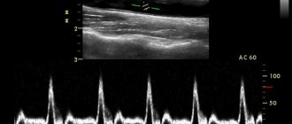
Pencil transducers, also called CW Doppler probes, are used to measure blood flow as well as the speed of sound in the blood.
This probe is small in size and uses a low frequency (typically 2 MHz–8 MHz).
Endocavital ultrasound sensor
In addition, there is an endocavitary type of ultrasonic sensor.
These probes give you the ability to perform internal examinations on the patient.
Therefore, they are designed to be installed in specific housing openings.
Endocavital ultrasound sensor
Endocavital transducers include endovaginal, endorectal and endocavital transducers.
As a rule, they have small traces and their frequency ranges from 3.5 MHz to 11.5 MHz.
Transesophageal ultrasound probe
In addition, there is a transesophageal (TEE) tube.
Like the previously mentioned probes, it is small in size and is used for internal examinations.
It is often used in cardiology to obtain high-quality ultrasound images of the heart through the esophagus.
The frequency is average, in the range of 3 MHz-10 MHz.