EEG is one of the main methods of testing the central nervous system. The use of computer processing allows neurophysiologists at the Yusupov Hospital to diagnose paroxysmal activity and gross changes in brain function, identify initial changes in brain function, determine the location of the focus of pathological activity in epilepsy, and accurately determine the area of brain damage. The principle of the method is based on recording and studying electrical potentials that arise during brain activity. In the presence of pathological processes in the nervous tissue, brain waves are changed or distorted.
EEG studies at the Yusupov Hospital are carried out using modern diagnostic equipment in accordance with the international protocol. This guarantees compliance with international quality standards. EEG analysis and interpretation of research results using a computer program are carried out by candidates of medical sciences, neurologists and neurophysiologists. If the EEG results are interpreted ambiguously, they are discussed at a meeting of the expert council with the participation of professors and doctors of the highest category. Doctors collectively establish a diagnosis based on the existing symptoms and EEG data and draw up a treatment plan. Complex therapy for the pathology of the central nervous system in the neurology clinic is carried out with effective and safe drugs that have been registered in the Russian Federation.
All conditions have been created for the treatment of patients at the neurology clinic:
- chambers of European comfort level;
- provision of individual personal hygiene products and dietary nutrition;
- Friendly service by staff with a high level of professional training.
Indications for EEG examination
If, according to the results of an EEG study, the patient does not need drug correction, neurologists observe him over time, perform repeated studies, conduct EEG video monitoring of day and night sleep, and use other neuroimaging methods.
An electroencephalogram provides the ability to:
- assess the nature and degree of brain dysfunction;
- determine the side and location of the pathological focus;
- study the changes in sleep and wakefulness;
- clarify the results of other types of diagnostics (computed tomography), when the patient has signs of damage to the central nervous system, and other research methods do not reveal a structural defect;
- monitor the effectiveness of medications;
- determine in which parts of the brain epileptic seizures begin;
- assess how the brain works between seizures;
- establish the causes of fainting, panic attacks, crises.
Neurologists at the Yusupov Hospital use EEG studies to distinguish a true visual or hearing disorder from a hysterical one, as well as from a simulation of such a condition. An EEG is performed if the following indications are present:
- epilepsy;
- insomnia, impaired sleep quality;
- sleep disorder (walking, talking in your sleep);
- night sleep apnea;
- seizures without an established cause;
- endocrine diseases;
- vascular pathology of the brain;
- traumatic brain injuries;
- inflammatory diseases of the central nervous system.
For frequent headaches, vegetative-vascular dystonia, and dizziness, an EEG is also performed. The study is indicated for patients who constantly feel tired, have had a stroke or mini-stroke, or have undergone neurosurgical surgery. The procedure is necessary for patients who have had more than one episode of fainting. An EEG is done for panic attacks and brain damage that developed before or after childbirth. An electroencephalogram helps to identify brain dysfunction in cases of delayed speech development, stuttering, and autism.
Diseases detected by EEG
But why is EEG included in the list of mandatory medical examinations when receiving a medical certificate? As already mentioned, EEG can indicate numerous pathologies. First of all, of a neurological nature. And the main place here is occupied by epilepsy. Since epilepsy is a common medical reason for many types of work: with large construction or industrial equipment, with high-tech equipment, with potentially dangerous technologies (for example, working as a nuclear reactor operator). The potential danger is that a person may have an epileptic attack while working, and this will lead to an emergency situation.
Most people have undergone an EEG at least once in their lives. And everyone remembers how you had to look at the rapidly blinking light bulb. This is a kind of test for epilepsy. In epileptics, exposure to flickering light can lead, for example, to generalized bilaterally synchronous arrhythmic peak-wave complexes. On the graph of electrical rhythms, this is clearly visible even to a non-specialist: a relatively uniform curve turns into a spasmodic one. True, there are types of epilepsy that are not associated with a reaction to flickering light. However, EEG still turns out to be a fairly highly accurate method for identifying this pathology.
In addition to epilepsy, EEG can reveal:
- various sleep pathologies (numerous types of insomnia - chronic and situational);
- inflammation of brain tissue (meningitis and encephalitis);
- chronic mental disorders - depression, neuroses, OCD, panic attacks, etc.
In this regard, an EEG can also become part of a psychiatric examination of a patient.
Contraindications and preparation for EEG
There are no absolute contraindications for performing electroencephalography. If a patient has seizures, suffers from coronary heart disease, arterial hypertension, or suffers from mental disorders, an anesthesiologist is present during the EEG procedure in the neurology clinic of the Yusupov Hospital. He provides emergency assistance in case of unusual situations.
Preparing for an EEG is quite simple. On the eve of the procedure, you must decide with your doctor whether or not to cancel the planned medication intake. 12 hours before the EEG study, it is recommended that the patient stop taking products that contain caffeine (coffee, tea, chocolate, cola) and energy drinks. The head should be thoroughly washed with shampoo, without applying any cosmetics (conditioners, varnishes, oils, masks) to the hair after washing, as this will not ensure sufficient contact of the electrodes with the scalp. No need to do your hair. Before the procedure, metal jewelry (earrings, piercings) should be removed.
You need to eat 2 hours before the procedure. The EEG is performed in a calm state, so you should not worry or be nervous during the study. If the doctor needs to detect seizure activity in the brain, he will ask the patient to get some sleep before the test. It is not recommended to get to a medical facility while driving. EEG is not performed on patients with signs of acute respiratory viral infection. The study is not contraindicated for children and pregnant women. During pregnancy, EEG is performed without functional tests.
Rules for patients when conducting video-EEG monitoring
- No special preparation is required before conducting overnight video-EEG monitoring.
- Before conducting daytime video-EEG monitoring, to facilitate falling asleep during the study, the patient is recommended to reduce the time of night sleep on the eve of the study.
In some cases, your doctor may recommend sleep deprivation before your daytime test. Sleep deprivation involves reducing nighttime sleep to 3-4 hours. For example, you go to bed at one in the morning, get up at four in the morning and do not go to bed until video-EEG monitoring is carried out. Sleep deprivation is carried out with the aim of provoking pathological (epileptiform) activity on the electroencephalogram. Since lack of sleep can provoke an epileptic attack, sleep deprivation can only be carried out on the recommendation of the attending physician!
- In some cases, the attending physician may recommend that you change the regimen of taking antiepileptic drugs before conducting video-EEG monitoring (reducing the dose, skipping a single, daily dose, etc.). This is done with the aim of provoking pathological (epileptiform) activity on the electroencephalogram. In the absence of such instructions from your attending physician, the regimen for taking antiepileptic drugs remains the same.
- In order to provoke pathological (epileptiform) activity on the electroencephalogram, the research protocol included provocative tests - a hyperventilation test (deep rapid breathing) and a photostimulation test (flickering light). Carrying out provocative tests can provoke an epileptic attack. Therefore, you have the right to refuse testing. But in this case, the information content of the study is reduced.
- It is not recommended to take psychotropic, sedative and hypnotic drugs for two days before video-EEG monitoring. Taking such drugs reduces the effectiveness of the study. If you take these medications regularly, discuss with your doctor the possibility of temporarily stopping them two days before the test.
- You should come to video-EEG monitoring with a clear head. It is not recommended to use gel, varnish, balm, wax and other cosmetics for hair styling.
- Tell the researcher in advance about the factors that provoke your seizures (flickering lights, working at a computer, fear, etc.)
- If during the study you feel the approach or onset of an attack, immediately notify the researcher.
- If during the study you have an attack that is invisible to others, immediately notify the researcher.
- A patient of any age can be in the study room with one accompanying person. Accompanying persons should not interfere with the research (obstruct the view of video cameras, etc.).
- During the study, you can lead a normal lifestyle within the ward in which the study is being conducted (in the field of view of video cameras): read, watch TV, work on a laptop, play board games, eat (bring food and drinks with you).
- During the study, you should not cover yourself with a blanket, as this may interfere with video recording of the attack.
- During video-EEG monitoring, try not to make sudden movements, as they cause interference (artifacts) on the electroencephalogram.
- You cannot remove or adjust the cap with electrodes yourself.
How is an EEG of the brain performed?
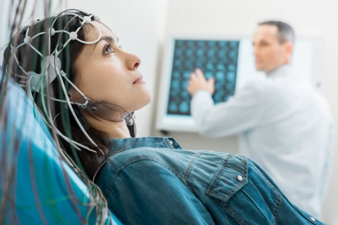
An EEG is usually performed during the daytime. In some cases, a sleep EEG is more informative. The patient goes to the neurology clinic in a special room that is isolated from light and sound. A functional diagnostics doctor puts a special cap with electrodes on his head. The subject sits in a comfortable chair or lies down on a couch. He is the only one left in the room. Communication with doctors is maintained using a camera and microphone.
The neurophysiologist asks the patient to close and open his eyes several times. This allows us to evaluate artifacts that appear on the EEG during blinking. The eyes remain closed during the diagnostic procedure. If at any point during the EEG study a person needs to change position or visit the toilet, he informs the functional diagnostics doctor about this, and the procedure is suspended.
The presence of modern equipment, compliance with the parameters of the international protocol, compliance with world standards, a high level of training and experience of specialists allows us to conduct high-quality EEG at the Yusupov Hospital. EEG is a reliable way to diagnose pathology of the central nervous system. It is absolutely harmless and safe, does not require external intervention, and does not cause discomfort or pain. In order to do an EEG, make an appointment with a neurologist-neurophysiologist by calling the Yusupov Hospital. Contact center specialists will answer all your questions; the clinic is open daily, 24 hours a day.
What is cerebral electroencephalography (EEG)?
Bioelectric activity of the brain can be recorded using electrodes placed on the surface of the patient's head. This study was called electroencephalography, abbreviated EEG (electro- + Greek encephalos - brain + Greek grapho - writing).
The set of visual curves (patterns or patterns) obtained during an electroencephalographic study is called an electroencephalogram (EEG)
. This diagnostic neurophysiological technique makes it possible to understand whether the brain is functioning normally or whether it has paroxysmal and/or pathological activity.
Electroencephalography is used as an additional diagnostic method for a wide range of diseases of the central nervous system, endocrine pathology, and even for diseases of internal organs (for example, for differential diagnostic purposes, for bronchial asthma and diabetes mellitus).
This method is widely used when conducting initial and periodic medical examinations in certain types of professions associated with a high risk of occupational injuries (aviation, railway transport, road transport, etc.).
For epilepsy and epileptic syndromes, EEG is
the main method of functional diagnosis.