Histological examination of neoplasms is an analysis of tissue taken from the patient under a microscope. This procedure allows you to make the most accurate diagnosis if the disease is associated with the occurrence of neoplasms.
Consequently, the effectiveness of the selected treatment depends on the correct diagnosis. For example, a visually ordinary mole does not cause concern, but it can become malignant at any time, therefore, in order to determine the type of mole and understand what to do with it next, a histological examination is prescribed.
Purpose of prescribing histological examination
Content:
- Purpose of prescribing histological examination
- Indications and contraindications for the procedure
- Algorithm for histology of neoplasms
- What can the analysis show?
Histological examination of neoplasms is prescribed to analyze the dynamics of the growth of a malignant tumor, changes in tissues that occurred after therapy (the effectiveness of treatment is checked), establishing indications for radical surgery, differentiating malignant and benign neoplasms, the presence of a tumor in the early stages of its development, staging the maximum accurate diagnosis.
If the results of the study demonstrate the presence of a pathological process, the doctor selects an effective treatment package in accordance with the diagnosis. It is important to remember that timely diagnosis is the key to successful and short-term treatment, which is possible without surgical intervention.
Two methods are used for research: studying tissues that were obtained during surgery and biopsy (targeted excision of a small piece of tissue for research).
It is important to understand that any wart can be cancer, and a mole can be a malignant tumor, and only histological diagnosis will determine the type of tumor.
Can histological examination be erroneous?
People who receive an examination report indicating the presence of a cancerous tumor dream that there is an error in it. It's a pity, but there are never any mistakes in such research. If this happens, it is very rare.
This method is considered the most accurate, and therefore it allows you to detect cancer cells and the reasons why they arose. Despite the fact that the method is the most accurate, doctors say that there is a small percentage of erroneous, incorrect results. But if the sample collection and examination process are followed correctly, such errors are simply excluded.
Indications and contraindications for the procedure
Among the contraindications it is worth noting:
- the presence of blood diseases (including poor blood clotting);
- diabetes;
- acute viral and infectious diseases;
- serious central nervous system disorders (congenital or acquired);
- stage 4 cancer.
It is also necessary to tell your doctor about the presence of drug treatment and allergies to anesthesia.
This type of diagnosis is prescribed if the presence of a tumor has already been established and it is necessary to determine its type and stage, if cosmetic removal of tumors on the skin is necessary, as well as to determine the etiology of tumors, etc. In other words, histological examination of neoplasms makes it possible to establish the diagnosis as accurately as possible. An analysis is also prescribed if there is a suspicion of polyposis. Polyps can form in the body of the uterus, small and large intestines, stomach, nose, and other places where there are mucous membranes.
Nervous tissue.
Nervous tissue consists of highly specialized cells - neurons, concentrated mainly in the gray matter of the brain and spinal cord. The long process of a neuron (axon) extends long distances from the place where the nerve cell body containing the nucleus is located. The axons of many neurons form bundles that we call nerves. Dendrites also extend from neurons - shorter processes, usually numerous and branched. Many axons are covered with a special myelin sheath, which consists of Schwann cells containing fat-like material. Adjacent Schwann cells are separated by small gaps called nodes of Ranvier; they form characteristic grooves on the axon. Nerve tissue is surrounded by a special type of supporting tissue known as neuroglia.
Algorithm for histology of neoplasms
The first thing you need to do is consult a doctor (gynecologist, gastroenterologist, ENT specialist, therapist, dermatologist, surgeon) - the choice of doctor depends on the symptoms. The doctor collects the necessary biological material (aspiration, suction, puncture, collection of secretions from the tumor, mucosal smear).
The resulting biological material is placed in formaldehyde (the solution in volume should be ten times the volume of the material being tested).
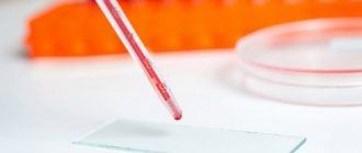
The jar with the formaldehyde solution is marked, indicating the patient's data, the date and area of collection of the histological sample. The doctor issues a referral for histology, in which he indicates all the necessary data, including bad habits. On the day of tissue collection, after all documentation has been completed, the excised tissues in formaldehyde solution are transferred to the pathologist. The doctor first stains the obtained samples (special dyes are used), then the process of complete dehydration occurs. After dehydration, the samples are placed in paraffin. The cube of the resulting paraffin is cut into thin slices, which are then placed on glass and examined by a specialist under a powerful microscope.
If no errors were made during the collection of tissues and cells, as well as in the histology algorithm, the results are as accurate as possible and allow you to diagnose the type and stage of the tumor. Taking materials for research will in no way speed up or stop the development of the tumor. A great danger to the human body is the constant traumatization of the neoplasm, as well as late diagnosis.
The average duration of histology of neoplasms after tissue sampling is three days. The doctor may also prescribe an emergency diagnostic test. It is usually prescribed during the operation.
Best materials of the month
- Coronaviruses: SARS-CoV-2 (COVID-19)
- Antibiotics for the prevention and treatment of COVID-19: how effective are they?
- The most common "office" diseases
- Does vodka kill coronavirus?
- How to stay alive on our roads?
Often the outcome of the operation will depend on the result obtained. Emergency diagnostics do not involve drying samples; they are simply frozen. The patient can receive the results in the form of a conclusion, or they will be transferred directly to the attending physician.
In any case, the attending doctor will decipher the conclusion in order to decide on further actions. Often, after receiving the results, the patient is referred for additional studies. The doctor can also refer you to other specialists in this field.
Histology analysis: interpretation
The conclusion (deciphering the histology analysis) indicates all the results of the histology analysis, but only specialists can correctly decipher the medical terms. By contacting a doctor, the patient can receive complete information about their health status, and if dangerous diseases are detected, receive qualified assistance.
How much does it cost to decipher a histology analysis?
Of course, the price of deciphering histological analysis depends on the clinic where you conduct it. If you want to get a high-quality second opinion from an oncologist, then you need to pick up the blocks with histological material and bring them or send them to another clinic. If a repeated conclusion and interpretation of the histology analysis is carried out without histological material, this means that this is not a “second opinion” or a “histology revision”, but a confirmation of the diagnosis of one doctor by another doctor.
When it comes to such an important area of medicine as oncology, deciphering the histology analysis can significantly improve the course of treatment, and sometimes even save the patient’s life.
Our company BK Medical Logistic offers a complete revision and interpretation of histology analysis after a special study in an Israeli clinic specialized in oncology and oncological diseases, with high-tech laboratories and competent doctors known all over the world.
The price of deciphering a histology analysis may vary, but on average it is 550-650 US dollars.
How is repeated histology analysis performed?
At the moment, only our company can deliver histological material from Ukraine or Russia to Israel for medical examination. We use special refrigerators and packaging to ensure that the results of the histology analysis reach the clinic in Israel safe and sound.
After the blocks are delivered to Israel, the histology tests are re-checked and studied in the laboratory, after which the doctor prepares a special report with the results and a transcript of the histology.
How to apply for a repeat histology analysis?
Even if you just want to ask questions and get information about the revision of histology tests, you can contact us by phone listed at the top of the site. CALLS AND CONSULTATION ON THIS NUMBER IS FREE!
What can the analysis show?
Histology analysis is not prescribed in all cases. Histology of neoplasms is necessary to determine the inflammatory process in the gastrointestinal tract, to diagnose pathological changes in the female genital organs, to determine the causes of infertility, and to diagnose cancer.
In rare cases, analysis shows false results; according to statistics, 98% of histology shows true results, which makes it possible to establish a diagnosis and determine further actions.
More fresh and relevant information about health on our Telegram channel. Subscribe: https://t.me/foodandhealthru
We will be grateful if you use the buttons:
Mechanical damage.
In case of mechanical damage (cut or fracture), the tissue reaction is aimed at filling the resulting gap and reuniting the edges of the wound. Poorly differentiated tissue elements, in particular fibroblasts, rush to the site of rupture. Sometimes the wound is so large that the surgeon must insert pieces of tissue into it to stimulate the initial stages of the healing process; For this purpose, fragments or even whole pieces of bone obtained during amputation and stored in a “bone bank” are used. In cases where the skin surrounding a large wound (for example, with burns) cannot provide healing, transplants of healthy skin flaps taken from other parts of the body are resorted to. In some cases, such transplants do not take root, since the transplanted tissue does not always manage to form contact with those parts of the body to which it is transferred, and it dies or is rejected by the recipient.
Foreign objects.
A very characteristic reaction occurs in response to the penetration of foreign objects into the tissue. If, for example, a bullet hits a part of the body that is not of vital importance, it soon becomes walled off from adjacent tissue by a deposit of fibrous tissue that forms around it. A similar reaction occurs when certain parasites penetrate tissue. So, trichinella ( Trichinella spiralis
), entering the human body with poorly cooked contaminated pork, penetrates the muscles, where the fibrous tissue forms a capsule around it. In the lungs of tuberculosis patients, tubercles consisting of concentric layers of tissue form around accumulations of pathogenic bacteria. In these and other cases, the body's tissues try to create a barrier between the foreign body, whether living or non-living, and the body's own tissues.
Where to get training
In addition to higher education, there are a number of short-term training on the market, usually lasting from a week to a year.
The Medical University of Innovation and Development invites you to take distance courses for retraining or advanced training in the field of “Histology” to obtain a diploma or state certificate. Training lasts from 16 to 2700 hours, depending on the program and your level of training.
The Institute of Professional Education "IPO" invites you to take distance courses in the field of "Histology" (there are options of 256, 512 and 1024 academic hours) to receive a diploma or state-issued certificate. We have trained more than 8,000 graduates from almost 200 cities. You can undergo external training and receive interest-free installments.
Differences between the studies
If we talk about the named scientific directions, the difference between them will be immediately clear:
- Histology is a scientific field that deals with the analysis of tissue structure;
- Cytology is a scientific field whose subject of study is cells.
Cytology and histology have certain differences in terms of analysis.
The first is a method for assessing the characteristics of the cell elements of the biomaterial under study. The method is applicable to detect tumors and non-tumor pathologies.
The second is a laboratory diagnostic method, the subject of research of which is biopsy samples, that is, tissue elements taken during manipulation.
Both directions are important from a purely practical point of view: they give the doctor a picture for making a diagnosis, preliminary or final, for various pathologies. They are also widely used in oncology.
Excisional and incisional biopsies
Excisional biopsy
– a method of taking pathological material by completely removing an organ or tumor during a surgical operation. It is the largest of all types of biopsy and, in addition to diagnostic purposes, is often performed for treatment purposes.
Incisional biopsy
– a method of taking pathological material by removing only the affected area of an organ or part of a neoplasm. It is also performed during surgery, but usually serves only diagnostic purposes.
Cytology
It is carried out to evaluate the morphological specifications of the elements of tissue cells of various organs, as well as fluids and various types of body secretions.
The procedure helps doctors understand the essence of dysplastic and malignant transformations of specific cells and, based on the collected information, make a preliminary diagnosis.
The method has its advantages:
- It is available;
- Economical for the patient;
- Does not require complex expensive equipment;
- The duration is 60 minutes;
- Non-invasive or minimally invasive (here it all depends on the method of sampling the material chosen by the doctor).
Agree, the listed advantages explain the wide popularity of the study; it is readily used, for example, as part of the initial examination of various diseases.
The specifics of the material removal became the basis for the following classification of the study:
- Preoperative;
- Intraoperative.
The first has the following types:
- Exfoliative. Here, the object of analysis is the discharge of the genitourinary organs, sputum, sweat, urine, etc. This type has one significant drawback - the safety of the material leaves much to be desired.
- Abrasive. The object of study is scrapings from areas of internal organs. To remove the material, the doctor uses special tools. This approach allows the material to be well preserved, which means the result will be clearer.
- Aspiration. A fine-needle biopsy technique is used. This gives the doctor the widest possible access to any organ. This method has found widespread use in the diagnosis of breast tumors at an early stage of their development.
Cytological analysis alone, with all its undeniable advantages, is not enough to perform a final analysis. Using the method, the doctor can determine the presence of a malignant neoplasm, nothing more. There are modern techniques, thanks to which a preliminary diagnosis is made more accurately and, based on it, the doctor can plan treatment tactics.
A few words must be said about cytological examination in gynecology. Here it manifests itself as a highly informative, absolutely safe method that gives the doctor the necessary picture to begin treatment in the presence of an identified pathology.
Cytology of a cervical smear is a necessary and mandatory measure in terms of diagnosing diseases of a woman’s genital area.
The procedure itself does not cause any inconvenience or anxiety to the patient; it is painless and does not take a long time.
Core biopsy
Core biopsy
(from the English
core
- core), which is also called
core
or
cutting biopsy
- a method of taking pathological material in the form of a tissue column using a special needle similar to a harpoon and a biopsy gun. Unlike a punch biopsy, which allows you to take material from the surface of the body, a core biopsy makes it possible to take full histological samples from deep-lying tissues and internal organs. In this case, the diameter of the needle is the same as for cytopuncture, which allows for minimal trauma to the animal.
The principle of the method is that the gun shoots the harpoon part of the needle into a given area at high speed. The harpoon has a groove in which a sample of soft tissue is fixed. After this, the frayed part of the needle is fired at the same high speed, which with its cutting edge cuts off the tissue column located in the harpoon. Thus, it is possible to obtain complete tissue of a tumor or internal organ for subsequent histological examination.
To ensure the accuracy and correctness of the biopsy, it is performed under the control of an ultrasound machine.
Punch biopsy
Punch biopsy
(from the English
punch
- hole puncher, composter) - a method of taking pathological material, mainly skin, using a special tubular scalpel - punch. This method allows you to obtain a cylindrical sample of fabric of a given height and diameter.
The choice of biopsy site depends on the type of lesion. Therefore, before taking material, it is better to consult a morphologist.
When taking the material, the punch is directed perpendicular to the surface of the skin, stretching the skin to the sides. At the same time, lightly press the punch from top to bottom and rotate it clockwise and counterclockwise until it is immersed to the required depth. Afterwards the punch is carefully removed. Using a needle, the biopsy is removed from the scalpel cavity and, using sharp scissors or a blade, the lower part of the sample is separated from the patient's body. If necessary, stitches and a bactericidal patch are applied to the resulting wound.
The sooner a skin lesion is biopsied, the better. The optimal time to take an inflammatory element for biopsy is 24-36 hours.
Before fixing the taken material, make a neat imprint on the glass slide, placing the selected biopsy specimen with its side to the surface of the glass. Under no circumstances make smearing movements on the glass, so as not to cause crushing of the cells and not to smear the fat cells of the subcutaneous tissue on the surface of the glass, which will prevent the adhesion of other cells to the slide.