child development during pregnancy
Obstetric ultrasound, first trimester (from 3 to 9 weeks)
Baby Fetal development at 9 weeks of gestation The small head remains disproportionately large
Prenatal screening of the first trimester (11-13 weeks) (venous blood) in Moscow
KTR of the fetus by week of pregnancy: table When a woman registers for pregnancy, the doctor
Ultrasound of early pregnancy in St. Petersburg
Ultrasound during pregnancy: how often can it be done, is it harmful, explanation
Ultrasound during pregnancy Ultrasound during pregnancy is a planned diagnostic procedure that needs to be done
Clinical and morphological approach to the diagnosis of breast cancer metastases to the lungs
In medical terminology, metastases are secondary foci of growth (dropouts) of malignant tumors. Viable cancer cells
Types of Doppler examination: wave Doppler, color Doppler mapping, tissue Doppler. Concept, diagnostic information received.
Ultrasound diagnostics is one of the safest and most effective ways to detect various diseases.
embryo in egg
Fertilization and development of the fertilized egg: stages, structure and possible anomalies
What is a fertilized egg? A fertilized egg consists of embryonic membranes and an embryo. This period
Heel spur
X-ray of the calcaneus: indications for the procedure, how is it done?
Heel spur is a disease characterized by inflammation of the plantar fascia, resulting in the accumulation of osteophytes in
Corpus luteum cyst of the ovary: symptoms, diagnosis and treatment in Moscow
Corpus luteum cyst of the ovary (luteal cyst) is a benign neoplasm that matures against the background of non-regressed
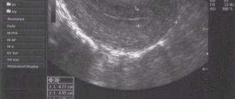
Ultrasound: normal uterus
Ultrasound of the uterus and appendages: how they do it, interpretation of the result and normal indicators
How is an ultrasound of the uterus and appendages performed? Ultrasound of the uterus and appendages does not cause pain
The structure of the liver and its location in the body
Ultrasound of the liver - description of the technique and clinical case
In short: Do an ultrasound of the liver once a year to notice possible abnormalities in time. Before the procedure
Для любых предложений по сайту: [email protected]